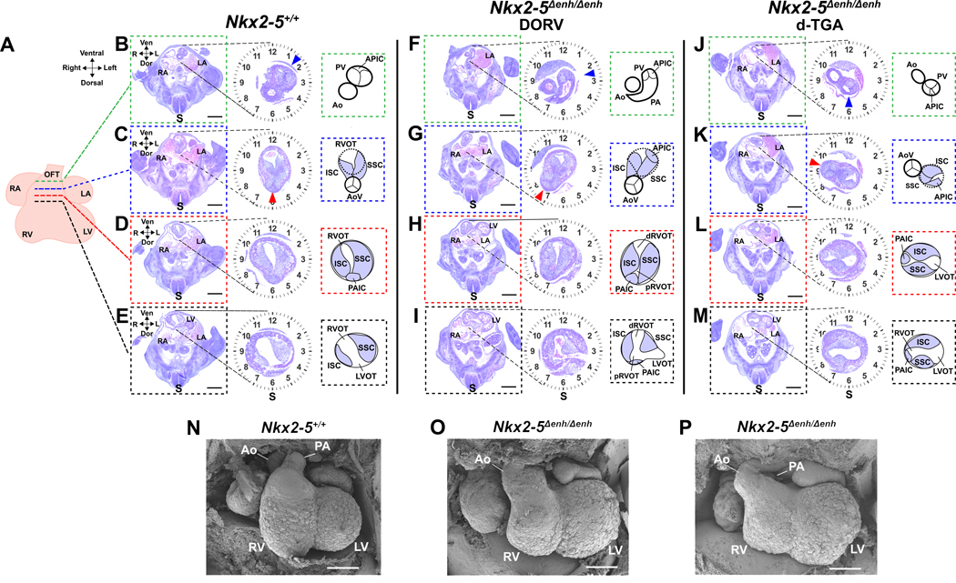
Figure 3. Histological and electron micrographic assessment of Nkx2–5∆enh/∆enh embryonic hearts.
(A) Schematic of an embryonic day E11.5 heart with corresponding sections through the outflow tract (OFT) of Nkx2–5+/+ (B−E) and Nkx2–5∆enh/∆enh (F−I and J−M) hearts at select levels: distal level (green hashed lines)(B, F, J), aortic valve (AoV) level (blue hashed lines)(C, G, K), sub-AoV level (red hashed lines)(D, H, L), and proximal level (black hashed lines)(E, I, M). Magnified view of the OFT for each section is shown with a clockface demarcating the spinal cord (S) axis at 6 o’clock. Schematic diagrams show labeled anatomical structures of the OFT to the right of each magnified section. In the Nkx2–5+/+ heart (B−E), the superior and inferior septal cushions (SSC and ISC) are oriented along the axis of the spinal cord so that the posterior aortic intercalated cushion (PAIC)(panel D), which becomes the non-coronary cusp of the AoV (panel C, red arrowhead), is situated at 6 o’clock in the OFT. This allows for the AoV to connect with the left ventricular outflow tract (LVOT) below (panel E). More distally, the aorta (Ao) remains in the 6 o’ clock position with the right ventricular outflow tract (RVOT) looping around the aorta clockwise, ending with pulmonary valve (PV) (panel B, blue arrowhead) positioned at 1:30 on the clockface. Two Nkx2–5∆enh/∆enh mutant hearts are shown with different severities of rotation defects. The first mutant (F−I) shows insufficient counterclockwise rotation so that axis of the SSC, ISC, and PAIC (non-coronary cusp) are shifted rightward by 45 degrees to 7:30 on the clockface (panel G, red arrowhead), displacing the AoV off the LVOT and onto the proximal RVOT (pRVOT)(panel I). The anterior pulmonary intercalated cushion (APIC), which becomes the anterior valve (AV) of the PV, is also shifted clockwise but remains attached to the distal RVOT (dRVOT)(panel F, blue arrowhead). This results in double-outlet right ventricle (DORV) phenotype where the Ao is laterally displaced to the right and lies anteriorly with the pulmonary artery (PA) (panel F). The second mutant (J−M) shows more severe insufficient counterclockwise rotation so that SSC, ISC, and PAIC (non-coronary cusp) are shifted rightward by 105 degrees to 9:30 on the clockface (panel K, red arrowhead), allowing for connection of the AoV to the RVOT. The APIC and PV now sit at the 6 o’clock position (panel J and K, blue arrowhead), connecting the PV to the LVOT. This results in dextro-transposition of great arteries (d-TGA) phenotype where the Ao is anterolaterally displaced relative to the PA (panel J). Scale bars: 500 µm. RA, right atrium; LA, left atrium; RV, right ventricle; LV, left ventricle; Ven, ventral; Dor, dorsal. (N–P) Scanned electron microscopic images of embryonic hearts at E12.5 in Nkx2–5∆enh/∆enh and wildtype littermates. Nkx2–5∆enh/∆enh hearts display rotational defects of the OFT. Scale bars: 200 µm.

