



Abstract
Background/Aim
The multiparametric magnetic resonance imaging (mpMRI)–ultrasound (US) fusion targeted biopsy (TB) is a useful diagnostic device for men with suspected prostate cancer (PC) and can increase the detection rate for clinically significant PCs (csPC). However, few studies have shown pathological findings of undetectable csPCs on the prostate mpMRI.
Patients and Methods
This study investigated the growth patterns of csPC undetected in prostate mpMRI. The study enrolled 248 patients with suspected PCs and ≥PI-RADS 2 lesions, who then underwent mpMRI–US fusion TB and nearly prostate-mapping systematic biopsies (SB). A total 248 biopsies included 404 regions of interest in TB and 2976 mapping-regions in SB.
Results
The detection rates of csPC, defined as PC grade group (GG) ≥2, were 42% in TB and 44% in SB, and the highest detection rate was 50%, using both TB and SB. Approximately 79% of PI-RADS 3/4/5 with any PC showed csPC. A total 201 PI-RADS 3/4/5 lesions showed benign prostatic hyperplasia, lymphocytic prostatitis, or fibromuscular stroma only in the core tissues. Notably, 22 csPCs detected in SB but undetected in prostate mpMRI preferentially showed a pattern of mixed well-formed and fused PC glands. The other patterns including cribriform glands and poorly formed glands with intracytoplasmic vacuoles were also seen. Approximately 85% of the 22 csPCs showed tumor volume less than 50% of core tissues.
Conclusion
Changes in prostatic stroma amounts, inflammation severity, tumor volume and growth patterns of PC glands affected the detectability of prostate mpMRI.
Keywords: Multiparametric MRI, ultrasound, fusion, biopsy, pathology
The prevalence of prostate cancer (PC) is increasing worldwide, including Taiwan (1,2). PC manifestations include indolent cancers and aggressive metastatic potential diseases, which are defined as very low-risk to very high-risk disease categories (3). Routine PC screening includes serial serum levels of prostate-specific antigen, digital rectal examination, and systematic transrectal ultrasonography-guided (TRUS) random biopsy (4).
The International Society of Urological Pathology (ISUP) and the World Health Organization (WHO) introduced a new grade grouping system for prostate adenocarcinoma in 2014, stratified into grade groups (GGs) 1 to 5 based on Gleason scores, to improve the correlation between histological grading and clinical outcomes (4). GG 1 (Gleason score 3+3=6) indicates an indolent disease with no significant difference in mortality rates between patients who underwent surgery and those under observation only (5-7). Epstein et al. (2016) defined clinically significant PC (csPC) as GG2 or higher (Gleason scores ≥3+4). However, the accuracy of TRUS random biopsy is limited due to the presence of multifocal PCs with little clinical significance and the small size of the csPC, which makes it difficult to detect (8).
Some studies have provided evidence suggesting that prostate multiparametric magnetic resonance imaging (mpMRI) is a useful diagnostic tool for the detection, localization, and size measurement of PC (9). Targeted biopsy (TB) is performed under the guidance of the mpMRI, including T2-weighted imaging (T2WI), diffusion-weighted imaging (DWI), and dynamic contrast-enhanced (DCE) imaging, to reduce the misleading problems of TRUS random biopsy (10). The mpMRI report is based on the Prostate Imaging Reporting and Data System (PI-RADS) with scores 1-5 (11,12), associated with PC GG and Gleason scores (13).
The imaging fusion of mpMRI and ultrasound (US) is better than either mpMRI or US at detecting and localizing PC. However, benign histological findings of TB are sometimes found in men with suspicious lesions on mpMRI (9,14-17). Accordingly, histological differences in PC exhibit one of the confounding factors that affect the detectability of csPC. For the past 4 years, we correlated pathological results of TB by the mpMRI–US fusion biopsy at regions of interest (ROI) with SB by the US-guided nearly prostate-mapping SB at 12 regions in 248 patients, where 22 cases showed csPC-negative TB but csPC-positive SB. This study examined the association of PC GG with the PI-RADS score and found undetectable csPC lesions on the mpMRI. The recognition of undetectable PCs may improve the detection power of the mpMRI.
Patients and Methods
Subjects. In this prospective observational study, we collected data on a total of 248 patients with suspected PC from June 2019 to February 2023 at the China Medical University Hospital, a tertiary referral medical center in Taiwan. This study was approved by the Research Ethics Committee of China Medical University Hospital, Taichung, Taiwan (protocol number: CMUH109-REC1-045).
The inclusion criteria involved men with a serum prostate-specific antigen (PSA) level ≥3 ng/ml or an abnormal digital rectal examination. The ROI was target biopsied at PI-RADS score ≥2 using a 3T mpMRI, and 12 regions were systematically biopsied based on the Ginsburg protocol (18) to get a prostate map for SB. The 12 systematic regions for mapping the prostate included the anterior medial, anterior lateral, middle medial, middle lateral, posterior medial and posterior lateral regions of the prostate bilateral lobes. However, men with a history of PC graded or not by the Gleason score, bacterial prostatitis within 3 months, or an inability to sign informed consent were excluded from this study.
mpMRI–US fusion biopsy protocol. All mpMRI scans were performed using a 3T scanner (Signa HDxt, GE Healthcare, Milwaukee, WI, USA) with an eight-channel high-resolution cardiac array coil. However, no endorectal coil was used. The scanning protocol was performed as described previously (19,20), and images were interpreted by two radiologists. The T2WI was used for contouring the prostate and targeted lesions, and then a 3-D model of the prostate targeted lesions was built using a BioJet (D&K Technologies GmbH, Barum, Germany) or bkFusion (BK Medical, Herlev, Denmark) system. Briefly, three T2WI, DWI, and DCE images showed the ROIs, and each was scored based on the PI-RADS category.
All TBs and SBs were performed by a urologist (Po-Fan Hsieh), and the detailed operation procedure was performed as described previously (19,20). Briefly, a transrectal probe (BK 8848, BK Medical, Peabody, MA, USA) was used to obtain an ultrasound (US) scan of the prostate. The segmented 3T mpMRI images were then overlaid on the real-time US images on the fusion platform using a rigid or elastic registration. The biopsy specimens were obtained using an 18G biopsy gun with a 22-mm specimen size (Bard Magnum, Bard Medical, Covington, KY, USA). At least two cores were sampled for each TB ROI, and at least 12 cores were sampled for each SB ROI.
Histology evaluation. All core tissues of TB and SB were sliced and stained with routine hematoxylin & eosin (H&E) reagents. Two pathologists (Kai-Po Chang and Han Chang) interpreted and reviewed all H&E slides. PC was graded in accordance with the 2014 International Society of Urological Pathology Consensus Conference guidelines (4). The PC GG of each ROI and the composite GG of each SB were recorded. The specimen length of each biopsy core and the length and percentage of PC involvement were measured. A multidisciplinary team meeting with the pathologists, urologists, and radiologists was held every 2 weeks, reviewing the procedural videos, imaging details on mpMRI, biopsy trajectories recorded on US and mpMRI, as well as histopathology of the PC GG, cell morphology, and growth architecture in each case. Then, we discussed the human factors, histopathological features, and other variables affecting the results. Furthermore, mpMRI-detectable and -undetectable lesions correlated with TB and SB were assessed. Negative and positive TB or SB were defined as the absence and presence of PC or csPC in core tissues, respectively.
Statistical analysis. Continuous variables were reported as means with standard error (SE), and categorical variables were reported as proportions. Pearson chi-square or Spearman correlation were used for correlation analysis. The study population was divided into five cohorts by the year the biopsy was taken (2019, 2020, 2021, 2022, and 2023), and the trend association was assessed using linear-by-linear association testing. The sensitivity, specificity, positive predictive value (PPV), and negative predictive value (NPV) of PI-RADS scores were calculated for detecting csPC using the SB as a referent test. All statistical analyses were performed using SPSS version 22 (IBM Corp., Armonk, NY, USA), assuming a two-sided test of Pearson chi-square and Spearman correlation with a p-Value of 0.05 for statistical significance.
Results
Distribution of 248 Cases with TB and SB. The prostate MRI–US fusion biopsies (n=248) included 404 ROIs of TB and 2976 regions of SB. The positive rate of the ROIs was 43% (172/404), and that of regions was 14% (403/2976), as shown in Table I. Among 404 ROI lesions, 172 ROIs showed PC GG ≥1, of which 137 were csPC. Of the total 248 cases, the detection rate of csPC with composite GG ≥2 using TB showed an increasing trend along with year cohorts, ranging from 3-50% (linear-by-linear association p=0.021, Table I).
Table I. Distribution of 248 prostate mpMRI-US fusion targeted and nearly-prostate-mapping systematic biopsies from June 2019 to February 2023.

mpMRI US, Multiparametric magnetic resonance imaging- ultrasound; TB, mpMRI-US fusion targeted biopsy; SB, nearly-prostate-mapping systematic biopsy; PC: prostatic adenocarcinoma; GG: grade group; ROI, region of interest biopsied by TB; region: biopsied by SB; +: presence of PC; –: absence of PC. #Regardless of TB ROI or/and SB region numbers for any case; *Spearman correlation for PC GG ≥2 of TB vs. SB, p<0.001.
The positive rates of SB showed 47-75% for detecting any PC and 35-69% for detecting csPC when stratified according to yearly cohorts (Table I). Briefly, the prostate mapping regions of SB showed 137 PC GG ≥1, of which 110 were csPC lesions. Twenty-two cases of csPC were undetected by mpMRI, leading to false-negative cases for detecting csPC using TB. Of all 248 cases, the detection rate of csPC was 42% using TB and 44% using SB. The highest detection rate of csPC was 50%, occurring when TB and SB were combined (Table I).
Correlation of PI-RADS Lesions and PC. The PI-RADS score was found to be positively associated with PC GG (Spearman correlation, p<0.001, Table II). When stratified by PI-RADS score, the detection rate of csPC with PI-RADS scores 2, 3, 4, and 5 was 3%, 12%, 35%, and 70%, respectively. Approximately 79% of PI-RADS 3/4/5 with any PC showed csPC, implying that prostate mpMRI was favorable in the detection of the csPC. Only three PI-RADS 2 showed two PC GG 1 and one PC GG 3. The PPVs of PI-RADS scores 2, 3, 4, and 5 were 0%, 75%, 83%, and 90%, respectively (Table II). However, the highest NPV (97%) occurred in the PI-RADS 2 ROI.
Table II. Size and PC GG in 404 ROIs stratified by the PI-RADS score.

*Spearman correlation for PC GG vs. PI-RADS score, p<0.001; #Systematic biopsy for detecting PC GG ≥2 as a reference. PPV, positive predictive value; NPV, negative predictive value.
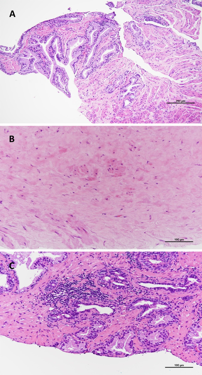
Pathological findings of 22 cases with undetectable PC in prostate mpMRI. Regardless of the PIRADS 3/4/5 with high PPV, 27% of the total 404 mpMRI ROIs showed benign prostate tissues. These included nodular hyperplasia (Figure 1A), fibromuscular stroma only (Figure 1B), and lymphocytic prostatitis (Figure 1C, benign ROI features). Benign nodular hyperplasia was frequently observed in PI-RADS-scored lesions.
Figure 1. Histopathology of Prostate Imaging–Reporting and Data System (PI-RADS) 4/5 lesions in targeted biopsies with absence of prostatic cancers. (A) nodular hyperplasia; (B) fibromuscular stroma only; (C) lymphocytic prostatitis. Sections were stained with hematoxylin and eosin.
As correlated TB and SB, a total of 22 cases showed csPC-negative TB but csPC-positive SB (Table I); that is, they had undetectable PC on the prostate mpMRI. These 22 PC cases revealed PC with 17 GG 2, 16 GG 3, 4 GG 4 and 1 GG 5, in which all of them were csPC (shown in Table III). PC detected in SB disclosed variable PC histopathologies including well- or -poorly formed glands, such as fused, cribriform and glomeruloid patterns, and the intracytoplasmic vacuoles in PC cells arranged singly (Table III and Figure 2). Gleason 4 or 5 patterns were observed in cribriform glands (Figure 2A, 2B), ductal adenocarcinoma (Figure 2C), fused glands (Figure 2D), glomeruloid glands (Figure 2E) and lack of glandular formation (Figure 2F). The most common pattern among these cases was well-formed glands (Gleason pattern 3) mixed with fused glands (Gleason pattern 4). Tumor volume was less than 50% in 85% of 48 core tissues.
Table III. Cases with prostatic adenocarcinomas undetected by the prostate MRI.


PC, Prostatic adenocarcinoma; GG, Grade Group; GS, Gleason score; LT, left; RT, right; ant., anterior; mid., middle; post., posterior; med., medial; lat., lateral.
Figure 2. Pathological findings of undetectable Prostate Imaging–Reporting and Data System (PI-RADS) lesions. (A, B) cribriform glands; (C) ductal adenocarcinoma; (D) fused gland; (E) glomeruloid glands; (F) Gleason pattern 5 tumor, with lack of gland formation. Sections were stained with hematoxylin and eosin.

Discussion
Despite the high detection rates and PPVs of PI-RADS 3/4/5, 22 cases (totally 38 cores) showed mpMRI-undetectable csPC lesions, but csPC was detected by SB in this study. These 22 csPC cases consisted of 16 cores with GG 3, 4 cores with GG 4, and 1 core with GG 5. The core with PC GG 5 showed poorly formed glands with intracytoplasmic vacuoles or arranged singly. Hoffmann and colleagues have demonstrated that high-grade PC (GG ≥3) shows a higher sensitivity than low-grade PC (GG <3) when corresponded to PI-RADS 4 or 5 (21). However, serial reports of high-grade PC were not detected in mpMRI but identified in the radical prostatectomy specimens; accordingly, histological characteristics in these studies included reduced tumor cellularity, increased stromal amount of tumor areas, and increased luminal spaces of PC glands, which affected the reading of PI-RADS categories in mpMRI (15,16,22,23). Our data showed similar findings, i.e. cases with a number of csPC glands lower than that of benign glands and increased prostatic stroma were mpMRI-undetectable. The evidence also supported that tumor volume affected csPC detectability on mpMRI when PC volume was less than two biopsy cores and <50% of any length (24). Similar conditions in this study showed tumor volume less than 50% in 85% of 48 core tissues among 22 mpMRI-undetectable cases. The quantity of Gleason pattern 4, particularly the cribriform pattern, contradicted the detectability of the prostate mpMRI. There was no significant difference in distribution of cribriform glands between mpMRI-detectable and MRI-undetectable PC (15). In contrast, cribriform glands showed lower detectability by mpMRI (22,23). In our data, four of the 22 cases disclosed cribriform glands undetected in mpMRI.
In this study, the growth patterns of the 48 undetectable mpMRI PC cores included clustered PC glands mixed with benign hyperplastic glands or prostatic fibromuscular stroma and non-circumscribed or loosely arranged PC glands. These non-circumscribed and loosely arranged PC glands, which went undetected on mpMRI, usually showed neither a markedly low signal intensity on an ADC map nor a moderate homogenous hypodensity on T2WI because the water of these lesions was not scarce. A possible key to identifying these lesions is referring to the DCE image when PC with neoangiogenesis could cause a faintly focal early enhancement, in contrast to the background of benign hyperplastic glands and stromal components (15). Accordingly, despite its importance, this finding was not addressed in the PI-RADS guideline (25).
Data showed that PC-negative PI-RADS 3/4/5 lesions included nodular hyperplasia, lymphocytic prostatitis, and prostatic stroma with or without hemorrhage. This was similar to a recent study by Yamanaka and colleagues (26), which showed that stromal hyperplasia, basal cell hyperplasia, and inflammation were linked to false-positive findings on mpMRI. As correlated with mpMRI, cases with predominant stroma showed homogenous hypointense areas between nodules that might mimic PC on T2WI. Lymphocytic prostatitis exhibits areas of lymphoid aggregates in the prostate, usually in the non-PC region (26). These aggregated lymphocytes might show water restriction on the DWI/ADC map because lymphocytes have scarce cytoplasm, causing intracellular restricted diffusion and hindering extracellular water diffusion. Further, inflammation focally induced the increase of blood flow, causing the early enhancement on DCE images, leading to difficulty separating lymphocytic prostatitis from PC.
Our data showed PI-RADS scores to be positively associated with PC GGs and detection rates of csPC. PPVs were also positively correlated with the PI-RADS scores, and the PPVs of PI-RADS 3, 4, and 5 were 75%, 83%, and 90%, respectively, for detecting csPC. The results were consistent with earlier meta-analysis research showing a correlation between PC GG and PI-RADS 3, 4, and 5, in PPV of 12%, 48%, and 72%, respectively (14). Studies using PI-RADS lesions on mpMRI mapping the radical prostatectomy specimens showed that accurate rates of PI-RADS for detecting csPC ranged from 76-92% (27,28). In contrast to PI-RADS 3/4/5, 34 PI-RADS 2 lesions in prostate mpMRI ROIs only showed two PC with GG 1 and only one PC with GG 3, consistent with the low clinical significance in this PI-RADS 2 category (6,7). Note that PIRADS 3 lesions presented 19 PC, of which 12 (63%) were csPC, and PPV was 75% in our data. In other words, the PI-RADS-3 lesions should be routinely biopsied and warrant follow-up clinically.
A study by Ahmed and colleagues (2017) assessing the diagnostic accuracy of mpMRI showed csPC defined as a Gleason score ≥4+3 or a maximum cancer core length of 6 mm or longer. Several studies defined the csPC as Gleason score ≥7 (16,29), similarly to our study, where the csPC was defined as GG ≥2 regardless of the maximum cancer core length. To date, the definitions of csPC have been variable, with no consensus. To further determine the clinical significance of biopsy tumor volume on PC may necessitate further studies on the relationship between recurrence rate, lymph node metastasis risk, and tumor volume on biopsy. In order to understand the relationship between recurrence rate and biopsy tumor volume, a prospective study may be conducted, but it will take a follow-up time of ten or more years. Alternatively, a retrospective study may be conducted on carefully selected cases. For the relationship between lymph node metastasis risk and tumor volume, a study on cases in which radical prostatectomy is performed may be conducted.
Study limitations. Firstly, the experience levels of the radiologist for interpreting the mpMRI and the urologist for taking the TB demonstrated a learning trend, as evidenced by the positive rates of TB for detecting PC spanning from 29% to 56%, with an increase as yearly cohorts increased (19). Secondly, we performed a comparison between mpMRI-US TB and SB but not using prostatectomy specimens to assess the mpMRI detectability; this mpMRI undetectable case might be a false negative. Finally, using SB as a referent test is not reasonable because it lacks accuracy due to low sensitivity and specificity (30). Therefore, we used the SB as a reference method to investigate detection rate, PPV and NPV, given the absence of other ethical specimens. We also persistently collected the prostatectomy specimens performed for other diseases, such as urinary bladder urothelial carcinoma, with a prior negative PC of TB for further investigation.
Conclusion
Changes in the amounts of prostatic stroma, the severity of inflammation, and the growth patterns of PC glands can affect the PC detectability of prostate mpMRI. The PI-RADS 3/4/5 might warrant a TB routinely. A combination of TB and SB could get the highest detection rate of csPC. It is important to recognize PC growth patterns that are undetectable in prostate mpMRI and to provide the histopathological data for improving PC diagnosis on mpMRI. A recent report showed that uptake of prostate specific membrane antigen (PSMA)-11 was stronger in Gleason patterns 3 and 4 than pattern 5 of PC in PET/MRI (31); further, our data exhibits that undetected PC in mpMRI was often Gleason pattern 3 or 4. Thus, the use of 68Ga-PSMA-11 PET/MRI alone or combined with mpMRI could be an alternate tool to reduce the undetectable csPC in mpMRI.
Conflicts of Interest
The Authors declare no conflicts of interest.
Authors’ Contributions
Han Chang designed the study. Han Chang and Kai-Po Chang wrote the article. Han Chang and Po-Fan Hsieh collected the data. Wei-Ching Lin offered scientific advice. Han Chang and Kai-Po Chang revised the manuscript. Han Chang supervised, critically revised the manuscript, and was the supervisor.
Acknowledgements
This research was supported by China Medical University Hospital: DMR-112-074.
References
- 1.Bell KJ, Del Mar C, Wright G, Dickinson J, Glasziou P. Prevalence of incidental prostate cancer: A systematic review of autopsy studies. Int J Cancer. 2015;137(7):1749–1757. doi: 10.1002/ijc.29538. [DOI] [PMC free article] [PubMed] [Google Scholar]
- 2.Pu Y. Prostate cancer in Taiwan: epidemiology and risk factors. Int J Androl. 2000;23(S2):34–36. doi: 10.1046/j.1365-2605.2000.00012.x. [DOI] [PubMed] [Google Scholar]
- 3.Cooperberg MR, Pasta DJ, Elkin EP, Litwin MS, Latini DM, Du Chane J, Carroll PR. The University of California, San Francisco cancer of the prostate risk assessment score: A straightforward and reliable preoperative predictor of disease recurrence after radical prostatectomy. J Urol. 2005;173(6):1938–1942. doi: 10.1097/01.ju.0000158155.33890.e7. [DOI] [PMC free article] [PubMed] [Google Scholar]
- 4.Epstein JI, Egevad L, Amin MB, Delahunt B, Srigley JR, Humphrey PA, Grading Committee The 2014 International Society of Urological Pathology (ISUP) consensus conference on Gleason grading of prostatic carcinoma. Am J Surg Pathol. 2016;40(2):244–252. doi: 10.1097/PAS.0000000000000530. [DOI] [PubMed] [Google Scholar]
- 5.Hassan O, Han M, Zhou A, Paulk A, Sun Y, Al-Harbi A, Alrajjal A, Baptista Dos Santos F, Epstein JI. Incidence of extraprostatic extension at radical prostatectomy with pure Gleason score 3+ 3=6 (grade group 1) cancer: Implications for whether Gleason score 6 prostate cancer should be renamed “not cancer” and for selection criteria for active surveillance. J Urol. 2018;199(6):1482–1487. doi: 10.1016/j.juro.2017.11.067. [DOI] [PubMed] [Google Scholar]
- 6.Miyamoto H, Hernandez DJ, Epstein JI. A pathological reassessment of organ-confined, Gleason score 6 prostatic adenocarcinomas that progress after radical prostatectomy. Human Pathol. 2009;40(12):1693–1698. doi: 10.1016/j.humpath.2009.05.001. [DOI] [PubMed] [Google Scholar]
- 7.Wilt TJ, Jones KM, Barry MJ, Andriole GL, Culkin D, Wheeler T, Aronson WJ, Brawer MK. Follow-up of prostatectomy versus observation for early prostate cancer. N Engl J Med. 2017;377(2):132–142. doi: 10.1056/NEJMoa1615869. [DOI] [PubMed] [Google Scholar]
- 8.Ahmed HU, El-Shater Bosaily A, Brown LC, Gabe R, Kaplan R, Parmar MK, Collaco-Moraes Y, Ward K, Hindley RG, Freeman A, Kirkham AP, Oldroyd R, Parker C, Emberton M, PROMIS study group Diagnostic accuracy of multi-parametric MRI and TRUS biopsy in prostate cancer (PROMIS): a paired validating confirmatory study. Lancet. 2017;389(10071):815–822. doi: 10.1016/S0140-6736(16)32401-1. [DOI] [PubMed] [Google Scholar]
- 9.Gayet M, van der Aa A, Beerlage HP, Schrier BP, Mulders PF, Wijkstra H. The value of magnetic resonance imaging and ultrasonography (MRI/US)-fusion biopsy platforms in prostate cancer detection: a systematic review. BJU Int. 2016;117(3):392–400. doi: 10.1111/bju.13247. [DOI] [PubMed] [Google Scholar]
- 10.Marks L, Young S, Natarajan S. MRI-ultrasound fusion for guidance of targeted prostate biopsy. Curr Opin Urol. 2013;23(1):43. doi: 10.1097/MOU.0b013e32835ad3ee. [DOI] [PMC free article] [PubMed] [Google Scholar]
- 11.Kasel-Seibert M, Lehmann T, Aschenbach R, Guettler FV, Abubrig M, Grimm MO, Teichgraeber U, Franiel T. Assessment of PI-RADS v2 for the detection of prostate cancer. Eur J Radiol. 2016;85(4):726–731. doi: 10.1016/j.ejrad.2016.01.011. [DOI] [PubMed] [Google Scholar]
- 12.Turkbey B, Rosenkrantz AB, Haider MA, Padhani AR, Villeirs G, Macura KJ, Tempany CM, Choyke PL, Cornud F, Margolis DJ, Thoeny HC, Verma S, Barentsz J, Weinreb JC. Prostate imaging reporting and data system version 2.1: 2019 update of prostate imaging reporting and data system version 2. Eur Urol. 2019;76(3):340–351. doi: 10.1016/j.eururo.2019.02.033. [DOI] [PubMed] [Google Scholar]
- 13.Mehralivand S, Bednarova S, Shih JH, Mertan FV, Gaur S, Merino MJ, Wood BJ, Pinto PA, Choyke PL, Turkbey B. Prospective evaluation of PI-RADS™ version 2 using the international society of urological pathology prostate cancer grade group system. J Urol. 2017;198(3):583–590. doi: 10.1016/j.juro.2017.03.131. [DOI] [PMC free article] [PubMed] [Google Scholar]
- 14.Barkovich EJ, Shankar PR, Westphalen AC. A systematic review of the existing prostate imaging reporting and data system version 2 (PI-RADSv2) literature and subset meta-analysis of PI-RADSv2 categories stratified by Gleason scores. Am J Roentgenol. 2019;212(4):847–854. doi: 10.2214/AJR.18.20571. [DOI] [PubMed] [Google Scholar]
- 15.Mikoshi A, Miyai K, Hamabe F, Edo H, Ito K, Matsukuma S, Tsuda H, Shinmoto H. MRI-detectability and histological factors of prostate cancer including intraductal carcinoma and cribriform pattern. Prostate. 2022;82(4):452–463. doi: 10.1002/pros.24291. [DOI] [PubMed] [Google Scholar]
- 16.Miyai K, Mikoshi A, Hamabe F, Nakanishi K, Ito K, Tsuda H, Shinmoto H. Histological differences in cancer cells, stroma, and luminal spaces strongly correlate with in vivo MRI-detectability of prostate cancer. Mod Pathol. 2019;32(10):1536–1543. doi: 10.1038/s41379-019-0292-y. [DOI] [PubMed] [Google Scholar]
- 17.Westphalen AC, McCulloch CE, Anaokar JM, Arora S, Barashi NS, Barentsz JO, Bathala TK, Bittencourt LK, Booker MT, Braxton VG, Carroll PR, Casalino DD, Chang SD, Coakley FV, Dhatt R, Eberhardt SC, Foster BR, Froemming AT, Fütterer JJ, Ganeshan DM, Gertner MR, Mankowski Gettle L, Ghai S, Gupta RT, Hahn ME, Houshyar R, Kim C, Kim CK, Lall C, Margolis DJA, McRae SE, Oto A, Parsons RB, Patel NU, Pinto PA, Polascik TJ, Spilseth B, Starcevich JB, Tammisetti VS, Taneja SS, Turkbey B, Verma S, Ward JF, Warlick CA, Weinberger AR, Yu J, Zagoria RJ, Rosenkrantz AB. Variability of the positive predictive value of PI-RADS for prostate MRI across 26 centers: Experience of the society of abdominal radiology prostate cancer disease-focused panel. Radiology. 2020;296(1):76–84. doi: 10.1148/radiol.2020190646. [DOI] [PMC free article] [PubMed] [Google Scholar]
- 18.Kuru TH, Wadhwa K, Chang RT, Echeverria LM, Roethke M, Polson A, Rottenberg G, Koo B, Lawrence EM, Seidenader J, Gnanapragasam V, Axell R, Roth W, Warren A, Doble A, Muir G, Popert R, Schlemmer HP, Hadaschik BA, Kastner C. Definitions of terms, processes and a minimum dataset for transperineal prostate biopsies: a standardization approach of the Ginsburg Study Group for enhanced prostate diagnostics. BJU Int. 2013;112(5):568–577. doi: 10.1111/bju.12132. [DOI] [PubMed] [Google Scholar]
- 19.Hsieh PF, Li TR, Lin WC, Chang H, Huang CP, Chang CH, Yang CR, Yeh CC, Huang WC, Wu HC. Combining prostate health index and multiparametric magnetic resonance imaging in estimating the histological diameter of prostate cancer. BMC Urol. 2021;21(1):161. doi: 10.1186/s12894-021-00928-y. [DOI] [PMC free article] [PubMed] [Google Scholar]
- 20.Hsieh PF, Li PI, Lin WC, Chang H, Chang CH, Wu HC, Chang YH, Wang YD, Huang WC, Huang CP. Learning curve of transperineal MRI/US fusion prostate biopsy: 4-year experience. Life (Basel) 2023;13(3):638. doi: 10.3390/life13030638. [DOI] [PMC free article] [PubMed] [Google Scholar]
- 21.Hoffmann MA, Taymoorian K, Ruf C, Gerhards A, Leyendecker K, Stein T, Jakobs FM, Schreckenberger M. Diagnostic performance of multiparametric magnetic resonance imaging and fusion targeted biopsy to detect significant prostate cancer. Anticancer Res. 2017;37(12):6871–6877. doi: 10.21873/anticanres.12149. [DOI] [PubMed] [Google Scholar]
- 22.Tonttila PP, Ahtikoski A, Kuisma M, Pääkkö E, Hirvikoski P, Vaarala MH. Multiparametric MRI prior to radical prostatectomy identifies intraductal and cribriform growth patterns in prostate cancer. BJU Int. 2019;124(6):992–998. doi: 10.1111/bju.14812. [DOI] [PubMed] [Google Scholar]
- 23.Truong M, Feng C, Hollenberg G, Weinberg E, Messing EM, Miyamoto H, Frye TP. A comprehensive analysis of cribriform morphology on magnetic resonance imaging/ultrasound fusion biopsy correlated with radical prostatectomy specimens. J Urol. 2018;199(1):106–113. doi: 10.1016/j.juro.2017.07.037. [DOI] [PubMed] [Google Scholar]
- 24.Matoso A, Epstein JI. Defining clinically significant prostate cancer on the basis of pathological findings. Histopathology. 2019;74(1):135–145. doi: 10.1111/his.13712. [DOI] [PubMed] [Google Scholar]
- 25.Purysko AS, Baroni RH, Giganti F, Costa D, Renard-Penna R, Kim CK, Raman SS. PI-RADS version 2.1: A critical review, from the AJR special series on radiology reporting and data systems. Am J Roentgenol. 2021;216(1):20–32. doi: 10.2214/AJR.20.24495. [DOI] [PubMed] [Google Scholar]
- 26.Yamanaka R, Sekino Y, Babasaki T, Kobatake K, Kitano H, Ikeda K, Goto K, Hayashi T, Teishima J, Takeshima Y, Honda Y, Awai K, Hinata N. Histopathological analysis of false-positive lesions in mpMRI/TRUS fusion prostate biopsy. In Vivo. 2022;36(1):496–500. doi: 10.21873/invivo.12730. [DOI] [PMC free article] [PubMed] [Google Scholar]
- 27.Blumenfeld W, Tucci S, Narayan P. Incidental lymphocytic prostatitis: Selective involvement with nonmalignant glands. Am J Surg Pathol. 1992;16(10):975–976. doi: 10.1097/00000478-199210000-00008. [DOI] [PubMed] [Google Scholar]
- 28.Lashay A, Gholivandan J, Sehri Y, Elahian A, Ghafari M. Correlation between mpMRI staging and final surgical pathology in prostate cancer. JMBR. 2020;10(1):6. doi: 10.5539/jmbr.v10n1p6. [DOI] [Google Scholar]
- 29.Radtke JP, Schwab C, Wolf MB, Freitag MT, Alt CD, Kesch C, Popeneciu IV, Huettenbrink C, Gasch C, Klein T, Bonekamp D, Duensing S, Roth W, Schueler S, Stock C, Schlemmer HP, Roethke M, Hohenfellner M, Hadaschik BA. Multiparametric magnetic resonance imaging (MRI) and MRI–transrectal ultrasound fusion biopsy for index tumor detection: Correlation with radical prostatectomy specimen. Eur Urol. 2016;70(5):846–853. doi: 10.1016/j.eururo.2015.12.052. [DOI] [PubMed] [Google Scholar]
- 30.Pripatnanont W, Opanuraks J, Prasopsanti K, Santi-ngamkun A, Ratchanon S, Tantiwongse K, Panumatrassamee K, Sowanthip D. A correlation of PI-RADS score and pathological grading outcome post radical prostatectomy: A retrospective review. Insight Urol. 2021;42:110–116. [Google Scholar]
- 31.Ferda J, Hes O, Hora M, Ferdová E, Pernický J, Rudnev V, Pecen L, Topolčan O, Mírka H. Assessment of prostate carcinoma aggressiveness: Relation to 68Ga-PSMA-11-PET/MRI and Gleason score. Anticancer Res. 2023;43(1):449–453. doi: 10.21873/anticanres.16181. [DOI] [PubMed] [Google Scholar]