Abstract
Background
Mitochondrial neurogastrointestinal encephalopathy syndrome (MNGIE) is an autosomal recessive disease caused by thymidine phosphorylase deficiency leading to progressive gastrointestinal dysmotility, cachexia, ptosis, ophthalmoparesis, peripheral neuropathy and leukoencephalopathy. Although liver transplantation corrects thymidine phosphorylase deficiency, intestinal deficiency of the enzyme persists. Retrospective chart review was carried out to obtain clinical, biochemical, and pathological details.
Case Report
We present a case of liver and subsequent intestine transplant in a 28-year-old man with MNGIE syndrome with gastrointestinal dysmotility, inability to walk, leukoencephalopathy, ptosis, cachexia, and elevated serum thymidine. To halt progression of neurologic deficit, he first received a left-lobe partial liver transplantation. Although his motor deficit improved, gastrointestinal dysmotility persisted, requiring total parenteral nutrition. After exhaustive intestinal rehabilitation, he was listed for intestine transplantation. Two-and-half years after liver transplantation, he received an intestine transplant. At 4 years after LT and 20 months after the intestine transplant, he remains off parenteral nutrition and is slowly gaining weight.
Conclusions
This is the first reported case of mitochondrial neurogastrointestinal encephalomyopathy to undergo successful sequential liver and intestine transplantation.
Keywords: Liver Transplantation; Parenteral Nutrition, Total; Thymidine Phosphorylase; Transplantation; Visceral Myopathy Familial External Ophthalmoplegia
Background
MNGIE is a rare autosomal recessive disease that primarily affects the gastrointestinal (GI) and nervous systems; it is caused by mutations in the TYMP gene which encodes thymidine phosphorylase (TP) [1]. Morbidity involves progressive GI dysmotility leading to cachexia, ptosis, opthalmoplegia, leukoencephalopathy, and peripheral neuropathy. [2] Gastrointestinal dysmotility is usually the most debilitating and life-limiting aspect of MNGIE. The prognosis of MNGIE is remains poor and inevitably fatal, with an average life expectancy of 37.5 years [3]. Gastrointestinal dysmotility is one of the most common features of MNGIE, eventually leading to malnutrition and cachexia [3,4]. Intestinal smooth muscle dysfunction caused by mitochondrial defects, and enteric nervous system dysfunction lead to GI pathogenesis [5]. As the disease progresses, the gastrointestinal symptoms worsen, with patients dying from severe malnutrition and gastrointestinal complications [6]. Unfortunately, affected patients often experience diagnostic delay. Allogeneic haemopoietic stem cell transplantation (HSCT) has been tried, with significant morbidity and mortality [7]. Liver transplantation (LT) has been successful and is considered to be safer than HSCT [8]. The first case of LT for MNGIE was reported in 2016 from the University of Bologna, Italy [9]. Since then, 7 cases of LT of MNGIE have been reported, 3 cases from Italy [8] and the other 4 cases from the United States [10]. LT immediately corrects TP levels, halting the progression or even improvement of some of the clinical features, and is associated with lower mortality compared to HSCT. Recently, advances have been made in the field of hematopoietic gene therapy for MNGIE, but initial studies suggest that gene therapy could be effective in the future [10]. Thus, for now, clinical application of these strategies remains experimental. Improvement in clinical features depends on cellular damage already incurred. This brings up the role of intestine transplantation [IT] in cases with damage to the GI system. IT in MNGIE has not been previously described and in conjunction with LT has potential to improve quality of life and prolong survival. Here, we describe the first case of MNGIE with sequential liver and intestine transplantation in a patient with significant intestinal damage.
Case Report
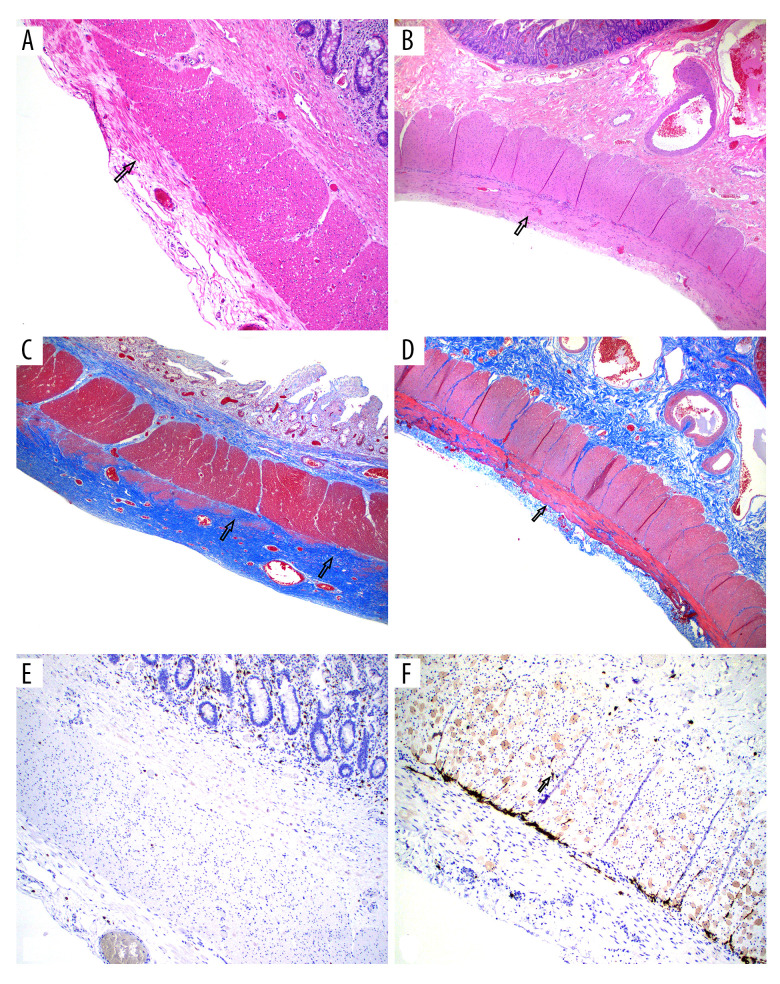
A 28-year-old man with neurologic symptoms, elevated serum thymidine, and biallelic missense pathogenic variants in TYMP (p.G311R and p.M294T) was diagnosed with MNGIE. He had gastrointestinal dysmotility since adolescence, but by the time of referral, he had cachexia and major weight loss. Initially, a liver–intestine combined (multivisceral) transplantation (MVT) was considered. MVT is a more major operation and is associated with significant physiologic stress on the recipient. Since the recipients’ ejection fraction was only 40% and he could barely walk at this time point, MVT was considered to be higher risk. To halt the progression neurologic deficit, he was listed for a LT. He had preserved synthetic liver function and transaminases were mildly elevated, resulting in a low MELD score. Given a MELD score of 6, a left-lobe LT was performed when right lobe from the same deceased donor was used for the index patient with MELD 24. The explanted liver showed moderate macrosteatosis with extensive perisinusoidal fibrosis. On post-transplant day 2, thymidine levels were normalized. The patient received steroid induction recovered well after LT. One-month after LT, he had biopsy-proven acute cellular rejection, which was treated with anti-thymocyte globulin, rituximab, and a steroid pulse. After LT, his motor deficit improved and he started to walk without support, but his GI dysmotility did not improve and he was placed on TPN. He was admitted to the hospital 4 times for malnutrition-related concerns. Two months after LT, he was admitted for ileus, which was treated conservatively. At 5, 11, and 20 months after LT he was hospitalized for dehydration, fevers, weight loss and diarrhea, and bloody diarrhea, respectively. Two years after LT, his LFTs were elevated, and a liver biopsy showed sinusoidal congestion. A venogram showed hepatic vein narrowing and was treated successfully with hepatic vein stents. He was then able to regain some level of independence, was able to drive with special disability-focused adaptations, and work as an athletic trainer, However, he remained TPN-dependent. With all efforts of GI rehabilitation exhausted, he was listed for an isolated IT. Six months later, he received IT. The explant intestine biopsies showed marked thinning of the muscle layers along with fibrosis and loss of interstitial cells of Cajal (Figure 1) At 12 months after IT, he was off TPN and tolerating an enteral diet. He has had no rejection episodes of either organ and remains on maintenance immunosuppression consisting of tacrolimus, prednisone, azathioprine, and monthly basiliximab. He has maintained normal thymidine levels and there has been no evidence of recurrent neuromuscular symptoms. The surveillance intestine allograft biopsies have been normal. At 4 months after IT, he underwent ileostomy takedown, and full-thickness biopsies from this specimen showed normal muscle layers without thinning and with normal distribution of interstitial cells of Cajal. At a follow-up 4 years after LT and 20 months after IT, he remains off TPN and is gaining weight and is now working a full-time job. To the best of our knowledge, this is the only reported patient with MNGIE to undergo successful sequential liver and intestine transplantation [11].
Figure 1. Native intestine.
(A) H&E-stained full-thickness native jejunum showing thinned external layer of muscularis propria with muscle loss and fibrosis on the left (arrow points to remaining bundle of muscle in the midst of fibrosis). (B) Normal intestine biopsy for comparison showing normal external layer of muscularis propria (100×). (C) Trichrome stain showing replacement of most of the external layer of muscularis propria by fibrosis (blue color, arrows). (D) Normal muscularis propria for comparison. (40×). (E) CD117 stain for interstitial cells of Cajal (stain cells brown to black, arrow) showing marked reduced number of cells in the myenteric plexus and inner circular layer (100×). (F) normal distribution of cells of Cajal for comparison.
Discussion
In this case report we demonstrate the utility of IT in restoration of intestinal function and nutritional independence. It has been shown that HSCT or LT, if performed late in the course of disease, may not restore GI function. The Milan Group reported no improvement in GI symptoms after LT [8]. Therefore, liver–intestine or MVT can potentially address the pathogenesis of MNGIE and restore GI function. Halter et al showed the mortality rate after HSCT reached to 62% [7]. LT has been shown to have lasting benefit in this condition. Since the GI tract is also significantly affected in this syndrome, most patients remain nutritionally challenged. In fact, many patients receiving HSCT for this condition had significant morbidity and mortality related to GI complications [12]. Furthermore, histologic studies have shown that in patients receiving HSCT, gut tissue changes do not revert to normal. Thus, in patients with significant GI damage, IT is a reasonable therapeutic option, especially when immunosuppression is already given for LT. Furthermore, in patients with significant GI dysfunction requiring parenteral nutritional support at presentation, a liver–intestine transplant/multivisceral transplant (MVT) may be an even more appropriate option, as the liver allograft provides immunoprotection to the intestine graft from the same donor [11].
Intestinal transplantation is performed in select centers in the world and majority in the U.S. [13] In the U.S. providing access to a deceased donor liver remains a challenge in patients without liver disease except for certain approved conditions. The current deceased donor liver allocation policy provides no feasible pathway for transplantation for MNGIE, whether LT or MVT. While living donor liver transplantation is an attractive option, living donor IT is uncommon, and the parents of an affected individual are obligate heterozygotes. Within organ allocation system, a pathway for exceptional cases such as this exists, however for our patient, an appeal for additional MELD points to access liver allograft was unsuccessful. For this reason, we performed a left-lobe graft where right lobe graft was allocated to an index patient with higher MELD score. One of the reasons for the denial of our MELD exception request was lack of evidence that LT benefits patients with MNGIE. We believe that awareness of therapies for MNGIE is important as early referrals are crucial for successful management of these patients.
Conclusions
Liver–intestine transplantation can be considered for late referrals of MNGIE presenting with irreversible damage to small intestine requiring parenteral nutrition.
Footnotes
Conflict of interest: None declared
Declaration of Figures’ Authenticity: All figures submitted have been created by the authors who confirm that the images are original with no duplication and have not been previously published in whole or in part.
Financial support: None declared
References
- 1.Filosto M, Piccinelli SC, Caria F, et al. Mitochondrial neurogastrointestinal encephalomyopathy (MNGIE-MTDPS1) J Clin Med. 2018;7(11):389. doi: 10.3390/jcm7110389. [DOI] [PMC free article] [PubMed] [Google Scholar]
- 2.Yadak R, Breur M, Bugiani M. Gastrointestinal dysmotility in MNGIE: From thymidine phosphorylase enzyme deficiency to altered interstitial cells of Cajal. Orphanet J Rare Dis. 2019;14(1):33. doi: 10.1186/s13023-019-1016-6. [DOI] [PMC free article] [PubMed] [Google Scholar]
- 3.Nishino I, Spinazzola A, Papadimitriou A, et al. Mitochondrial neurogastrointestinal encephalomyopathy: An autosomal recessive disorder due to thymidine phosphorylase mutations. Ann Neurol. 2000;47(6):792–800. [PubMed] [Google Scholar]
- 4.Garone C, Tadesse S, Hirano M. Clinical and genetic spectrum of mitochondrial neurogastrointestinal encephalomyopathy. Brain. 2011;134(Pt 11):3326–32. doi: 10.1093/brain/awr245. [DOI] [PMC free article] [PubMed] [Google Scholar]
- 5.Verma A, Piccoli DA, Bonilla E, et al. A novel mitochondrial G8313A mutation associated with prominent initial gastrointestinal symptoms and progressive encephaloneuropathy. Pediatr Res. 1997;42(4):448–54. doi: 10.1203/00006450-199710000-00005. [DOI] [PubMed] [Google Scholar]
- 6.Pacitti D, Levene M, Garone C, et al. Mitochondrial neurogastrointestinal encephalomyopathy: into the fourth decade, what we have learned so far. Front Genet. 2018;9:669. doi: 10.3389/fgene.2018.00669. [DOI] [PMC free article] [PubMed] [Google Scholar]
- 7.Halter JP, Michael W, Schüpbach M, et al. Allogeneic haematopoietic stem cell transplantation for mitochondrial neurogastrointestinal encephalomyopathy. Brain. 2015;138(Pt 10):2847–58. doi: 10.1093/brain/awv226. [DOI] [PMC free article] [PubMed] [Google Scholar]
- 8.D’Angelo R, Boschetti E, Amore G, et al. Liver transplantation in mitochondrial neurogastrointestinal encephalomyopathy (MNGIE): Clinical long-term follow-up and pathogenic implications. J Neurol. 2020;267(12):3702–10. doi: 10.1007/s00415-020-10051-x. [DOI] [PubMed] [Google Scholar]
- 9.De Giorgio R, Poironi L, Rinaldi R, et al. Liver transplantation for mitochondrial neurogastrointestinal encephalomyopathy. Ann Neurol. 2016;80(3):448–55. doi: 10.1002/ana.24724. [DOI] [PubMed] [Google Scholar]
- 10.Kripps KA, Nakayuenyongsuk W, Shayota BJ, et al. Successful liver transplantation in mitochondrial neurogastrointestinal encephalomyopathy (MNGIE) Mol Genet Metab. 2020;30(1):58–64. doi: 10.1016/j.ymgme.2020.03.001. [DOI] [PMC free article] [PubMed] [Google Scholar]
- 11.Selvaggi G, Gaynor JJ, Moon J, et al. Analysis of acute cellular rejection episodes in recipients of primary intestinal transplantation: A single center, 11-year experience. Am J Transplant. 2007;7(5):1249–57. doi: 10.1111/j.1600-6143.2007.01755.x. [DOI] [PubMed] [Google Scholar]
- 12.Zaidman I, Elhasid R, Gefen A, et al. Hematopoietic stem cell transplantation for mitochondrial neurogastrointestinal encephalopathy: A single-center experience underscoring the multiple factors involved in the prognosis. Pediatr Blood Cancer. 2021;68(5):e28926. doi: 10.1002/pbc.28926. [DOI] [PubMed] [Google Scholar]
- 13.Grant D, Abu-Elmagd K, Mazariegos G, et al. Intestinal transplant registry report: Global activity and trends. Am J Transplant. 2015;15(1):210–19. doi: 10.1111/ajt.12979. [DOI] [PubMed] [Google Scholar]


