ABSTRACT
Research in model organisms is central to the characterization of signaling pathways in multicellular organisms. Here, we present the comprehensive and systematic curation of 17 Drosophila signaling pathways using the Gene Ontology framework to establish a dynamic resource that has been incorporated into FlyBase, providing visualization and data integration tools to aid research projects. By restricting to experimental evidence reported in the research literature and quantifying the amount of such evidence for each gene in a pathway, we captured the landscape of empirical knowledge of signaling pathways in Drosophila.
Keywords: Signaling pathways, Drosophila, FlyBase, Gene Ontology, Network, Biocuration
Summary: Comprehensive curation of Drosophila signaling pathways and new visual displays of the pathways provide a new FlyBase resource for researchers, and new insights into signaling pathway architecture.
INTRODUCTION
Signaling pathways are vital to life, allowing cells to respond to cues such as extracellular messengers, nutrient levels, tissue damage and infection, to maintain homeostatic control and to execute developmental programs. Development of a multicellular organism is a complex, co-ordinated interplay of factors using precise spatial and temporal deployment. In Drosophila, spatial information inherited from asymmetrically deposited maternal mRNAs in the oocyte leads to pattern formation via hierarchical transcriptional control within the syncytial embryo (Perrimon et al., 2012). Cellularization marks the point at which cell-cell communication becomes the driver of development and, from what are just a small number of pathways, multiple cell and tissue types are generated and co-ordinated to form complex organs and systems (Li and Elowitz, 2019). Research using Drosophila was central to the discovery and elucidation of many key signaling pathways, such as Notch, Hedgehog, Decapentaplegic (Dpp/BMP), Hippo and Toll, with the genetic approaches possible in Drosophila synergizing with biochemical approaches using vertebrate cells in culture. Using Drosophila to study pathways is no less popular today; searching the abstract/title of papers in PubMed with the terms ‘signaling’ and ‘Drosophila’ show the publication of >500 papers/year since the year 2000. Importantly, although the main ligands and receptors have been identified for each pathway, numerous regulators and effectors continue to be discovered. For example, the post-transcriptional regulatory networks of miRNAs is still a relatively new field of study. In a recent paper, He et al. (2022) described seven miRNAs that regulate the Hedgehog pathway, such as miR-958 that downregulates two key signaling components, Smoothened and Fused. Furthermore, many newly described regulatory interactions show cross-species conservation. Orthologs human CORO7 and Drosophila Pod1 modulate Hippo signaling by promoting the assembly of the Hippo kinase complex (Park et al., 2021).
Knowledge of signaling pathways can be recorded and integrated in a variety of ways. In review-style representations, signaling pathways are often presented as a sequential procession of events, simplified so that only the main components are shown. But signaling pathways are seldom, if ever, linear. They are complex, branching and can be regulated at multiple points and in context-dependent manners (Pires-daSilva and Sommer, 2003). Furthermore, it can be difficult to distinguish between gene products whose function is solely involved in a particular signaling pathway and those that are more promiscuous. Online resources such as Reactome (Gillespie et al., 2022) and KEGG (Kanehisa and Goto, 2000) provide valuable bioinformatic summaries of pathways but are restricted to the main players. Given the volume of active research on pathways, it would be valuable to provide a resource that could be rapidly updated in response to new discoveries, including the numerous single observations that may not be followed up by subsequent studies, providing comprehensive coverage and allowing users to see that the evidence supporting some components is much stronger than for others.
We therefore set out to design a curation strategy which would capture the richness of the experimental research landscape on Drosophila signaling pathways, that could be maintained and scaled with ease. The widely used Gene Ontology (GO), a structured vocabulary used to annotate gene/gene product biological function (Ashburner et al., 2000; Gene Ontology Consortium, 2021), was used to assign genes as either core components or regulators of a particular pathway. An experimental-evidence-weighted pathway resource was designed based on counting research papers with GO annotations supported by experimental evidence codes as a proxy for the strength of support for a gene's involvement in a pathway. This data was assimilated into the Drosophila database, FlyBase (Gramates et al., 2022), in a way that can be easily sustained by future curators and presented with the aim of facilitating and seeding future research. Importantly, this resource displays the different pathways in the same format, highlighting the differences and similarities between these pathways. Comparison of pathway make-up reflects their deployment in response to stimuli and in development.
RESULTS AND DISCUSSION
Curation of signaling pathways using annotation with the Gene Ontology to quantify experimental support
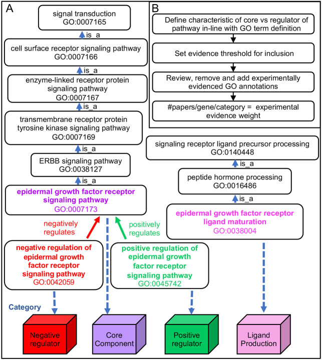
As a first step toward developing a signaling pathway resource for FlyBase, we sought to review and improve the curation of signaling pathway genes using the GO. The GO consists of a controlled vocabulary of specific biological terms linked in a hierarchical manner (Ashburner et al., 2000; Gene Ontology Consortium, 2023). Terms in the ‘signal transduction’ (GO:0007165) branch of GO biological processes are used to annotate genes that encode members of pathways using the relationship ‘is_a’ (Fig. 1A). In the GO, processes may also have the relationship type ‘regulates’, and therefore genes are also labeled with terms that reflect this relationship to the pathway. Taking as an example the Epidermal Growth Factor Receptor (EGFR) signaling pathway, the receptor, Egfr, and the Erk mitogen-activated protein kinase, Rolled, which are both essential for the execution of the pathway, are annotated with ‘epidermal growth factor receptor signaling pathway’ (GO:0007173), whereas Argos, an antagonist of the pathway, is annotated with ‘negative regulation of epidermal growth factor receptor signaling pathway’ (GO:0042059). For some pathways, such as the EGFR pathway, very specific gene products are required for the biogenesis of pathway ligand, such as Rhomboid family proteases, required for the production of soluble, secreted EGFs (Urban et al., 2002). The biological role of such components can be annotated with terms that capture this e.g. epidermal growth factor receptor ligand maturation (GO:0038004). Thus, GO terms can be used as a handle to place pathway members and their regulators into simple categories, in our case to define a gene as either a core pathway member, a positive or negative regulator or involved in ligand production (Fig. 1A).
Fig. 1.
Strategy for experimental evidence-weighted curation of signaling pathways, using the EGFR signaling pathway as an example. (A) How use of the GO to curate pathway research papers allows the terms to be used as labels to place pathway members in different categories. The structure of the GO relevant to the EGFR signaling pathway is used as an example. (B) Schematic of the signaling pathway review, curation process and calculation of evidence weight for inclusion.
A standard GO annotation links a GO term to a gene and includes an evidence code and the data source (e.g. publication) for that specific assertion. Evidence codes are used to distinguish the provenance of the assertion. For example, experimental evidence codes include ‘inferred from direct assay’ and ‘inferred from mutant phenotype’ and computational inference evidence codes include ‘inferred from electronic annotation’. Curating multiple examples of the same assertion from different research publications can quantify the support for a given gene's involvement in a pathway. In FlyBase, GO annotations can be seen in the Function section of a gene page e.g. for the gene page for hpo, http://flybase.org/reports/FBgn0261456#function. These annotations are searchable in FlyBase via the Vocabularies tool (Gramates et al., 2022) and via numerous sites such as AmiGO, QuickGO and via GO enrichment tools (as detailed in Munoz-Torres and Carbon, 2017).
The set of signaling pathways that are presented in this paper was chosen based on literature review and consultation with Drosophila signaling researchers. Our initial focus was on well-characterized cell-surface-receptor-mediated signaling pathways with major roles in development, comprising Hedgehog, canonical Wnt (Wnt-TCF), transforming growth factor-β (TGFβ, encompassing BMP and Activin signaling in Drosophila; Upadhyay et al., 2017), Notch, JAK/STAT and the main receptor tyrosine kinase signaling pathways (Mele and Johnson, 2019; Perrimon et al., 2012). Hippo signaling was added due to its fundamental importance in controlling tissue size and development (Pan, 2022). As Toll and JAK-STAT also have key roles in innate immunity, we completed the set by adding the Immune deficiency (Imd) and Tumor necrosis factor α (TNFα) pathways (Mussabekova et al., 2017). Thus, this set of 16 signaling pathways covers the best characterized pathways involved in developmental decisions and innate immunity, covering two very active areas of research in the Drosophila field and, for which, providing a comprehensive and up-to-date resource would be valuable for comparison and discovery. We used this set of pathways to test the utility of our model of data collation and presentation to draw inferences about the nature of pathway control.
An important aspect of this review was developing a clear set of rules that can be consistently followed to curate the pathways (see Materials and Methods). Each pathway often has its own set of assays used as a readout of pathway activation, such as: expression of particular target genes (native or transgenic reporters, e.g. 10xSTAT92E-luciferase for JAK-STAT; Chen et al., 2014); signature phenotype(s) (e.g. border follicle cell migration for the Pvr pathway; Schober et al., 2005); or other readouts [e.g. the translocation of Armadillo (β-catenin) into the nucleus for Wnt-TCF signaling; Tolwinski et al., 2003]. Thus, although there is variation in the way pathways are studied, standards were established for a particular pathway and applied consistently during curation. Application of these rules also involved reviewing existing GO annotations and either validating them or removing them if incorrect. We then counted the number of papers associated with each gene annotated to a pathway that was supported by an experimental evidence code to give the ‘weighted’ evidence (Fig. 1B).
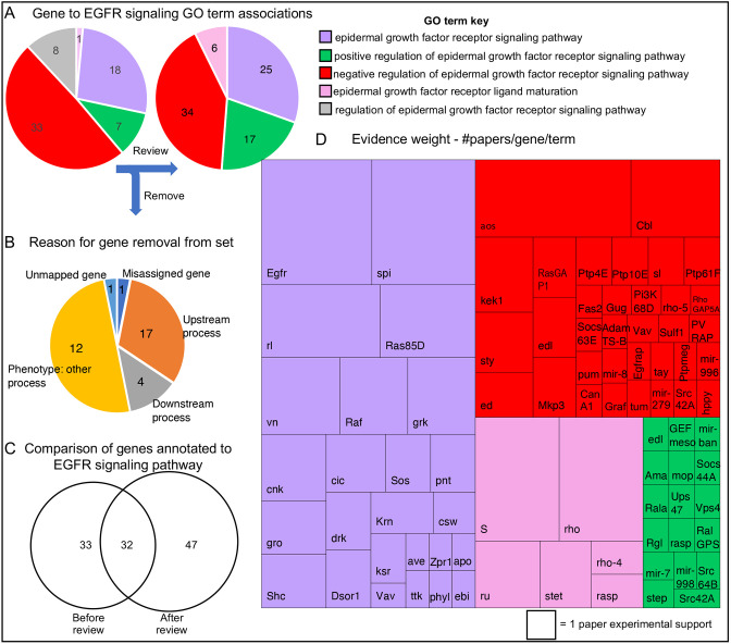
As an example, we describe how this protocol impacted on the genes included in the EGFR signaling pathway. At the start, 65 genes were already annotated to GO terms related to the pathway using both experimental and non-experimental evidence codes. Following our review of the literature, 32 genes were removed that did not meet our criteria, 33 were retained, 47 were added (Fig. 2A-C; note that Fig. 2A reports gene-GO term associations, and as a gene may be associated with more than one GO term, e.g. Src42A is annotated with both positive and negative regulatory terms for EFGR signaling, the numbers shown are greater than the number of genes). Many of the genes removed contributed to processes that indirectly affect EGFR signaling and were initially inferred to be part of the pathway based on experiments using a specific assay in which they phenocopied EGFR signaling mutants and/or had a genetic interaction with pathway members. For example, when disrupted, components of the multivesicular-body sorting pathway ESCRT complexes, such as Vps28, Vps2 and Chmp1, affect ligand-dependent internalization and destruction of Egfr but, as this is a separate biological process that acts on many proteins, upon review these genes were no longer considered to be specific to the signaling pathway itself (Fig. 2B). Genes meeting the criteria to be components of the EGFR pathway were mapped to one of four categories depending on their GO annotation (Figs 1A and 2A). By a concerted effort to cover the research literature, we were able to capture many single studies where a gene was shown to have a role in the context of EGFR signaling. Curating each example that met the annotation criteria, and counting the number of research papers per gene per term, revealed the extent to which experimental research supports the role of each gene, thus building a picture of the experimental landscape of EGFR signaling research (Fig. 2D). Over half of the genes in the EGFR pathway group are supported by only one experimental observation, and many of these genes are categorized as regulatory components. Our comprehensive identification of these single papers will help researchers find experimental results that may confirm their own observations and avoid duplication of effort.
Fig. 2.
Evidence-weighted curation of the EGFR signaling pathway. (A) The number of genes associated with each EGFR-related GO term before (left pie chart) and after (right pie chart) review. The key is shown to the right. During the review process, all regulatory components were classed as positive or negative regulators and therefore there were no direct annotations to ‘regulation of epidermal growth factor receptor signaling pathway’ (grey segment) after review. (B) Pie chart displaying the reasons for removal of 32 genes previously annotated to the EGFR pathway, but which did not pass our curation criteria. (C) A Venn diagram of GO annotated gene sets, before and after review, summarizes the extent of revision. (D) A treemap chart displaying experimental evidence weight. The block size is proportional to the number of research papers supporting the role of each gene in the EGFR signaling pathway. The corresponding gene symbols are shown. The term key is the same as in A.
Comparison of the regulatory landscape of pathways from the results of experimental evidence-weighted curation
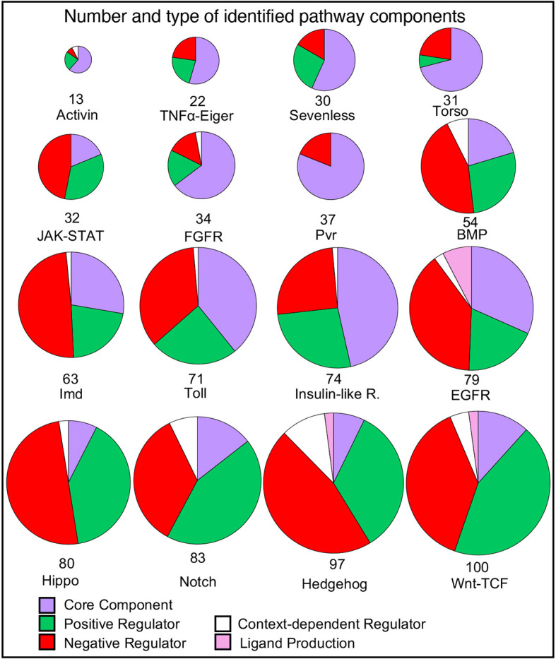
The number of genes associated with each pathway varies widely, from 13 (Activin) to 100 (Wnt-TCF) genes (Fig. 3). We wondered whether this reflected the inherent complexity of the pathway or was an indirect consequence of the length of time the pathway had been studied, i.e. more accumulated research. The correlation between the number of curated pathway members and the year a selected defining/prototypic member gene of the pathway (e.g. dome for JAK-STAT; dpp for BMP; sev for Sevenless) was first described was only moderate (Fig. S1; R2=0.40, P-value=8.81×10⁻³). For the four pathways with the largest number of members [Wnt-TCF (100 genes), Hedgehog (96), Notch (83) and Hippo (80)] the date of the first publication on prototypic genes spanned nine decades [wg (1923), hh (1950), N (1916) and hpo (2002), respectively]. Therefore, the number of components in each pathway appears to reflect some intrinsic features of the pathway, which may include the diversity of its roles and associated regulation, which have in turn contributed to the extent of the literature for each pathway.
Fig. 3.

Distribution of types of components in each pathway. The number of genes associated by GO annotations for each pathway are illustrated as pie charts. Each pie chart is broken down into the categories indicated by the key. The total number of genes in the pathway is illustrated by the diameter of the circle and the number at the base. The pathways are ordered by the number of component genes. Where genes have different regulatory effects in different contexts, they are classed as context-dependent regulators. Note that for this figure, in the Toll signaling pathway, which has a more complex extracellular activation cascade, those genes annotated to ‘negative regulation of Toll receptor ligand protein activation cascade’ (GO:0160035) are included in the ‘Negative Regulator’ category and genes annotated to ‘Toll receptor ligand protein activation cascade’ (GO:0160032) are included in the ‘Core Component’ category.
Looking at the classification of pathway components, for most of the pathways (10/16) the number of regulatory components exceeded the number of core components (Fig. 3) and for six pathways (JAK-STAT, BMP, Hippo, Notch, Hedgehog, Wnt-TCF) more than 80% of the components are regulators. A general observation is that pathways that are used most frequently in development and those that are involved with immune processes are more heavily weighted towards regulators. This is elaborated on with examples below.
The innate immunity pathways – JAK-STAT, Toll and Imd – display a high proportion of negative regulators (37-49% of components). Unchecked immune response can lead to damaging inflammation, as such downregulation of these pathways after activation is a vital part of their circuitry (Stramer and Dionne, 2014). For example, both the Toll and Imd pathways can be negatively regulated by the removal of extracellular stimuli. For Toll, the serine protease inhibitors (serpins) dampen the ligand-activating zymogen cascade (Fullaondo et al., 2011) and, for the Imd pathway, bacterial peptidoglycans are removed by enzymatically active peptidoglycan recognition proteins (Costechareyre et al., 2016).
In Drosophila, the Hedgehog, Wnt-TCF, Notch, BMP and EGFR pathways are repeatedly used in development in numerous contexts. The portion of regulators curated for these pathways is high (91-61%). First, as these pathways are used in so many different developmental contexts, it would be reasonable to assume that the range of regulators encountered, in terms of context such as tissue- and temporal-specific or precision control needed for patterning, would be correspondingly diverse. An example of the complex interplay in context-dependent control is that of Numb and Sanpodo, which only regulate Notch signaling during asymmetric cell division. In the absence of Numb, Sanpodo positively regulates Notch signaling, but in the presence of Numb, Sanpodo inhibits Notch signaling. Numb, which is inhibitory, can only act in the presence of Sanpodo (Babaoglan et al., 2009). Thus, the asymmetric distribution of Numb drives the acquisition of different cell fates. Second, the strict control of a signaling window is essential for the execution of developmental programs – when a pathway is switched off is as important as when it is switched on (Perrimon et al., 2012). Some pathways need to be continuously repressed in the absence of signal, notably the Wnt-TCF (Roberts et al., 2012) and Hedgehog pathways (Han et al., 2015), which are de-repressed by the presence of ligand. Pathways such as the receptor tyrosine kinase (RTK) pathways are principally controlled by the availability of receptor ligands rather than having to be actively repressed. The EGFR pathway has a high proportion of negative regulators, 39% of components, in comparison with other RTK pathways, which most likely represents its widespread usage in development. It has a similar regulatory profile to BMP signaling, which has 44% negative regulators (Shilo, 2005). Thus, precise spatial and temporal control is required to build complex systems and structures.
Comparing the RTK pathways in order of number of members – Torso, Sevenless, FGFR, Pvr, Insulin-like receptor, EGFR (from 30 to 79 genes) – reflects the scope of their use. Of the RTK pathways, Torso and Sevenless have the smallest number of components (31 and 30 genes, respectively). Both pathways are restricted in their use – Torso signaling is principally involved in the development of embryonic termini and Sevenless for the specification of the R7 photoreceptor cells. The lack of complexity of these pathway reflects their extremely limited developmental deployment. The Pvr pathway is an interesting contrast in having a large proportion of genes (81%) characterized as core components. Of all signaling pathway curated, the Pvr pathway appears to be the most branched, capable of employing several intracellular cassettes such as the Erk kinase cascade, PI3K/TORC1, Rac/Rho signaling and the JNK cascade. It is associated with inducing a number of processes, such as border follicle cell migration, epidermal wound healing, hemocyte spreading and cell proliferation (Tsai et al., 2022; Mues et al., 2023).
In contrast to the Pvr pathway, the Hippo pathway has a particularly high proportion of regulators (92%), which may reflect the substantial difference in logic of this pathway. It is an intracellular cassette in which Hippo kinase phosphorylates Warts kinase which in turns phosphorylates the transcriptional coactivator Yorkie leading to its cytoplasmic retention (Huang et al., 2005). The regulatory components include many membrane proteins and scaffolding proteins, as the Hippo pathway integrates mechanical cues to manage cell growth and survival and its balance is key in determining organ size and tumor suppression (Chang et al., 2019). Pathway components are therefore heavily weighted towards regulation (Pires-daSilva and Sommer, 2003).
Developing a research-based pathway resource
The next step was to develop a signaling pathway resource within FlyBase to facilitate future research. Pathway Reports were generated using a similar architecture to the FlyBase Gene Group resource (detailed in Attrill et al., 2016), with key differences to maximize usefulness to pathway research. Pathway Reports are organized in a hierarchical fashion, with a top-level parent report and sub-groups, and we divided pathway components into the sub-groups of ‘core members’, ‘positive regulators’, ‘negative regulators’ and, in some cases, specific ‘ligand production’ sub-groups, mirroring the curation scheme in Fig. 1A. The reports can be queried using the FlyBase QuickSearch ‘Pathways’ tab on the homepage (http://flybase.org/) and a list is available at http://flybase.org/lists/FBgg/pathways. As described more fully below, the pathway reports contain: (1) a customizable table of pathway members; (2) a visual summary of the GO annotation (the GO ribbon stack); (3) two types of pathway diagrams, static thumbnails and dynamic physical interaction networks; and (4) links to other resources.
A customizable table of pathway members as a resource and information hub
Each gene belonging to a pathway sub-group is represented as a row in the table, and by default the columns contain the following data: Gene Symbol, Gene Name, Gene Group Membership, GO Molecular Function (Experimental) and # Pathway Refs (Fig. S2). Additional columns can be selected: # All Research Refs, Also Known As, Antibody, Classical/Insertion Alleles, Transgenic Constructs, Disease Models (Experimental), Potential Disease Models, Other Pathways and Human Orthologs. The user can select which columns are shown or hidden, order based on the column value or filter the entries by text match. The ‘# Pathway Refs’ column contains the number of GO-curated papers that provide direct experimental evidence for a gene's role in a pathway. Clicking the hyperlinked number in this column generates a FlyBase ‘HitList’ of these references, which can be downloaded or analyzed further. Thus, the user has access to the molecular function of each gene product, the literature, the experimental reagents used in the study of each gene and links to human disease and fly models of those diseases.
GO ribbon stack
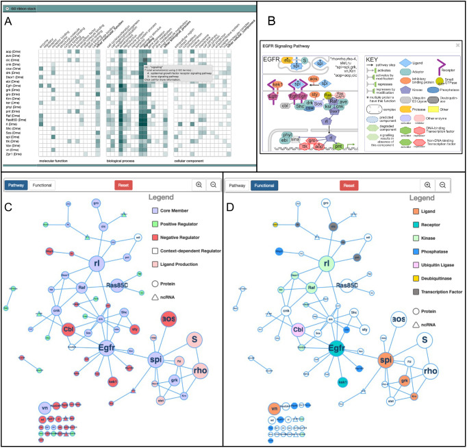
To enable users to compare the biological functions of the pathway components, we chose to display a stack of GO summary ribbons (Fig. 4A). These summaries take advantage of the hierarchical nature of the GO to group the detailed GO annotations into broad categories, each represented as a square within the ribbon; the presence of GO annotations in each category colors them, from faint to dark blue, depending on the number of annotations. Clicking on each colored square displays more detail, as indicated in Fig. 4A. In this way, a user can easily get an idea of which genes encode enzymes or extracellular proteins and whether the pathway has more prominent roles in, for example, the immune, nervous or reproductive systems and quicky see the more detailed information.
Fig. 4.
FlyBase Pathway Report pages. (A-D) Three visual summaries are presented on the Pathway pages, exemplified with the EGFR Signaling Pathway. (A) The GO ribbon stack is a graphical summary of the GO annotations for each gene, with annotations grouped under high-level categories. The color intensity of each cell indicates how many unique terms are grouped in that particular category. The unique terms that are grouped under a particular cell can be seen by hovering over or clicking on the cell, as shown for one of the cells of cic. (B) A thumbnail review-style image for the EGFR Signaling Pathway. (C,D) A physical interaction network in which the nodes represent genes, the sizes of which are proportional to the number of supporting papers, up to ≥10. C shows the ‘Pathway view’ and D shows the ‘Functional view’ (for visual simplicity, we selected fewer higher level categories compared with the thumbnails). The edges represent physical interactions between components, from FlyBase. Unconnected nodes are also displayed.
Pathway diagrams
To display a visual summary of each pathway, two contrasting approaches were developed. To give a relatively simply summary of how each pathway works, a static ‘Thumbnail’ for each pathway is provided, using a standard review-type representation that shows the position and role of the components in the cell, and gives a sense of the route of the pathway from outside the cell into the nucleus (an example, the EGFR signaling pathway thumbnail is shown in Fig. 4B and all thumbnails are shown in Fig. S3). So they did not become too complex, the thumbnails focus on the core pathway members and major regulators. Experts on each pathway were consulted to ensure that each pathway was represented correctly (see acknowledgements). By using a uniform format for depicting the different types of molecules, the molecule architecture of the different pathways can be easily compared. The Thumbnails are available within FlyBase as downloadable SVG images, so they can be modified by researchers if desired. They are displayed next to a concise textual summary of the pathway.
We also sought a dynamic way of visualizing all the pathway members with information about their function and the weight of evidence for their inclusion in the pathway, generated computationally from the data in FlyBase so it would be automatically updated to include new data. For this, each pathway member is displayed as a node in a network of physical interactions (Fig. 4C,D), generated with Cytoscape.js (Franz et al., 2016). The size of each node is a function of the number of curated papers that report experimental evidence for the membership of the gene in the pathway (with a maximum size at ≥10 papers). The edges between nodes are derived from physical interaction data curated in FlyBase. Post-transcriptional regulation by non-coding RNAs (triangular nodes) is integrated into the interaction network to show where regulatory intersections may occur. Users can switch between two color-coded schemes: a ‘Pathway’ view (Fig. 4C), where the nodes are colored according to high-level roles in the pathway (Core Member; Positive Regulator; Negative Regulator; Context-dependent Regulator; Ligand Production) and a ‘Functional’ view, with the node color corresponding to selected molecular function classes (Fig. 4D). The networks can be rearranged by users by manually dragging nodes, which will maintain their position between views, or by resetting the view. As these networks are updated each FlyBase release through in-house computational pipelines they reflect the current state of curated knowledge.
Links to other resources
The Pathway Reports are designed to act as a knowledge hub. The customizable table provides links to other data and resources in FlyBase. In addition, links are provided to relevant FlyBase Gene Groups e.g. links from the ‘Wnt-TCF Signaling Pathway Core Components’ page to the ‘FRIZZLED-TYPE RECEPTORS’, ‘WNTs’, ‘WNT ENHANCEOSOME’ Gene Groups pages. In the External Data section, linkouts are provided to external resources, such as Reactome, KEGG, Wikipathways and The Interactive Fly.
A scalable model – adding a newly described antiviral pathway
One of the core strengths of this pathway resource is that the underlying model is scalable and easy to maintain. A common addition to our set of well-characterized pathways is the description of new regulatory interactions. However, we also intend to grow this resource with more signaling pathways – a recent example of which is the addition of the cGAS/STING signaling pathway. In vertebrates, STING (stimulator of interferon genes) was first shown as a component of innate immunity, involved in the induction of interferon (Ishikawa and Barber, 2008). STING is activated by cyclic GMP-AMP (cGAMP), which is produced by cGAMP synthase (cGAS) in response to cytosolic DNA, e.g. viral DNA. STING then activates IKK and TBK1 and, further downstream, the activation of the transcription factors IRF3 and NF-κB leads to the transcription of immune response genes. In insects, although Sting had been linked to defense response (Goto et al., 2018; Martin et al., 2018) and NF-κB (Rel) activation, the cGAS component remained elusive until Slavik et al. (2021) and Holleufer et al. (2021) identified two cytosolic cGAS-like genes that respond to viral infection. It was therefore apparent that the cGAS-STING module was an evolutionarily conserved pathway with the potential to respond to other pathogen-associated molecular patterns such as dsRNA. As such, a new GO term ‘cGAS/STING signaling pathway’ (GO:0140896) was created in 2022, allowing pathway components to be specifically annotated to this pathway rather than to a more general terms related to ‘defense response to virus’ (GO:0051607) or canonical NF-kappaB signal transduction (GO:0007249). Given the relative novelty of this pathway, so far only six core components have been annotated to it. FlyBase curators will continue to populate this page as more experimental information arises, keeping pace with research. The importance of this pathway is highlighted as Drosophila grows as a popular model for virus infection, including viruses with a direct impact on human health such as EBV, HIV-1, SARS-CoV-1 and -2, Zika, dengue and West Nile fever viruses (Hughes et al., 2012). Together with Toll, JAK-STAT, Imd and TNFα, cGAS-STING completes the current set of Drosophila innate immune signaling pathways.
Conclusions
This paper describes a novel experimental-evidence weighted approach to assembling a signaling pathway resource. Using GO curation to redundantly curate the same experimental conclusion from multiple research papers provides a mechanism for measuring the strength of characterization of a gene to pathway relationship. By presenting this information in a tabular and graphical way in dedicated pages in FlyBase, researchers can easily distinguish ‘central players’ from single observations identifying new components. From a curatorial perspective, this is easy to update, maintain and scale beyond the initial focused effort. As the GO is machine-readable, widely used and distributed with many existing tools, this work adheres to FAIR (Findability, Accessibility, Interoperability and Reusability) principles (Wilkinson et al., 2016). The pathway knowledge we have curated has already been incorporated by two bioinformatic projects: FlyPhoneDB, a tool used to predict cell-cell communication between cell types from Drosophila single-cell RNA-sequencing data (Liu et al., 2022), and PANGEA, a Gene Set Enrichment Tool (Hu et al., 2023), demonstrating the utility of the resource beyond FlyBase.
The approach to curating and presenting information described in this paper is applicable across organisms and, as it is based on the GO, to any other term in the biological process branch. Integrating the GO curation of pathways into dedicated pages in FlyBase allows users to see other data side-by-side and make their own observations. An example of this is the link between genes annotated to ‘BMP signaling pathway’ (GO:0030509) and disease models for ‘juvenile polyposis syndrome’ (DOID:0050787) (Fig. S4). To further improve this integrated system, we aim to impart directionality to some of the edges in the pathway networks by annotating which gene products are the targets in various interactions.
By a clear and simple division of components into core, regulatory and, in some cases, ligand production, we have been able to observe differences in the diversity and scale of regulatory interactions between pathways deployed in different contexts. An important aspect of our curation effort has been the inclusion of post-transcriptional regulation by ncRNAs – an element that is missed by other pathway modelling approaches. In Drosophila, ∼2.7% of genes (485/17,896 mapped genes) encode miRNAs, which play a crucial role in the regulation of cellular processes by directing the activity of the RNA-induced silencing complex. In this project, we have associated 31 unique miRNA genes with pathways. Some miRNAs regulate multiple pathways – for example mir-8, mir-ban, mir-279, mir-958 and mir-7 have curated interactions with three or more pathways.
Most pathway resources must be, by pragmatic design, limited to an orthodox set of genes, thereby missing new and incremental knowledge. Repetition is often seen as the gold-standard for measuring confidence in a result. The evidence-weighted model for curation presented here allows a measure of reproducibility i.e. how often the observation has been made by separate studies. We believe that the dynamic model of pathway curation we have outlined here, and its integration with other data curation streams, shows how a simple curation framework can be used produce a rich, up-to-date resource which can further feed back into research.
MATERIALS AND METHODS
Curation strategy
For each pathway, an initial list of genes to review was downloaded from FlyBase using the FlyBase Vocabularies term tool queried with the appropriate pathway term (including child and regulatory terms). In concert with current reviews, papers and the relevant GO term descriptions, we established the definitions of what constitutes the core pathway boundaries. The initial list of genes was reviewed and annotations that did not fit the curation criteria were removed/replaced. The annotations for each pathway were made consistently at an appropriate level – for example, with genes annotated to ‘Wnt signaling pathway’ (GO:0016055), we replaced the term with ‘canonical Wnt signaling pathway’ (GO:0060070) where this was appropriate, and this term was used as the handle to compile the list of Wnt-TCF Signaling Pathway Core Components. For this initial pass, we also focused on identifying research papers reporting experimental evidence for well-established pathway members that did not have any such evidence annotated to them. Further to this, we reviewed early the key papers that demonstrated the involvement of certain genes in pathways, including papers which had been previously missed as they pre-dated the introduction of GO. As we reviewed papers, we established curation benchmark standards for pathway membership inclusion and recorded these as a Drosophila pathway curation guide (https://wiki.flybase.org/mediawiki/images/9/9f/FB_Pathway_Curation_Manual.pdf). Many pathways, especially those with extensive characterization, were subject to further rounds of curation after the establishment of pathway pages in FlyBase. Pathway pages were constructed by using the GO terms as markers of the inclusion of papers in a Signaling Pathway group (see Table S1 for mapping).
Construction of networks
The pathway networks are generated for every FlyBase release using Cytoscape.js (Franz et al., 2016) and an in-house pipeline that takes as input the FlyBase physical interaction file in MITAB format (Perfetto et al., 2019) and an internal FlyBase gene group membership reporting file, to produce a JavaScript file per pathway. The layout of the networks is established using the fCoSE algorithm (Genc and Dogrusoz, 2016; Balci and Dogrusoz, 2022), with the following parameters: “directed: true”, “nodeRepulsion: 450000”, “idealEdge- Length: 100”, “gravity: −500”. The networks are then integrated into the Cytoscape.js widget developed by FlyBase, available at https://github.com/FlyBase/cytoscape-widget.
GO stacked ribbons
The GO annotation ribbon stacks are generated using the GO Consortium Web Component (available at github.com/geneontology/wc-ribbon) with some in-house modifications and configuration, with the input of a FlyBase-generated file of gene IDs and FlyBase GO annotation data. The FlyBase GO annotation ribbon mapping categories used can be found at https://wiki.flybase.org/wiki/FlyBase:Gene_Report#Function and are labelled as subset: goslim_flybase_ribbon in GO downloads (http://geneontology.org/docs/download-ontology/). These FlyBase data are publicly available via the FlyBase API at http://flybase.org/cgi-bin/getRibbonJSON_agr-like.pl, with a ‘subject’ parameter consisting of a list of FlyBase IDs. Please note that this API address may change in the future, but any changes will be documented at https://flybase.github.io/api/swagger-ui/, in the FlyBase GitHub repository.
Version information
The data used was from FlyBase release FB_2023_01, GO version 2023-01-01. For comparison of before review, data from release FB_2017_02 was used.
Supplementary Material
An Excel spreadsheet summary of the Signaling Pathway page content in FlyBase release version FB_2023_01. The name and unique FlyBase Gene Group ID (FBgg#) are given in the first column. The second column is the GO term and ID that is mapped to the FBgg. The number of genes and papers linked to the pathway are given into the last two columns. Note that the sum of the numbers for the member groups is often different than for the top-level group as papers and genes may be shared between more than 1 subgroup. For each pathway group, except cGAS/STING, the table also includes the gene selected as the prototypic representative and year of publication of first research paper on the prototype.
Acknowledgements
We thank David Strutt (University of Sheffield, UK), Julius Mieszczanek (MRC Laboratory of Molecular Biology, Cambridge, UK), Alfonso Martinez-Arias (University of Cambridge, UK), Golnar Kolahgar (University of Cambridge, UK) and Nic Tapon (Francis Crick Institute, UK) for advice on the design of the Signaling Pathway pages. For reviewing the Pathway thumbnails, we thank Erika Bach (New York University, NY, USA), Sarah Bray (University of Cambridge, UK), Edan Foley (University of Alberta, Canada), Marc Furriols (Institute of Molecular Biology of Barcelona, Spain), Jin Jiang (University of Texas Southwestern Medical Center at Dallas, TX, USA), Bruno Lemaitre (EPFL-GHI-SV-UPLEM, Switzerland), Steven Marygold (FlyBase, University of Cambridge, UK), Ian Mcgough (Babraham Institute, UK), Gillian Millburn (FlyBase, University of Cambridge, UK), Arno Müller (Universität Kassel, Germany), Mike O'Connor (University of Minnesota, MN, USA), David Stein (University of Texas at Austin, TX, USA), Neal Silverman (UMass Chan Medical School, MA, USA), Hugo Stocker (Institut für Biochemie, Switzerland), Nic Tapon (Francis Crick Institute, UK), Jean-Paul Vincent (Francis Crick Institute, UK), Lei Xue (Tongji University, China). We also thank members of the GOC and the Alliance of Genome Resources for useful discussion and contribution to signaling pathway guidelines, in particular Pascale Gaudet (Swiss Institute of Bioinformatics, Switzerland), Ruth Lovering (UCL, UK), Paul Thomas (University of Southern California, CA, USA), Kimberly van Auken (Caltech, CA, USA), David Hill (Jackson Laboratory, USA) and, for the development of and help with implementing stacked GO ribbons, Laurent-Philippe Albou (Université Louis Pasteur, France). Members of the FlyBase Consortium: Norbert Perrimon (PI), Nicholas H. Brown (Co-PI), Brian Calvi (Co-PI), Richard Cripps (Co-PI), Thomas Kaufman (Co-PI), Susan Russo Gelbart (PD), Giulia Antonazzo, Helen Attrill, Kris Broll, Seth Campbell, Lynn Crosby, Gil dos Santos, Kathleen Falls, Josh Goodman, Damien Goutte-Gattat, L. Sian Gramates, Victoria Jenkins, Pravija Krishna, Aoife Larkin, Ian Longden, TyAnna Lovato, Steven Marygold, Beverley Matthews, Alex McLachlan, Gillian Millburn, Arzu Ozturk-Colak, Clare Pilgrim, Jolene Seme, Victor Strelets, Christopher J. Tabone, Jim Thurmond, Vitor Trovisco, Pinglei Zhou and Mark Zytkovicz.
Footnotes
Author contributions
Conceptualization: H.A., G.A., N.H.B.; Methodology: H.A., G.A.; Software: J.L.G., J.T., V.B.S.; Validation: G.A.; Formal analysis: H.A., G.A.; Investigation: H.A.; Resources: H.A., G.A., J.L.G., J.T., V.B.S.; Data curation: H.A., G.A.; Writing - original draft: H.A.; Writing - review & editing: G.A., N.H.B.; Visualization: G.A., J.L.G., J.T., V.B.S.; Supervision: N.H.B.; Funding acquisition: H.A., N.H.B.
Funding
This work was supported by UK Medical Research Council grants MR/N030117/1 and MR/W024233/1 and FlyBase grant (National Institutes of Health/National Human Genome Research Institute, U41HG000739). Open Access funding provided by University of Cambridge. Deposited in PMC for immediate release.
Data availability
GO data used for this analysis are available at https://zenodo.org/records/7504797. The data used in the analysis is deposited at https://doi.org/10.6084/m9.figshare.c.7029977.v1. This comprises the data files physical_interactions_mitab_fb_2023_01.tsv, pathway_group_data_fb_2023_01.tsv and gene_group_data_fb_2023_01.tsv. The current list of FlyBase Pathway Reports is available at http://flybase.org/lists/FBgg/pathways.
References
- Ashburner, M., Ball, C. A., Blake, J. A., Botstein, D., Butler, H., Cherry, J. M., Davis, A. P., Dolinski, K., Dwight, S. S., Eppig, J. T.et al. (2000). Gene Ontology: tool for the unification of biology. The Gene Ontology Consortium. Nat. Genet. 25, 25-29. 10.1038/75556 [DOI] [PMC free article] [PubMed] [Google Scholar]
- Attrill, H., Falls, K., Goodman, J. L., Millburn, G. H., Antonazzo, G., Rey, A. J. and Marygold, S. J.. and the FlyBase consortium. (2016). FlyBase: establishing a Gene Group resource for Drosophila melanogaster. Nucleic. Acids Res. 44, D786-D792. 10.1093/nar/gkv1046 [DOI] [PMC free article] [PubMed] [Google Scholar]
- Babaoglan, A. B., O'Connor-Giles, K. M., Mistry, H., Schickedanz, A., Wilson, B. A. and Skeath, J. B. (2009). Sanpodo: a context-dependent activator and inhibitor of Notch signaling during asymmetric divisions. Development 136, 4089-4098. 10.1242/dev.040386 [DOI] [PMC free article] [PubMed] [Google Scholar]
- Balci, H. and Dogrusoz, U. (2022). fCoSE: a fast compound graph layout algorithm with constraint support. IEEE Trans. Vis. Comput. Graph 28, 4582-4593. 10.1109/TVCG.2021.3095303 [DOI] [PubMed] [Google Scholar]
- Chang, Y.-C., Wu, J.-W., Wang, C.-W. and Jang, A. C.-C. (2019). Hippo Signaling-Mediated Mechanotransduction in Cell Movement and Cancer Metastasis. Front. Mol. Biosci. 6, 157. 10.3389/fmolb.2019.00157 [DOI] [PMC free article] [PubMed] [Google Scholar]
- Chen, Q., Giedt, M., Tang, L. and Harrison, D. A. (2014). Tools and methods for studying the Drosophila JAK/STAT pathway. Methods 68, 160-172. 10.1016/j.ymeth.2014.03.023 [DOI] [PubMed] [Google Scholar]
- Costechareyre, D., Capo, F., Fabre, A., Chaduli, D., Kellenberger, C., Roussel, A., Charroux, B. and Royet, J. (2016). Tissue-specific regulation of Drosophila NF-κB pathway activation by peptidoglycan recognition protein SC. J. Innate. Immun. 8, 67-80. 10.1159/000437368 [DOI] [PMC free article] [PubMed] [Google Scholar]
- Franz, M., Lopes, C. T., Huck, G., Dong, Y., Sumer, O. and Bader, G. D. (2016). Cytoscape.js: a graph theory library for visualisation and analysis. Bioinformatics 32, 309-311. 10.1093/bioinformatics/btv557 [DOI] [PMC free article] [PubMed] [Google Scholar]
- Fullaondo, A., García-Sánchez, S., Sanz-Parra, A., Recio, E., Lee, S. Y. and Gubb, D. (2011). Spn1 regulates the GNBP3-dependent Toll signaling pathway in Drosophila melanogaster. Mol. Cell. Biol. 31, 2960-2972. 10.1128/MCB.01397-10 [DOI] [PMC free article] [PubMed] [Google Scholar]
- Genc, B. and Dogrusoz, U. (2016). An algorithm for automated layout of process description maps drawn in SBGN. Bioinformatics 32, 77-84. 10.1093/bioinformatics/btv516 [DOI] [PMC free article] [PubMed] [Google Scholar]
- Gene Ontology Consortium (2021). The Gene Ontology resource: enriching a GOld mine. Nucleic Acids Res. 49, D325-D334. 10.1093/nar/gkaa1113 [DOI] [PMC free article] [PubMed] [Google Scholar]
- Gene Ontology Consortium, Aleksander, S. A., Balhoff, J., Carbon, S., Cherry, J. M., Drabkin, H. J., Ebert, D., Feuermann, M., Gaudet, P., Harris, N. L.et al. (2023). The Gene Ontology knowledgebase in 2023. Genetics 224, iyad031. 10.1093/genetics/iyad031 [DOI] [PMC free article] [PubMed] [Google Scholar]
- Gillespie, M., Jassal, B., Stephan, R., Milacic, M., Rothfels, K., Senff-Ribeiro, A., Griss, J., Sevilla, C., Matthews, L., Gong, C.et al. (2022). The reactome pathway knowledgebase 2022. Nucleic Acids Res. 50, D687-D692. 10.1093/nar/gkab1028 [DOI] [PMC free article] [PubMed] [Google Scholar]
- Goto, A., Okado, K., Martins, N., Cai, H., Barbier, V., Lamiable, O., Troxler, L., Santiago, E., Kuhn, L., Paik, D.et al. (2018). The kinase IKKβ regulates a STING- and NF-κB dependent antiviral response pathway in Drosophila. Immunity 49, 225-234.e4. 10.1016/j.immuni.2018.07.013 [DOI] [PMC free article] [PubMed] [Google Scholar]
- Gramates, L. S., Agapite, J., Attrill, H., Calvi, B. R., Crosby, M. A., Dos Santos, G., Goodman, J. L., Goutte-Gattat, D., Jenkins, V. K., Kaufman, T.et al. (2022). FlyBase: a guided tour of highlighted features. Genetics 220, iyac035. 10.1093/genetics/iyac035 [DOI] [PMC free article] [PubMed] [Google Scholar]
- Han, Y., Shi, Q. and Jiang, J. (2015). Multisite interaction with Sufu regulates Ci/Gli activity through distinct mechanisms in Hh signal transduction. Proc. Natl. Acad. Sci. USA 112, 6383-6388. 10.1073/pnas.1421628112 [DOI] [PMC free article] [PubMed] [Google Scholar]
- He, T., Fan, Y., Wang, Y., Liu, M. and Zhu, A. J. (2022). Dissection of the microRNA network regulating hedgehog signaling in Drosophila. Front. Cell Dev. Biol. 10, 866491. 10.3389/fcell.2022.866491 [DOI] [PMC free article] [PubMed] [Google Scholar]
- Holleufer, A., Winther, K. G., Gad, H. H., Ai, X., Chen, Y., Li, L., Wei, Z., Deng, H., Liu, J., Frederiksen, N. A.et al. (2021). Two cGAS-like receptors induce antiviral immunity in Drosophila. Nature 597, 114-118. 10.1038/s41586-021-03800-z [DOI] [PubMed] [Google Scholar]
- Hu, Y., Comjean, A., Attrill, H., Antonazzo, G., Thurmond, J., Chen, W., Li, F., Chao, T., Mohr, S. E., Brown, N. H.et al. (2023). PANGEA: a new gene set enrichment tool for Drosophila and common research organisms. Nucleic Acids Res. 51, W419-W426. 10.1093/nar/gkad331 [DOI] [PMC free article] [PubMed] [Google Scholar]
- Huang, J., Wu, S., Barrera, J., Matthews, K. and Pan, D. (2005). The Hippo signaling pathway coordinately regulates cell proliferation and apoptosis by inactivating Yorkie, the Drosophila Homolog of YAP. Cell 122, 421-434. 10.1016/j.cell.2005.06.007 [DOI] [PubMed] [Google Scholar]
- Hughes, T. T., Allen, A. L., Bardin, J. E., Christian, M. N., Daimon, K., Dozier, K. D., Hansen, C. L., Holcomb, L. M. and Ahlander, J. (2012). Drosophila as a genetic model for studying pathogenic human viruses. Virology 423, 1-5. 10.1016/j.virol.2011.11.016 [DOI] [PMC free article] [PubMed] [Google Scholar]
- Ishikawa, H. and Barber, G. N. (2008). STING is an endoplasmic reticulum adaptor that facilitates innate immune signalling. Nature 455, 674-678. 10.1038/nature07317 [DOI] [PMC free article] [PubMed] [Google Scholar]
- Kanehisa, M. and Goto, S. (2000). KEGG: kyoto encyclopedia of genes and genomes. Nucleic Acids Res. 28, 27-30. 10.1093/nar/28.1.27 [DOI] [PMC free article] [PubMed] [Google Scholar]
- Li, P. and Elowitz, M. B. (2019). Communication codes in developmental signaling pathways. Development 146, dev170977. 10.1242/dev.170977 [DOI] [PMC free article] [PubMed] [Google Scholar]
- Liu, Y., Li, J. S. S., Rodiger, J., Comjean, A., Attrill, H., Antonazzo, G., Brown, N. H., Hu, Y. and Perrimon, N. (2022). FlyPhoneDB: an integrated web-based resource for cell-cell communication prediction in Drosophila. Genetics 220, iyab235. 10.1093/genetics/iyab235 [DOI] [PMC free article] [PubMed] [Google Scholar]
- Martin, M., Hiroyasu, A., Guzman, R. M., Roberts, S. A. and Goodman, A. G. (2018). Analysis of Drosophila STING reveals an evolutionarily conserved antimicrobial function. Cell Rep. 23, 3537-3550.e6. 10.1016/j.celrep.2018.05.029 [DOI] [PMC free article] [PubMed] [Google Scholar]
- Mele, S. and Johnson, T. K. (2019). Receptor tyrosine kinases in development: insights from Drosophila. Int. J. Mol. Sci. 21, 188. 10.3390/ijms21010188 [DOI] [PMC free article] [PubMed] [Google Scholar]
- Mues, N., Hammer, K. and Leatherman, J. (2023). Pvr regulates cyst stem cell division in the Drosophila testis niche, and has functions distinct from Egfr. Cells Dev. 173, 203822. 10.1016/j.cdev.2022.203822 [DOI] [PMC free article] [PubMed] [Google Scholar]
- Munoz-Torres, M. and Carbon, S. (2017). Get GO! Retrieving GO data using AmiGO, QuickGO, API, files, and tools. Methods Mol. Biol. 1446, 149-160. 10.1007/978-1-4939-3743-1_11 [DOI] [PubMed] [Google Scholar]
- Mussabekova, A., Daeffler, L. and Imler, J.-L. (2017). Innate and intrinsic antiviral immunity in Drosophila. Cell. Mol. Life Sci. 74, 2039-2054. 10.1007/s00018-017-2453-9 [DOI] [PMC free article] [PubMed] [Google Scholar]
- Pan, D. (2022). The unfolding of the Hippo signaling pathway. Dev. Biol. 487, 1-9. 10.1016/j.ydbio.2022.04.001 [DOI] [PubMed] [Google Scholar]
- Park, J., Jun, K., Choi, Y., Yoon, E., Kim, W., Jang, Y.-G. and Chung, J. (2021). CORO7 functions as a scaffold protein for the core kinase complex assembly of the Hippo pathway. J. Biol. Chem. 296, 100040. 10.1074/jbc.RA120.013297 [DOI] [PMC free article] [PubMed] [Google Scholar]
- Perfetto, L., Acencio, M. L., Bradley, G., Cesareni, G., Del Toro, N., Fazekas, D., Hermjakob, H., Korcsmaros, T., Kuiper, M., Lægreid, A.et al. (2019). CausalTAB: the PSI-MITAB 2.8 updated format for signalling data representation and dissemination. Bioinformatics 35, 3779-3785. 10.1093/bioinformatics/btz132 [DOI] [PMC free article] [PubMed] [Google Scholar]
- Perrimon, N., Pitsouli, C. and Shilo, B.-Z. (2012). Signaling mechanisms controlling cell fate and embryonic patterning. Cold Spring Harb. Perspect Biol. 4, a005975. 10.1101/cshperspect.a005975 [DOI] [PMC free article] [PubMed] [Google Scholar]
- Pires-daSilva, A. and Sommer, R. J. (2003). The evolution of signalling pathways in animal development. Nat. Rev. Genet. 4, 39-49. 10.1038/nrg977 [DOI] [PubMed] [Google Scholar]
- Roberts, D. M., Pronobis, M. I., Alexandre, K. M., Rogers, G. C., Poulton, J. S., Schneider, D. E., Jung, K.-C., McKay, D. J. and Peifer, M. (2012). Defining components of the β-catenin destruction complex and exploring its regulation and mechanisms of action during development. PLoS One 7, e31284. 10.1371/journal.pone.0031284 [DOI] [PMC free article] [PubMed] [Google Scholar]
- Schober, M., Rebay, I. and Perrimon, N. (2005). Function of the ETS transcription factor Yan in border cell migration. Development 132, 3493-3504. 10.1242/dev.01911 [DOI] [PubMed] [Google Scholar]
- Shilo, B. Z. (2005). Regulating the dynamics of EGF receptor signaling in space and time. Development 132, 4017-4027. 10.1242/dev.02006 [DOI] [PubMed] [Google Scholar]
- Slavik, K. M., Morehouse, B. R., Ragucci, A. E., Zhou, W., Ai, X., Chen, Y., Li, L., Wei, Z., Bähre, H., König, M.et al. (2021). cGAS-like receptors sense RNA and control 3'2’-cGAMP signalling in Drosophila. Nature 597, 109-113. 10.1038/s41586-021-03743-5 [DOI] [PMC free article] [PubMed] [Google Scholar]
- Stramer, B. M. and Dionne, M. S. (2014). Unraveling tissue repair immune responses in flies. Semin. Immunol. 26, 310-314. 10.1016/j.smim.2014.04.004 [DOI] [PubMed] [Google Scholar]
- Tolwinski, N. S., Wehrli, M., Rives, A., Erdeniz, N., DiNardo, S. and Wieschaus, E. (2003). Wg/Wnt signal can be transmitted through arrow/LRP5,6 and Axin independently of Zw3/Gsk3beta activity. Dev. Cell 4, 407-418. 10.1016/s1534-5807(03)00063-7 [DOI] [PubMed] [Google Scholar]
- Tsai, C.-R., Wang, Y., Jacobson, A., Sankoorikkal, N., Chirinos, J. D., Burra, S., Makthal, N., Kumaraswami, M. and Galko, M. J. (2022). Pvr and distinct downstream signaling factors are required for hemocyte spreading and epidermal wound closure at Drosophila larval wound sites. G3 (Bethesda) 12, jkab388. 10.1093/g3journal/jkab388 [DOI] [PMC free article] [PubMed] [Google Scholar]
- Upadhyay, A., Moss-Taylor, L., Kim, M.-J., Ghosh, A. C. and O'Connor, M. B. (2017). TGF-β Family Signaling in Drosophila. Cold Spring Harb. Perspect Biol. 9, a022152. 10.1101/cshperspect.a022152 [DOI] [PMC free article] [PubMed] [Google Scholar]
- Urban, S., Lee, J. R. and Freeman, M. (2002). A family of Rhomboid intramembrane proteases activates all Drosophila membrane-tethered EGF ligands. EMBO J. 21, 4277-4286. 10.1093/emboj/cdf434 [DOI] [PMC free article] [PubMed] [Google Scholar]
- Wilkinson, M. D., Dumontier, M., Aalbersberg, I. J. J., Appleton, G., Axton, M., Baak, A., Blomberg, N., Boiten, J. W., da Silva Santos, L. B., Bourne, P. E.et al. (2016). The FAIR Guiding Principles for scientific data management and stewardship. Sci. Data 3, 160018. 10.1038/sdata.2016.18 [DOI] [PMC free article] [PubMed] [Google Scholar]
Associated Data
This section collects any data citations, data availability statements, or supplementary materials included in this article.
Supplementary Materials
An Excel spreadsheet summary of the Signaling Pathway page content in FlyBase release version FB_2023_01. The name and unique FlyBase Gene Group ID (FBgg#) are given in the first column. The second column is the GO term and ID that is mapped to the FBgg. The number of genes and papers linked to the pathway are given into the last two columns. Note that the sum of the numbers for the member groups is often different than for the top-level group as papers and genes may be shared between more than 1 subgroup. For each pathway group, except cGAS/STING, the table also includes the gene selected as the prototypic representative and year of publication of first research paper on the prototype.


