Abstract
Acute myeloid leukemia (AML) is a highly heterogeneous malignant disease of the blood cell. The current therapies for AML are unsatisfactory and the molecular mechanisms underlying AML are unclear. 5-methylcytosine (m5C) is an important posttranscriptional modification of mRNA, and is involved in the regulation of mRNA stability, translation, and other aspects of RNA metabolism. However, based on our knowledge of published literature, the role of the m5C regulators has not been explored in AML till date. In this study, we clarified the expression and gene variants of m5C regulators in AML and found that most m5C regulators were differentially expressed and correlated with disease prognosis. We also found that the methylation status of certain m5C regulators (e.g., DNMT3A, DNMT3B) affects the survival of AML patients. Two m5C modification subtypes, and high- and low-risk subgroups identified based on the expression of m5C regulators showed significant differences in the prognosis as well as immune cell infiltration. In addition, most of the m5C regulators were found to be correlated with miRNA expression in AML, as well as IC50 values of many drugs. The miRNA and GSVA analysis were used to identify the different miRNAs and KEGG or hallmark pathways between high- and low-risk subgroups. We also built a prognostic model based on m5C regulators, which was validated by two GSE databases. To verify the reliability of our analysis and conclusions, qPCR was used to identify the expressions of m5C regulators between normal and AML. In summary, we comprehensively explored the molecular characteristics of m5C regulators and built a prognostic model in AML. We proposed new mechanistic insights into the role of m5C in multiple databases and clinical data, which may pave novel ways for the development of therapeutic strategies.
Keywords: acute myeloid leukemia, 5-methylcytosine, immune infiltration, prognostic model, qPCR
INTRODUCTION
Acute myeloid leukemia (AML) is a fatal hematopoietic disease, which is characterized by excessive proliferation of hematopoietic progenitor cells during the differentiation and development of myeloid cells [1]. Clinical manifestations include infection, hemorrhage, anemia and infiltration of extramedullary tissues and organs, and the disease progresses rapidly with a natural course of only a few weeks to a few months [2]. The incidence rate increases with age and there are about 20,000 new cases of AML diagnosed each year in the USA [3]. AML places a significant financial burden on society and causes heavy emotional trauma to many families [4]. Despite advances in therapy (e.g., Venetoclax, Azacitidine, Gilteritinib, Glasdegib) [5–8], the AML patients remain have poor survival rates (5-year survival = 24%). AML pathogenesis is a complex multistep process: not only related to gene mutation, but also affected by epigenetic modifications (DNA methylation and histone modifications) [9] and posttranscriptional regulation (m6A methylation and alternative polyadenylation) [10–13]. Understanding the mechanisms of AML molecular pathogenesis can support the development of better therapeutic strategies.
5-methylcytosine (m5C) is a common RNA modification in eukaryotic cells [14, 15], which occurs in mRNAs, tRNAs, rRNAs and ncRNAs [16, 17]. The dynamic m5C regulation depends on three kinds of regulatory factors, including methyltransferases (“writers”), demethylases (“erasers”), and m5C binding proteins (“readers”). “Writer” includes 11 factors (NOP2, NSUN2, NSUN3, NSUN4, NSUN5, NSUN6, NSUN7, DNMT1, DNMT3A, DNMT3B, and TRDMT1), “Erasers” (TET1, TET2, TET3, and ALKBH1) change the status of m5C modification and “Readers” (ALYREF and YBX1) recognize and bind to the m5C modification sites [17–20]. The m5C modification plays a critical regulatory role in multiple aspects of biological processes, including RNA export, RNA stability, and translation [21]. Recent studies have shown that m5C modification plays an important role in the development and progression of a variety of tumors [22, 23]. However, the expression pattern and molecular mechanisms of m5C regulators remain unclear in AML.
In this study, we retrospectively analyzed 17 m5C regulators in AML based on their expression profiles, mutation annotation data and copy number alteration data collected from The Cancer Genome Atlas (TCGA) database. We found that these m5C regulators were differentially expressed in AML, and mutations and copy number variations (CNVs) of some of the m5C regulators were significantly associated with the survival of AML patients. Furthermore, based on these 17 m5C regulators, AML patients were classified into two clusters. Subsequently, we identified a 6-gene signature and constructed a m5C-regulators risk model for assessing the risk of AML patients. We also analyzed the differences in immune cell infiltration in high- and low-risk groups and explored the immunological mechanisms underlying differential prognosis of these groups. Finally, we analyzed the relationship of m5C regulators with miRNAs and drug sensitivity to provide a reference for exploring AML mechanisms and develop possible novel epigenetic therapies.
RESULTS
Identification of expression levels and clinical correlation of m5C regulators in acute myeloid leukemia (AML)
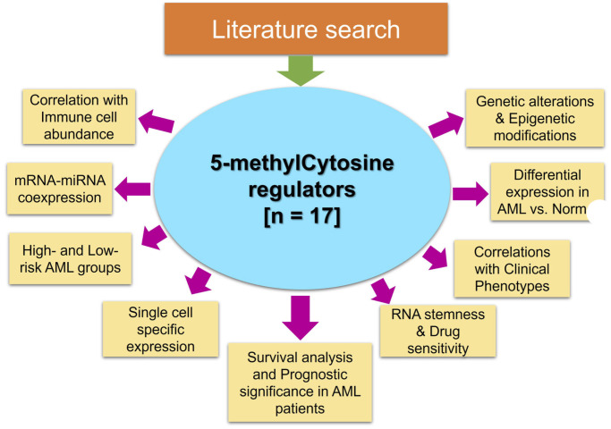
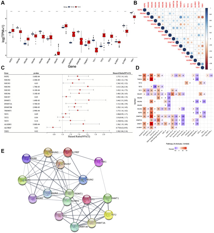
According to the existing literature reports [16, 17], seventeen m5C regulators (NOP2, NSUN2, NSUN3, NSUN4, NSUN5, NSUN6, NSUN7, DNMT1, DNMT3A, DNMT3B, TRDMT1, TET1, TET2, TET3, ALKBH1, ALYREF, YBX1) were identified and explored in this study through various approaches (Figure 1). We first used inSilicoMerging package to merge datasets from GTEx and TCGA databases followed by combat function in SVA package to remove the batch effects, and then compared the expression levels of these regulators between AML and normal samples. Interestingly, we found unexpectedly high expression of nine m5C regulators in the tumor group (NOP2, NSUN3, NSUN6, NSUN7, DNMT1, DNMT3A, TRDMT1, TET2, and TET3), indicating their essential role in AML pathogenesis (Figure 2A). Furthermore, correlation analysis revealed that most of the m5C regulators positively correlated with each other, while NSUN5, ALYREF and YBX1 had negative correlations with other m5C regulators (Figure 2B). To clarify whether m5C regulators could affect the prognosis of AML patients, we performed univariate Cox regression analysis. Our results demonstrated that higher expression of m5C regulators (except for TET1, TET2, TET3, ALYREF) was associated with worse prognosis (Figure 2C). Further, the pathway activity analysis showed that most of the studied m5C regulators activated apoptosis, cell cycle and DNA damage response signaling pathways, whereas inhibited EMT, hormone ER and RAS/MAPK signaling pathways (Figure 2D). Protein-protein interaction (PPI) analysis showed that there are implicated relationships among these m5C regulators (Figure 2E).
Figure 1.
Overall flow of the analysis.
Figure 2.
Expression, correlation, and function of m5C regulators. (A) Boxplot showing differential expression of m5C regulators in AML and normal samples. (B) Pearson correlation analysis, (C) univariate Cox regression analysis, (D) pathway activity analysis and (E) protein-protein interaction (PPI) analysis of m5C regulators. *p < 0.05, **p < 0.01, ***p < 0.001.
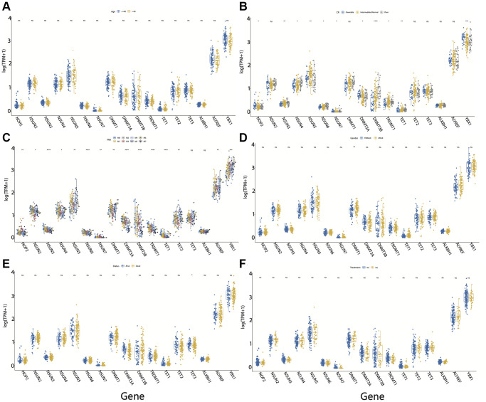
It is unclear whether different clinical indicators can affect the expression of m5C regulators. Our results suggested that the expression of m5C regulators did not differ across the age groups (Figure 3A). In different CR groups, NOP2, NSUN3, NSUN4, NSUN5, NSUN6, NSUN7, DNMT3A, DNMT3B, TRDMT1, TET1 and YBX1 were found to be significantly different (Figure 3B). Furthermore, most m5C regulators (except for TET3 and ALKBH1) were significantly differentially expressed in different FAB groups (Figure 3C). In terms of gender, we did not observe any significant difference in the expression of m5C regulators (except for DNMT1) between male and female patients. (Figure 3D). The expression of only DNMT3A and TET3 varied significantly between different survival statuses of AML patients (Figure 3E). We also reviewed the expression of m5C regulators based on treatment status and found that only TRDMT1 is differentially expressed before and after treatment (Figure 3F).
Figure 3.
Clinical characteristics of m5C regulators in acute myeloid leukemia (AML). Expression of m5C regulators between (A) age groups, (B) CR groups, (C) FAB groups, (D) male and female, (E) survival status, and (F) before and after treatment in AML patients. *p < 0.05, **p < 0.01, ***p < 0.001.
Genetic alterations associated with m5C regulators in AML
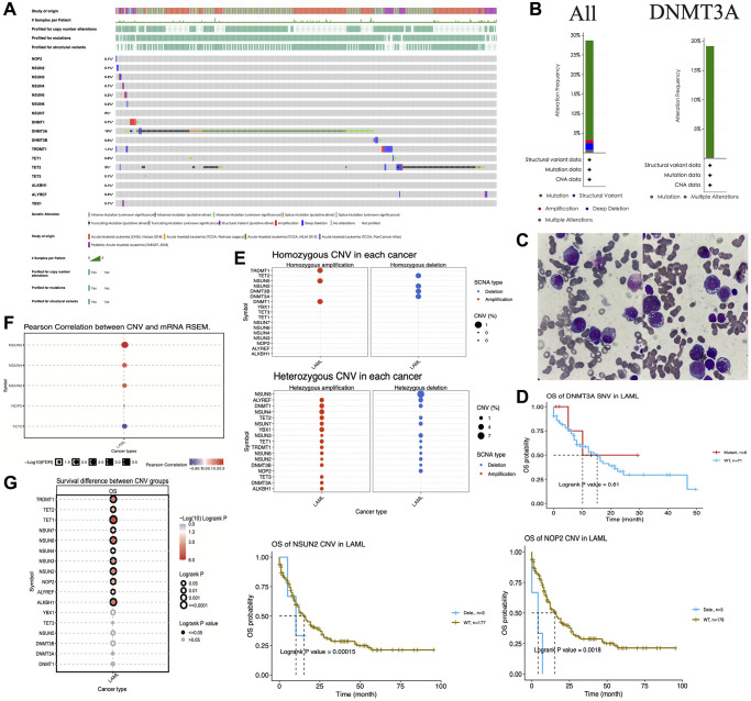
To elucidate the genetic alterations associated with m5C regulators, we integrated 4 datasets [24–26] from different studies using cBioPortal (http://www.cbioportal.org/) tools. We found that these regulators harbor a large number of mutations in AML patients (Figure 4A). Surprisingly, DNMT3A mutation rate was comparatively higher than other regulators and reached 19%, with missense mutations accounting for the majority of all mutations in this gene. TET2 was the next with ~9% mutations, and it mainly harbored truncating mutations (Figure 4A). Figure 4B shows the alteration frequency of all m5C regulators together and DNMT3A alone. The peripheral blood smears of two AML patients showed morphologically aberrant red blood cells (Figure 4C). Genome sequencing of these two patients also identified mutations in DNMT3A.
Figure 4.
Genetic alterations of m5C regulators in acute myeloid leukemia (AML). (A) Mutation analysis of m5C regulators in AML patients. (B) Alteration frequency of m5C regulators and DNMT3A. (C) The peripheral blood smears of two AML patients. (D) Kaplan–Meier curves showing the overall survival of AML patients with DNMT3A mutations. (E) Homozygous and heterozygous CNV analysis of m5C regulators in AML patients. (F) Pearson correlation analysis between CNV and mRNA expression of m5C regulators. (G) Association between overall survival and deletions in m5C regulators in AML patients.
Given this high mutation rate of DNMT3A, we performed Kaplan–Meier survival analysis to validate the effect of DNMT3A mutations. The findings suggested that DNMT3A mutation did not significantly affect the survival of AML patients (Figure 4D). The CNVs of m5C regulators were also analyzed in AML. We found that TRDMT1, NSUN5 and DNMT1 showed homozygous amplifications and TET2, NSUN2, DNMT3B and DNMT3A showed homozygous deletions in AML patients, while almost all m5C regulators showed heterozygous amplifications (except for NSUN5 and NOP2) and heterozygous deletions (except for NSUN4, YBX1, TET3, DNMT3A and ALKBH1) (Figure 4E). We further explored the correlation between the CNV and mRNA expression, and the result suggested that the CNVs of NSUN2, NSUN4, NUSN5 and NOP2 were positively correlated with their mRNA expression levels, whereas TET2 CNV was negatively correlated with TET2 mRNA expression (Figure 4F). We further investigated whether deletions in m5C regulators affect the survival of AML patients. Our results revealed that deletions in NSUN3, NOP2 and a few other regulators were significantly associated with worse prognosis of AML patients (Figure 4G).
DNA methylation of m5C regulators in AML
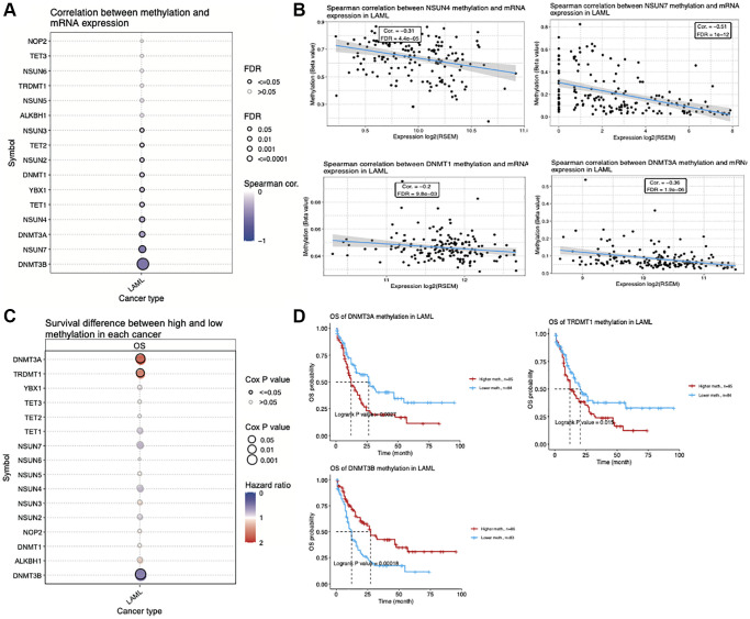
DNA methylation is an epigenetic modification that is involved in the regulation of gene expression [27, 28]. We found that TET2, NSUN2, DNMT1, YBX1, TET1, NSUN4, DNMT3A, NSUN7 and DNMT3B methylation levels were negatively correlated with their expression levels (Figure 5A). Figure 5B shows the Spearman’s correlation values between the methylation and expression levels of four representative genes (NSUN4: R = −0.31, FDR = 4.4e-05; NSUN7: R = −0.51, FDR = 1e-12; DNMT3A: R = −0.36, FDR = 1.9e-06; DNMT1: R = −0.20, FDR = 9.8e-03). Furthermore, the methylation levels of m5C regulators also affects the overall survival of AML patients (Figure 5C). Interestingly, we found that AML patients with high DNA methylation levels of DNMT3A and TRDMT1 have a significantly shorter overall survival, while those with high DNA methylation levels of DNMT3B have a significantly longer overall survival (Figure 5C, 5D).
Figure 5.
Correlation between DNA methylation status and expression of m5C regulators in acute myeloid leukemia (AML). (A) Correlation between the methylation status and mRNA expression of m5C regulators in AML. (B) Scatterplots showing correlation between methylation status and expression of four representative m5C regulators. (C) Association between overall survival of AML patients and methylation status of m5C regulators. (D) Kaplan–Meier curves showing the overall survival of AML patients with DNMT3A, DNMT3B and TRDMT1 methylation.
Evaluation of clinical values of m5C regulated gene subgroups in AML using consensus clustering
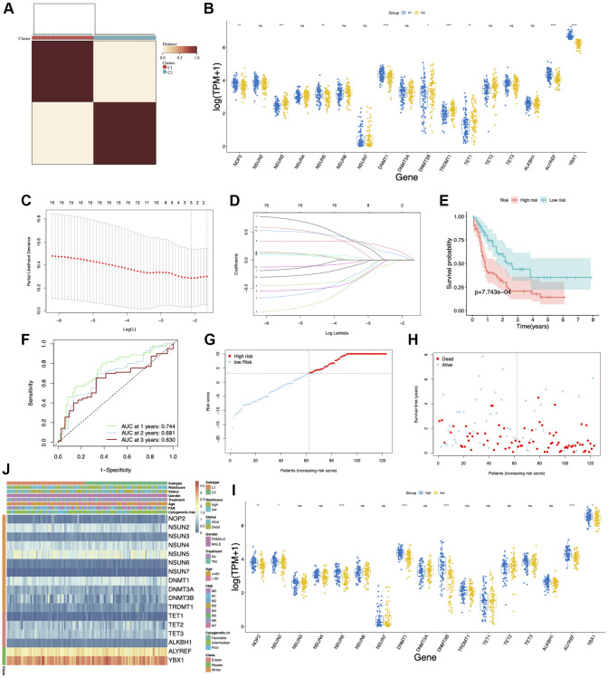
To clarify whether the AML patients can be divided into different subgroups according to the expression of m5C regulators, we performed unsupervised clustering by ConsensusClusterPlus based on the 17 m5C regulators. Two m5C modification patterns (C1 and C2) were finally identified in AML patients (Figure 6A). NOP2, NSUN3, NSUN5, DNMT1, DNMT3B, TRDMT1, TET1, ALYREF and YBX1 were differentially expressed in these two clusters (Figure 6B). The LASSO Cox regression algorithm was applied to these candidate genes in the AML cohort. Eventually, 11 genes were identified based on the criteria to construct the m5C modification signature prognostic model (Figure 6C, 6D). We further divided the AML patients into a high-risk score group and a low-risk score group based on the median risk scores. The K-M plots demonstrated that the low-risk score group survived longer than the high-risk score group (Figure 6E). The ROC curve analysis showed that the AUC of the prognostic model was 0.744, indicating that the model has a good predictive ability (Figure 6F). The risk score and clinical status from two risk groups are shown in Figure 6G, 6H. We also observed the expression of m5C regulators in the low-risk and high-risk group; NOP2, NSUN2, NSUN5, DNMT1, DNMT3B, ALYREF and YBX1 were found to be highly expressed in the high-risk group (Figure 6I). In addition, there was a difference between the AML patients in the two cluster groups in terms of status, treatment, and cytogenetic risk (Figure 6J).
Figure 6.
Consensus clustering and prognostic model of m5C regulators in acute myeloid leukemia (AML). (A) Consensus clustering matrix for k = 2. (B) Box plots showing the expression of m5C regulators in two clusters. (C, D) LASSO Cox regression algorithm was used for calculating the minimum criteria. (E) Kaplan–Meier curves showing the overall survival of AML patients with high- and low-risk scores. (F) ROC curve showing the AUC value of the model for different survival times. (G, H) Distribution of the risk score and survival status. (I) Box plots showing the expression of m5C regulators in high- and low-risk groups. (J) Heatmap and clinicopathological characteristics of AML molecular subtypes and high- and low-risk groups. *p < 0.05, **p < 0.01, ***p < 0.001.
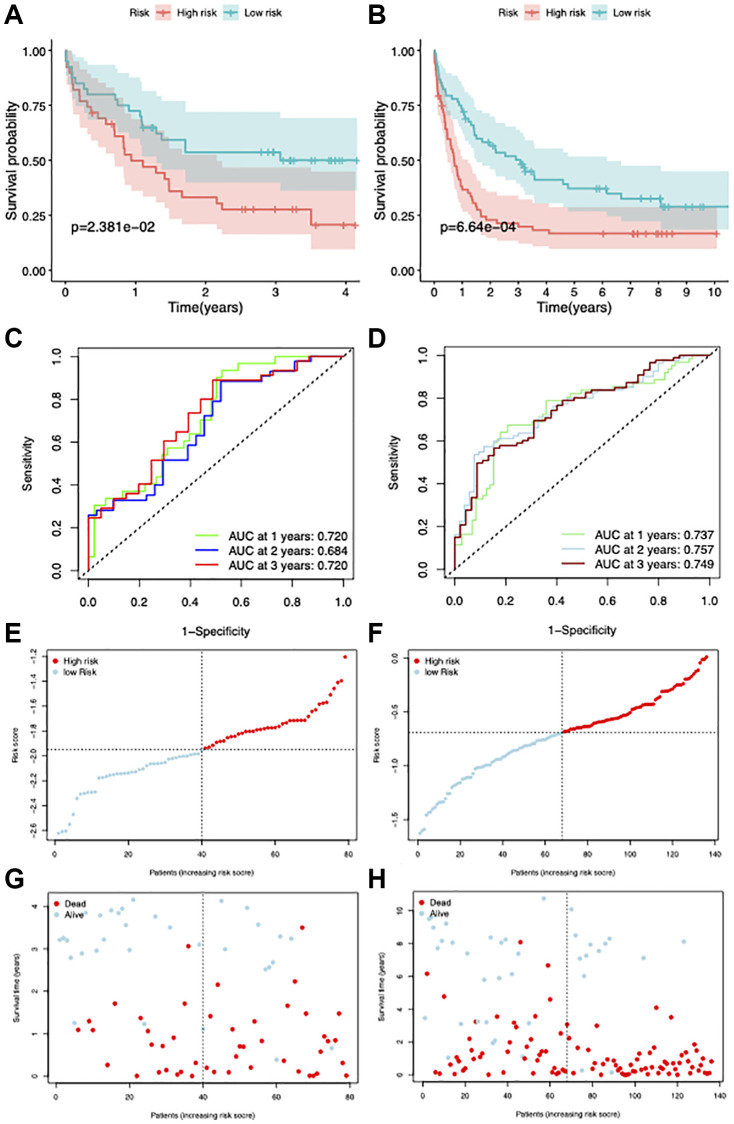
To validate the stability of our model, GSE12417 [29] and GSE37642 [30] datasets were used. The survival statistics for the high- and low-risk groups showed that the proportion of surviving patients was comparatively higher in the low-risk group, which had a better prognosis (Figure 7A, 7B). The AUC values of this model were 0.720 and 0.757 for GSE12417 and GSE37642 datasets, respectively (Figure 7C, 7D). The risk score and clinical status of each case from the two risk groups are shown in Figure 7E–7H.
Figure 7.
Validation of the prognostic model. (A, B) Survival curves of patients in high and low-risk groups in GSE12417 and GSE37642 datasets. (C, D) ROC curves showing the AUC value of the model for different survival times. (E–H) Distribution of the risk score and survival status.
Differential infiltration of immune cells in AML correlates with the cluster groups and risk model of m5C regulators
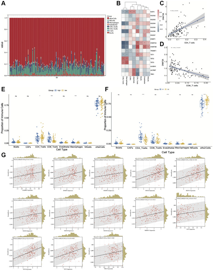
To clarify the relationship between immune cell infiltration and m5C clusters or risk grouping, we used the EPIC algorithm to quantify the proportions of immune cells (Figure 8A). Further, we analyzed the correlation between m5C regulators and the infiltrated immune cell types (Figure 8B–8D). NSUN4 and NSUN6 were negatively correlated with CD8 T-cells and macrophages, and positively correlated with CD4 T-cells. DNMT3A and DNMT3B were negatively correlated with CD8 T-cells and macrophages, and positively correlated with CD4 T-cells, endothelial and NK cells. TET1 and TET2 were positively correlated with CD8 T-cells and macrophages. Next, we wanted to know whether there is a difference in the infiltration of various immune cells between the low- and high-risk groups. The result showed that CD4 T-cells were enriched in the low-risk group, while CD8 T-cells and macrophages were enriched in the high-risk group (Figure 8E). For different m5C subtypes, CD4 T-cells and endothelial cells were enriched in C1 cluster, while CD8 T-cells were enriched in C2 cluster (Figure 8F). To gain more insight into the relationship between immune infiltration and m5C regulators, we calculated stromal score of AML microenvironment by using ESTIMATE and evaluated the correlation between m5C regulators and stromal score. Our results suggested that ALKBH1, DNMT1, DNMT3A, NSUN2, NSUN3, NSUN4, NSUN5, NSUN6, TRDMT1, TET1, TET2, TET3, YBX1 were positively correlated with the stromal score (Figure 8G). The above results suggest that infiltration of different immune cells does exist in high and low risk groups as well as in different m5C clusters. These differences in the proportion of immune cells may be an important factor affecting the prognosis of patients in different groups of AML patients.
Figure 8.
Correlation of the immune cell infiltration with high- and low-risk groups and m5C regulators-based subtypes. (A) The proportion of immune cells by EPIC algorithm. (B) The correlation between m5C regulators and different immune cell types. (C) DNMT3B is positively correlated with CD4 T-cells. (D) DNMT3A is negatively correlated with CD8 T-cells. (E) Differential infiltration of immune cell types between low- and high-risk groups. (F) Differential infiltration of immune cell types between C1 and C2 groups. (G) Correlation between m5C regulators and stromal score. *p < 0.05, **p < 0.01, ***p < 0.001.
Relationship between m5C regulators and RNA based stemness score, drug sensitivity and miRNA expression
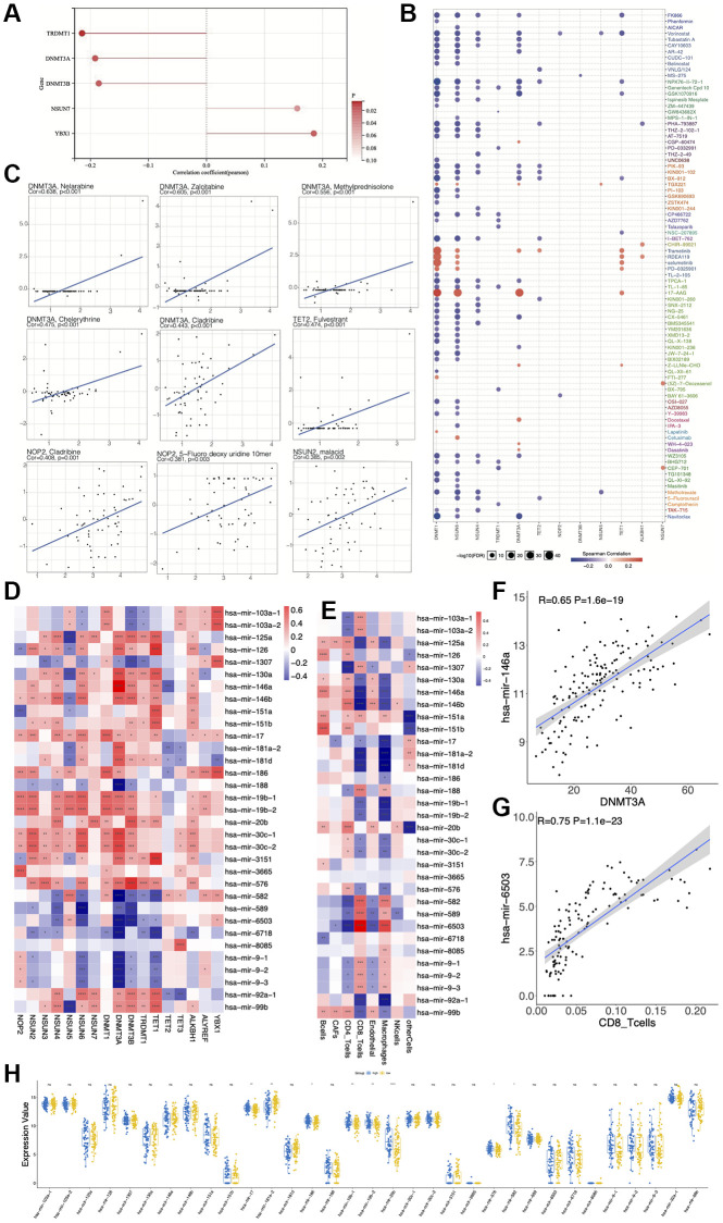
We also wondered whether those m5C regulators could affect other features of AML. Hence, we analyzed RNA Based Stemness Score (RNAss), miRNA expression and drug sensitivity. Our results suggested that TRDMT1, DNMT3A and DNMT3B were negatively correlated, while NSUN7 and YBX1 were positively correlated with the RNAss (Figure 9A). For the drug sensitivity analysis, we calculated the correlation between drug IC50 and gene expression. Our results revealed that m5C regulators were correlated with many drugs (Figure 9B). We found that DNMT3A was positively correlated with Nelarabine (R = 0.638, p < 0.001), Zalcitabine (R = 0.605, p < 0.001), Methylprednisolone (R = 0.556, p < 0.001), Chelerythine (R = 0.475, p < 0.001), Cladribine (R = 0.443, p < 0.001); TET2 was positively correlated with Fulvestrant (R = 0.474, p < 0.01); NOP2 was positively correlated with Cladribine (R = 0.408, p = 0.001), 5-Fluorodeoxyuridine 10mer (R = 0.381, p = 0.003); and NSUN2 was positively correlated with malacid (R = 0.385, p = 0.002) (Figure 9C). Furthermore, we performed correlation analysis between all miRNAs and m5C regulators and found 33 miRNAs to be significantly correlated with m5C genes. We found DNMT3A to play a significant role in this gene-miRNA network, which positively regulated 13 miRNAs (P < 0.0001) and negatively regulated 7 miRNAs (P < 0.0001) (Figure 9D). Further CD8 T-cells played a key role in the immune cell-miRNA network, and positively regulated 4 miRNAs (P < 0.0001), while negatively regulated 5 miRNAs (P < 0.0001) (Figure 9E). As shown in the scatter plot, DNMT3A is positively correlated with hsa-mir-146a (R = 0.65, P = 1.6e-19) (Figure 9F) and CD8-T cells are positively correlated with hsa-mir-6503 (R = 0.75, P = 1.1e-23) (Figure 9G). The differential expression analysis of miRNAs between the low- and high-risk groups revealed hsa-mir-17, hsa-mir-186, hsa-mir-19b-1, hsa-mir-19b-2, hsa-mir-20b, hsa-mir-576 and hsa-mir-582 to be highly expressed, while hsa-mir-181a-2 to be lowly expressed in the high-risk group (Figure 9H).
Figure 9.
Relationship between expression of m5C regulators and RNA based stemness score, miRNA expression and drug sensitivity. (A) Correlation between m5C regulators and RNA based stemness score (RNAss). (B, C) Correlation between drug IC50 and m5C regulators. (D) Correlation between expressions of miRNAs and m5C regulators. (E) Correlation between immune cells and expressions of miRNAs. (F) has-mir-146a is positively correlated with DNMT3A. (G) has-mir-6503 is positively correlated with CD8 T cells. (H) Differential expression of miRNAs between low- and high-risk groups.
Pathways different between the high- and low-risk groups
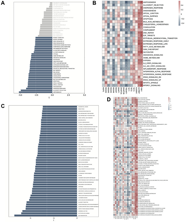
The GSVA tool was used to analyze the differences in the KEGG and hallmark pathways between the high- and low- risk groups. Almost all the hallmark and KEGG pathways were found to be enriched in the high-risk group (Figure 10A, 10C). Further, we performed correlation analyses between the gene expression values and pathway scores. For the hallmark pathways enriched in the high-risk group, we found NOP2, NSUN3, NSUN6, TRDMT1, and TET1 to be significantly negatively correlated with most pathways, while TET3, ALYREF, and YBX1 to be significantly positively correlated (Figure 10B). For KEGG pathways enriched in the high-risk group, we found TRDMT1 and TET1 to be significantly negatively correlated, while ALYREF, and YBX1 to be significantly positively correlated with most of the pathways (Figure 10D).
Figure 10.
GSVA analysis between high- and low-risk AML groups. (A) Enrichment of hallmark pathways between high- and low-risk groups. (B) Correlation between the hallmark pathway scores enriched in high-risk group and m5C regulators. (C) Enrichment of KEGG pathways between high- and low-risk groups. (D) Correlation between the KEGG pathway scores enriched in high-risk group and m5C regulators.
Expression levels of m5C regulators in specific cell types
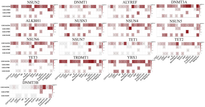
We downloaded the cell type specific expression data including scRNA-seq data from GSE116256 [31], GSE135851 [32], GSE147989 [33], and GSE154109 [34] datasets to illustrate the expression of m5C regulators in specific cells (Figure 11). Based on these four datasets, we identified the expression of the genes in the following cells: conventional CD4 T cells (referred as CD4Tconv), proliferating T cells (Tprolif), CD8 T cells (CD8T), exhausted CD8 T Cells (CD8Tex), natural killer cells (NK), B cells (B), plasma cells (Plasma), monocytes or macrophages (Mono/Macro), mast cells (Mast), erythroid progenitor cells (EryPro), granulocyte-macrophage progenitor cells (GMP), hematopoietic stem cells (HSC), progenitor cells (Progenitor), promonocytes (Promonocyte), endothelial cells (Endothelial) and malignant cells (Malignant). At single-cell level, the expressions of ALYREF, DNMT1, NSUN5, and YBX1 were higher than other genes. Furthermore, ALYREF was found to be enriched in EryPro, DNMT1 was enriched in Tprolif and Promonocyte, NSUN5 was enriched in HSC, while YBX1 was enriched in almost all the cells (Figure 11).
Figure 11.
Expression levels of m5C regulators in specific cell types based on single cell expression data.
RT-qPCR validation of the selected m5C regulators
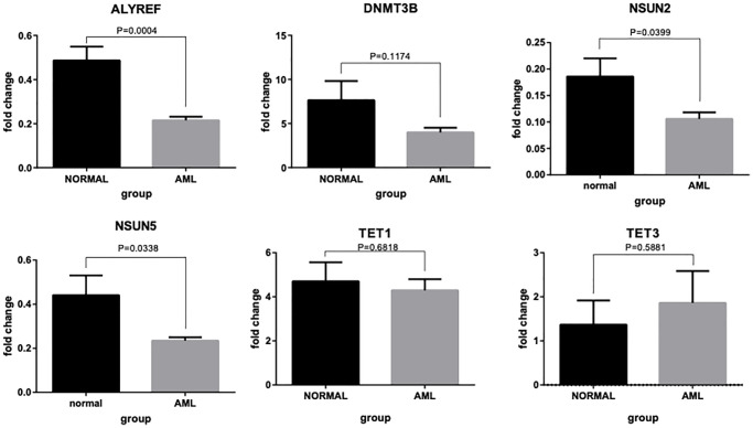
To verify the reliability of our analysis and conclusions, six molecules (ALYREF, DNMT3B, NSUN2, NUSN5, TET1 and TET3) were selected further to confirm their expression levels in AML versus normal tissues using qRT-PCR (Figure 12). The results showed that the expression levels of ALYREF, NSUN2, and NUSN5 were significantly higher in normal controls than in AML patients. This is in agreement with the results of our other analyses. Although we did not observe any significant differences in the expression levels of DNMT3B, TET1 and TET3 between the normal and AML samples, the trends in their expression levels were quite consistent with our other analysis results. Thus, the partial disagreement in the qRT-PCR results could be attributed to the heterogeneity of AML patients.
Figure 12.
RT-qPCR validation of six m5C regulator genes. Bar plots showing differential expressions of ALYREF, DNMT3B, NSUN2, NUSN5, TET1 and TET3 between AML patients and normal controls.
DISCUSSION
Recently, with the development of high-throughput sequencing technology, RNA modification has gradually become an important research area [22, 35]. (m6A) is the most abundant internal modification of mRNA and has been studied in depth [36–38]. Zhang et al. found that m6A reader IGF2BP3, which interacts with and enhances the RCC2 mRNA stability, is essential for AML cell survival [39]. Yankova et al. suggested that inhibition of METTL3 could be a potential target for the treatment of AML [40]. m5C modifications have also been found to play regulatory roles in a variety of biological processes, particularly in tumor progression. Compared to m6A modifications, m5C modifications have been less studied in AML. Cheng et al. reported the mechanism of m5C methylation in AML drug resistance [41]. Liu et al., [42] collected and analyzed multicenter AML data to suggest that not only AML diversity but also generation of complex tumor microenvironments are impacted by m5C modifications. Mutations in the well-known DNA methyltransferase gene DNMT3A are highly recurrent in patients with de novo AML and are known to be independently associated with a poor outcome [43]. More recently, 5-hydroxymethyl cytosine (5-hmC) has garnered lot of attention as a regulatory epigenetic modification with diagnostic and prognostic significance for several cancers [44]. TET family of proteins can oxidize m5C to 5hmC, which is known to play important roles in the pathogenesis of various tumors including AML [45, 46]. Both TET2 and IDH1/2 mutations can impair the production of 5hmC, thus decreasing 5hmC levels. Mutant IDH enzymes increase the production of oncometabolite (R)-2-hydroxyglutarate that competitively inhibits dioxygenase enzymes that are required for m5C to 5-hmC modification and histone tail methylation [47]. Mutations in both these genes have been observed in AML [48]. Overall, the role of m5C modifications in AML remains ambiguous and limited. The present study focuses on investigating the multifaceted characteristics of m5C regulators in AML with the aim of contributing new evidence to the field of AML research.
Network analysis of various molecular and regulatory factors [49, 50] is a strong and widely used approach for exploring the underlying mechanisms of any disease. It has not only been used in AML research [51–53] but also for several other diseases and physiological conditions [54–56]. In the current study, we constructed protein-protein interaction network and revealed the close relationships among the m5C regulators. Additionally, studying the interactions by combining various gene-regulatory molecules including TFs, non-coding RNAs, and m5C regulators could expand our understanding of the regulation of these factors. Furthermore, construction of the independent networks for high- and low-risk AML groups may help us in gaining better insights into the differential role of these m5C regulators as risk factors.
The analysis of molecular subtypes based on some features has been widely used in tumor research. The molecular subtypes can cluster cancer patients into different groups that have different characteristics (e.g., different sensitivity to drugs). Based on such subtypes, cancer patients can be diagnosed and treated more precisely. Jayavelu et al. classified AML into five subtypes based on proteogenomic features; they identified a “Mito-AML” group, which is characterized by high expression of mitochondrial proteins and poor prognosis [57]. Mou et al. identified molecular subtype- specific mRNA expression patterns and studied the characteristics of different subtypes in depth [58]. Mer et al. reported a subtype with NPM1 mutation, which was based on stemness, and suggested applied kinase inhibitors for the treatment of this subtype [59]. However, whether m5C regulators can classify AML patients into different subtypes has not been explored till date. In the present study, AML patients were classified into C1 and C2 subgroups using m5C regulators, and significant differential gene expression and prognosis between the subgroups could be observed. Therefore, it is of great interest to further explore the differences between these two subtypes.
In recent years, with the advancement of immunological research, immunotherapy has been applied to the treatment of AML. These patients have a comparable number of T cells in bone marrow (BM); the percentage of CD3+ and CD8+ T-cells in BM could predict response to the treatments [60]. Williams et al. showed that NK cell-based therapy has a promise to improve the drug response and survival of AML patients [61]. Myeloid-derived suppressor cells (MDSCs) have also been found to dramatically increase in AML patients [62, 63]. MDSCs are proliferated by inducing AML cells through the extracellular vesicle (EVs), which contain several proliferation factors (e.g., MUC1) [64]. In the present study, we were also curious about the changes in the proportion of immune cells in different AML patient subgroups. Therefore, we used the EPIC algorithm for deconvolution to infer immune cell infiltration. We found differences in the proportion of immune cells across molecular subtypes as well as high- or low- risk groups, suggesting that further exploration of the mechanisms of immune cell changes in m5C subtype may provide novel insights into the treatment of AML.
Our drug sensitivity analysis identified several drugs to be significantly correlated with the m5C regulators. For example, DNMT3A correlated with Nelarabine, which is an FDA approved drug and is used for the treatment of hematologic malignancies [65]. Zalcitabine, IC50 value of which was positively correlated with DNMT3A expression has been FDA approved for the treatment of HIV/AIDS [66], while Methylprednisolone is an FDA-approved medication for the management and treatment of allergic conditions and acts as an anti-inflammatory and immunosuppressive agent [67]. Fulvestrant, an antiestrogenic medication that is used for the treatment of receptor-positive metastatic breast cancer was found to be positively correlated with TET2 [68]. Cladribine, an FDA approved drug correlated with both DNMT3A and NOP2. It is currently used for the treatment of hematologic malignancies and a few other diseases [69, 70]. Malacid (Pyrimethamine), which significantly correlated with NSUN2 expression is mainly used for fungal and parasitic infection [71]. The strong positive correlation of these drugs with m5C regulators indicates that many of these that are currently used for other diseases and conditions can be explored as a therapeutic option for AML. Furthermore, the analysis of antagonistic and coordinated effects between drugs will provide references for precise medical treatment of LAML [72].
Certainly, although several meaningful results were obtained in the present study, there were a few limitations. Some of the important molecules we found (e.g., DNMT3A) require further validation using other datasets as well as experiments. Additional external validations based on other cohorts are needed to evaluate whether the m5C molecular subtype and risk-score still perform well in AML patients. In addition, this study indirectly revealed the role of m5C modification in AML. In the future, we will use m5C MeRIP-seq technology to globally map m5C modifications in AML and explore the changes in modification levels in different m5C subtypes as well as in high- and low-risk groups.
MATERIALS AND METHODS
Data source
In this study, we downloaded the standardized data from the UCSC (https://xenabrowser.net/) database. The mRNA expression profiles, mutation annotation data, CNV data and clinical metadata were also downloaded from the UCSC database. The samples with incomplete clinical data were removed.
Gene expression analysis of m5C regulators and survival analysis
Based on existing literature, we collected 17 m5C regulators. We employed the “inSilicoMerging” package to merge GTEx and TCGA database based on the matrixes of all gene expressions [73], and removed the batch effects by empirical bayes algorithm [74], and used the “t-test” algorithm to compare the differential expression of those m5C regulators. Pearson correlation analysis was used to measure the correlation among the m5C regulators. The univariate Cox regression was implemented to analyze the relationship of each m5C regulators’ expression with overall survival.
Analysis of the CNV data and mutation annotation data
The CNVs of m5C regulators, including homozygous and heterozygous amplifications and deletions, were visualized using the bubble plot. The mutation annotation data of AML was used for further analysis with the R package “maftools” [75].
Identification of m5C molecular clustering
We used the 17 m5C regulators for identifying m5C patterns in AML patients. Consensus clustering was performed by using R package “ConsensusClusterPlus” [76] to identify different clusters based on the differential expression of the m5C regulators. The number of classifications was chosen according to the area under the CDF curve and the k-value. The classification step was repeated 1000 times to increase the reliability.
m5C regulators risk model construction
We applied the “glmnet” (https://glmnet.stanford.edu/articles/glmnet.html) [77] and ‘survival’ (https://github.com/therneau/survival) [78] packages for the least absolute shrinkage and selection operator (LASSO) regression to further screen the candidate m5C regulators, and finally a new m5C regulator signature was established. Based on the results of LASSO regression, we developed a risk score formula, which was calculated as follows:
RiskScore = −0.3202 × NSUN3 − 0.2526 × NSUN4 + 0.0372 × NSUN5 − 0.3514 × NSUN6 + 0.3160 × NSUN7 + 0.3244 × DNMT1 −0.3648 × DNMT3A + 0.3476 × DNMT3B −0.0334 × TET2 − 0.0897 × TET3 + 0.2137 × ALKBH1.
In addition, we clustered AML patients into high-risk and low-risk groups based on the median risk score. Then, GSE12417 and GSE37642 datasets were used to validate the established risk model.
Assessment of infiltrated immune cells and correlation analysis
To clarify the immune infiltration differences between different m5C clusters and different risk groups, the “EPIC” algorithm [79] was used to evaluate the score of 8 immune cells. We further applied Wilcox test to compare the differences in immune cell score between different m5C clusters, and high and low risk score groups. Additionally, we applied the ESTIMATE algorithm to calculate the stromal infiltration and then correlation was calculated between m5C regulators and stromal score.
Analysis of miRNA co-expression and drug sensitivity of m5C regulators in AML
We obtained miRNA expression data of AML patients using the TCGA database. Candidate miRNA-m5C regulator pairs were obtained from the ENCORI database [80]. Pearson correlation analysis was performed to calculate the correlation between miRNA expression and that of m5C regulators. The drug sensitivity data were acquired from The Genomics of Drug Sensitivity in Cancer (GDSC) database (https://www.cancerrxgene.org/). We downloaded the IC50 values of each drug using the R package “pRRophetic” [24]. Then, correlation analysis was performed between the sensitivity and the expression of m5C regulators.
GSVA and correlation analysis
GSVA was used to analyze the KEGG and hallmark pathway scores between high-risk and low-risk subtypes [81]. We calculated the correlations between m5C regulators and KEGG or hallmark pathway scores.
Real-time quantitative reverse transcription polymerase chain reaction (RT-qPCR)
Peripheral blood mononuclear cells were isolated from AML patients and the patient’s healthy family members (n = 4) from the Affiliated Hospital of Nantong University for in vivo testing. RNA was extracted using the MiniBEST Universal RNA Extraction Kit (Takara Bio, USA), followed by reverse transcription using a HiScript III 1st Strand cDNA Synthesis Kit (gDNA digester plus) and RT-qPCR. The primers used for the amplification are the following:
5′-CGTGGAGACAGGTGGGAAAC-3′ (forward) 5′-GTTCCTAAGCTGCGACCAGA-3′ (reverse) for ALYREF, 5′-ACAGAAAAGGAATGTGTGAAGGA-3′ (forward) 5′-TGGAATAGGGGACCTCGTGT-3′ (reverse) for DNMT3B, 5′-GACATAGCCTGTCGCTTGGA-3′ (forward) 5′-ATCCGCATAAGACGATGGGAC-3′ (reverse) for NSUN2, 5′-CGTGGAGACAGGTGGGAAAC-3′ (forward) 5′-TCCAGCAACTTCCAGAACGTGA-3′ (reverse) for NSUN5, 5′-TCAAATCTGGGGCCATCGAG-3′ (forward) 5′-TCATCATCGCAGCCCTCTTC-3′ (reverse) for TET1, 5′-CCACCAGCCTCTTTTGGGAA-3′ (forward) 5′-GCTCTGCTACTTCTTTCCTTGC-3′ (reverse) for TET3.
The result of the experiment was represented by relative quantitative analysis of 2−ΔΔCT. ABL1 was used as control gene for normalizing the gene expression.
Data availability statement
Data of mRNA expression profiles, mutation annotation data, CNV data and clinical metadata were from TCGA-LAML in UCSC (https://xenabrowser.net/). RNA-seq data have been deposited in the Gene Expression Omnibus (GEO) with accession numbers GSE12417 and GSE37642. scRNA-seq data have been deposited in GEO with accession numbers GSE116256, GSE135851, GSE147989, and GSE154109.
Code availability statement
The full code used during the current study is available at https://github.com/NTUWangLab/M5C_LAML.
Footnotes
AUTHOR CONTRIBUTIONS: XW, LL and WL conceived and designed the study. YD and FW analyzed the data and drafted the initial manuscript. LX, JM, QL, AKB and QP participated in the analysis of data and experiment. AKB, LL and XW made the revision for the initial manuscript. All authors reviewed and approved the final manuscript.
CONFLICTS OF INTEREST: The authors declare no conflicts of interest related to this study.
FUNDING: This study was funded by Nantong Science and Technology Project (Grant No. JC12022095).
REFERENCES
- 1.Newell LF, Cook RJ. Advances in acute myeloid leukemia. BMJ. 2021; 375:n2026. 10.1136/bmj.n2026 [DOI] [PubMed] [Google Scholar]
- 2.Kantarjian H, Kadia T, DiNardo C, Daver N, Borthakur G, Jabbour E, Garcia-Manero G, Konopleva M, Ravandi F. Acute myeloid leukemia: current progress and future directions. Blood Cancer J. 2021; 11:41. 10.1038/s41408-021-00425-3 [DOI] [PMC free article] [PubMed] [Google Scholar]
- 3.Shallis RM, Wang R, Davidoff A, Ma X, Zeidan AM. Epidemiology of acute myeloid leukemia: Recent progress and enduring challenges. Blood Rev. 2019; 36:70–87. 10.1016/j.blre.2019.04.005 [DOI] [PubMed] [Google Scholar]
- 4.Smith MT, Zhang L. Biomarkers of leukemia risk: benzene as a model. Environ Health Perspect. 1998. (Suppl 4); 106:937–46. 10.1289/ehp.98106s4937 [DOI] [PMC free article] [PubMed] [Google Scholar]
- 5.DiNardo CD, Jonas BA, Pullarkat V, Thirman MJ, Garcia JS, Wei AH, Konopleva M, Döhner H, Letai A, Fenaux P, Koller E, Havelange V, Leber B, et al. Azacitidine and Venetoclax in Previously Untreated Acute Myeloid Leukemia. N Engl J Med. 2020; 383:617–29. 10.1056/NEJMoa2012971 [DOI] [PubMed] [Google Scholar]
- 6.Wei AH, Döhner H, Pocock C, Montesinos P, Afanasyev B, Dombret H, Ravandi F, Sayar H, Jang JH, Porkka K, Selleslag D, Sandhu I, Turgut M, et al. , and QUAZAR AML-001 Trial Investigators. Oral Azacitidine Maintenance Therapy for Acute Myeloid Leukemia in First Remission. N Engl J Med. 2020; 383:2526–37. 10.1056/NEJMoa2004444 [DOI] [PubMed] [Google Scholar]
- 7.Perl AE, Martinelli G, Cortes JE, Neubauer A, Berman E, Paolini S, Montesinos P, Baer MR, Larson RA, Ustun C, Fabbiano F, Erba HP, Di Stasi A, et al. Gilteritinib or Chemotherapy for Relapsed or Refractory FLT3-Mutated AML. N Engl J Med. 2019; 381:1728–40. 10.1056/NEJMoa1902688 [DOI] [PubMed] [Google Scholar]
- 8.Cortes JE, Heidel FH, Hellmann A, Fiedler W, Smith BD, Robak T, Montesinos P, Pollyea DA, DesJardins P, Ottmann O, Ma WW, Shaik MN, Laird AD, et al. Randomized comparison of low dose cytarabine with or without glasdegib in patients with newly diagnosed acute myeloid leukemia or high-risk myelodysplastic syndrome. Leukemia. 2019; 33:379–89. 10.1038/s41375-018-0312-9 [DOI] [PMC free article] [PubMed] [Google Scholar]
- 9.Sonnet M, Claus R, Becker N, Zucknick M, Petersen J, Lipka DB, Oakes CC, Andrulis M, Lier A, Milsom MD, Witte T, Gu L, Kim-Wanner SZ, et al. Early aberrant DNA methylation events in a mouse model of acute myeloid leukemia. Genome Med. 2014; 6:34. 10.1186/gm551 [DOI] [PMC free article] [PubMed] [Google Scholar]
- 10.Zheng X, Gong Y. Functions of RNA N6-methyladenosine modification in acute myeloid leukemia. Biomark Res. 2021; 9:36. 10.1186/s40364-021-00293-w [DOI] [PMC free article] [PubMed] [Google Scholar]
- 11.Sheng Y, Wei J, Yu F, Xu H, Yu C, Wu Q, Liu Y, Li L, Cui XL, Gu X, Shen B, Li W, Huang Y, et al. A critical role of nuclear m6A reader YTHDC1 in leukemogenesis by regulating MCM complex-mediated DNA replication. Blood. 2021; 138:2838–52. 10.1182/blood.2021011707 [DOI] [PMC free article] [PubMed] [Google Scholar]
- 12.Huang Y, Su R, Sheng Y, Dong L, Dong Z, Xu H, Ni T, Zhang ZS, Zhang T, Li C, Han L, Zhu Z, Lian F, et al. Small-Molecule Targeting of Oncogenic FTO Demethylase in Acute Myeloid Leukemia. Cancer Cell. 2019; 35:677–91.e10. 10.1016/j.ccell.2019.03.006 [DOI] [PMC free article] [PubMed] [Google Scholar]
- 13.Davis AG, Johnson DT, Zheng D, Wang R, Jayne ND, Liu M, Shin J, Wang L, Stoner SA, Zhou JH, Ball ED, Tian B, Zhang DE. Alternative polyadenylation dysregulation contributes to the differentiation block of acute myeloid leukemia. Blood. 2022; 139:424–38. 10.1182/blood.2020005693 [DOI] [PMC free article] [PubMed] [Google Scholar]
- 14.Zhao BS, Roundtree IA, He C. Post-transcriptional gene regulation by mRNA modifications. Nat Rev Mol Cell Biol. 2017; 18:31–42. 10.1038/nrm.2016.132 [DOI] [PMC free article] [PubMed] [Google Scholar]
- 15.Wiener D, Schwartz S. The epitranscriptome beyond m6A. Nat Rev Genet. 2021; 22:119–31. 10.1038/s41576-020-00295-8 [DOI] [PubMed] [Google Scholar]
- 16.Chen YS, Yang WL, Zhao YL, Yang YG. Dynamic transcriptomic m5 C and its regulatory role in RNA processing. Wiley Interdiscip Rev RNA. 2021; 12:e1639. 10.1002/wrna.1639 [DOI] [PubMed] [Google Scholar]
- 17.Zhang Q, Liu F, Chen W, Miao H, Liang H, Liao Z, Zhang Z, Zhang B. The role of RNA m5C modification in cancer metastasis. Int J Biol Sci. 2021; 17:3369–80. 10.7150/ijbs.61439 [DOI] [PMC free article] [PubMed] [Google Scholar]
- 18.Hussain S, Aleksic J, Blanco S, Dietmann S, Frye M. Characterizing 5-methylcytosine in the mammalian epitranscriptome. Genome Biol. 2013; 14:215. 10.1186/gb4143 [DOI] [PMC free article] [PubMed] [Google Scholar]
- 19.Nombela P, Miguel-López B, Blanco S. The role of m6A, m5C and Ψ RNA modifications in cancer: Novel therapeutic opportunities. Mol Cancer. 2021; 20:18. 10.1186/s12943-020-01263-w [DOI] [PMC free article] [PubMed] [Google Scholar]
- 20.Song H, Zhang J, Liu B, Xu J, Cai B, Yang H, Straube J, Yu X, Ma T. Biological roles of RNA m5C modification and its implications in Cancer immunotherapy. Biomark Res. 2022; 10:15. 10.1186/s40364-022-00362-8 [DOI] [PMC free article] [PubMed] [Google Scholar]
- 21.Cui L, Ma R, Cai J, Guo C, Chen Z, Yao L, Wang Y, Fan R, Wang X, Shi Y. RNA modifications: importance in immune cell biology and related diseases. Signal Transduct Target Ther. 2022; 7:334. 10.1038/s41392-022-01175-9 [DOI] [PMC free article] [PubMed] [Google Scholar]
- 22.Han X, Wang M, Zhao YL, Yang Y, Yang YG. RNA methylations in human cancers. Semin Cancer Biol. 2021; 75:97–115. 10.1016/j.semcancer.2020.11.007 [DOI] [PubMed] [Google Scholar]
- 23.Guo G, Pan K, Fang S, Ye L, Tong X, Wang Z, Xue X, Zhang H. Advances in mRNA 5-methylcytosine modifications: Detection, effectors, biological functions, and clinical relevance. Mol Ther Nucleic Acids. 2021; 26:575–93. 10.1016/j.omtn.2021.08.020 [DOI] [PMC free article] [PubMed] [Google Scholar]
- 24.Geeleher P, Cox N, Huang RS. pRRophetic: an R package for prediction of clinical chemotherapeutic response from tumor gene expression levels. PLoS One. 2014; 9:e107468. 10.1371/journal.pone.0107468 [DOI] [PMC free article] [PubMed] [Google Scholar]
- 25.Tyner JW, Tognon CE, Bottomly D, Wilmot B, Kurtz SE, Savage SL, Long N, Schultz AR, Traer E, Abel M, Agarwal A, Blucher A, Borate U, et al. Functional genomic landscape of acute myeloid leukaemia. Nature. 2018; 562:526–31. 10.1038/s41586-018-0623-z [DOI] [PMC free article] [PubMed] [Google Scholar]
- 26.Ley TJ, Miller C, Ding L, Raphael BJ, Mungall AJ, Robertson A, Hoadley K, Triche TJ Jr, Laird PW, Baty JD, Fulton LL, Fulton R, Heath SE, et al. , and Cancer Genome Atlas Research Network. Genomic and epigenomic landscapes of adult de novo acute myeloid leukemia. N Engl J Med. 2013; 368:2059–74. 10.1056/NEJMoa1301689 [DOI] [PMC free article] [PubMed] [Google Scholar]
- 27.Greenberg MVC, Bourc'his D. The diverse roles of DNA methylation in mammalian development and disease. Nat Rev Mol Cell Biol. 2019; 20:590–607. 10.1038/s41580-019-0159-6 [DOI] [PubMed] [Google Scholar]
- 28.Nishiyama A, Nakanishi M. Navigating the DNA methylation landscape of cancer. Trends Genet. 2021; 37:1012–27. 10.1016/j.tig.2021.05.002 [DOI] [PubMed] [Google Scholar]
- 29.Metzeler KH, Hummel M, Bloomfield CD, Spiekermann K, Braess J, Sauerland MC, Heinecke A, Radmacher M, Marcucci G, Whitman SP, Maharry K, Paschka P, Larson RA, et al. , and Cancer and Leukemia Group B, and German AML Cooperative Group. An 86-probe-set gene-expression signature predicts survival in cytogenetically normal acute myeloid leukemia. Blood. 2008; 112:4193–201. 10.1182/blood-2008-02-134411 [DOI] [PMC free article] [PubMed] [Google Scholar]
- 30.Herold T, Jurinovic V, Batcha AMN, Bamopoulos SA, Rothenberg-Thurley M, Ksienzyk B, Hartmann L, Greif PA, Phillippou-Massier J, Krebs S, Blum H, Amler S, Schneider S, et al. A 29-gene and cytogenetic score for the prediction of resistance to induction treatment in acute myeloid leukemia. Haematologica. 2018; 103:456–65. 10.3324/haematol.2017.178442 [DOI] [PMC free article] [PubMed] [Google Scholar]
- 31.van Galen P, Hovestadt V, Wadsworth Ii MH, Hughes TK, Griffin GK, Battaglia S, Verga JA, Stephansky J, Pastika TJ, Lombardi Story J, Pinkus GS, Pozdnyakova O, Galinsky I, et al. Single-Cell RNA-Seq Reveals AML Hierarchies Relevant to Disease Progression and Immunity. Cell. 2019; 176:1265–81.e24. 10.1016/j.cell.2019.01.031 [DOI] [PMC free article] [PubMed] [Google Scholar]
- 32.Guo M, Yu JJ, Perl AK, Wikenheiser-Brokamp KA, Riccetti M, Zhang EY, Sudha P, Adam M, Potter A, Kopras EJ, Giannikou K, Potter SS, Sherman S, et al. Single-Cell Transcriptomic Analysis Identifies a Unique Pulmonary Lymphangioleiomyomatosis Cell. Am J Respir Crit Care Med. 2020; 202:1373–87. 10.1164/rccm.201912-2445OC [DOI] [PMC free article] [PubMed] [Google Scholar]
- 33.Riether C, Pabst T, Höpner S, Bacher U, Hinterbrandner M, Banz Y, Müller R, Manz MG, Gharib WH, Francisco D, Bruggmann R, van Rompaey L, Moshir M, et al. Targeting CD70 with cusatuzumab eliminates acute myeloid leukemia stem cells in patients treated with hypomethylating agents. Nat Med. 2020; 26:1459–67. 10.1038/s41591-020-0910-8 [DOI] [PubMed] [Google Scholar]
- 34.Bailur JK, McCachren SS, Pendleton K, Vasquez JC, Lim HS, Duffy A, Doxie DB, Kaushal A, Foster C, DeRyckere D, Castellino S, Kemp ML, Qiu P, et al. Risk-associated alterations in marrow T cells in pediatric leukemia. JCI Insight. 2020; 5:140179. 10.1172/jci.insight.140179 [DOI] [PMC free article] [PubMed] [Google Scholar]
- 35.Barbieri I, Kouzarides T. Role of RNA modifications in cancer. Nat Rev Cancer. 2020; 20:303–22. 10.1038/s41568-020-0253-2 [DOI] [PubMed] [Google Scholar]
- 36.He L, Li H, Wu A, Peng Y, Shu G, Yin G. Functions of N6-methyladenosine and its role in cancer. Mol Cancer. 2019; 18:176. 10.1186/s12943-019-1109-9 [DOI] [PMC free article] [PubMed] [Google Scholar]
- 37.Liu L, Li H, Hu D, Wang Y, Shao W, Zhong J, Yang S, Liu J, Zhang J. Insights into N6-methyladenosine and programmed cell death in cancer. Mol Cancer. 2022; 21:32. 10.1186/s12943-022-01508-w [DOI] [PMC free article] [PubMed] [Google Scholar]
- 38.Wang T, Kong S, Tao M, Ju S. The potential role of RNA N6-methyladenosine in Cancer progression. Mol Cancer. 2020; 19:88. 10.1186/s12943-020-01204-7 [DOI] [PMC free article] [PubMed] [Google Scholar]
- 39.Zhang N, Shen Y, Li H, Chen Y, Zhang P, Lou S, Deng J. The m6A reader IGF2BP3 promotes acute myeloid leukemia progression by enhancing RCC2 stability. Exp Mol Med. 2022; 54:194–205. 10.1038/s12276-022-00735-x [DOI] [PMC free article] [PubMed] [Google Scholar]
- 40.Yankova E, Blackaby W, Albertella M, Rak J, De Braekeleer E, Tsagkogeorga G, Pilka ES, Aspris D, Leggate D, Hendrick AG, Webster NA, Andrews B, Fosbeary R, et al. Small-molecule inhibition of METTL3 as a strategy against myeloid leukaemia. Nature. 2021; 593:597–601. 10.1038/s41586-021-03536-w [DOI] [PMC free article] [PubMed] [Google Scholar]
- 41.Cheng JX, Chen L, Li Y, Cloe A, Yue M, Wei J, Watanabe KA, Shammo JM, Anastasi J, Shen QJ, Larson RA, He C, Le Beau MM, Vardiman JW. RNA cytosine methylation and methyltransferases mediate chromatin organization and 5-azacytidine response and resistance in leukaemia. Nat Commun. 2018; 9:1163. 10.1038/s41467-018-03513-4 [DOI] [PMC free article] [PubMed] [Google Scholar]
- 42.Liu Y, Chen Y, Cai M, Hong Y, Wu X, Li S. m5C methylation modification guides the prognostic value and immune landscapes in acute myeloid leukemia. Aging (Albany NY). 2023; 15:9858–76. 10.18632/aging.205059 [DOI] [PMC free article] [PubMed] [Google Scholar]
- 43.Ley TJ, Ding L, Walter MJ, McLellan MD, Lamprecht T, Larson DE, Kandoth C, Payton JE, Baty J, Welch J, Harris CC, Lichti CF, Townsend RR, et al. DNMT3A mutations in acute myeloid leukemia. N Engl J Med. 2010; 363:2424–33. 10.1056/NEJMoa1005143 [DOI] [PMC free article] [PubMed] [Google Scholar]
- 44.Bachman M, Uribe-Lewis S, Yang X, Williams M, Murrell A, Balasubramanian S. 5-Hydroxymethylcytosine is a predominantly stable DNA modification. Nat Chem. 2014; 6:1049–55. 10.1038/nchem.2064 [DOI] [PMC free article] [PubMed] [Google Scholar]
- 45.Xu T, Gao H. Hydroxymethylation and tumors: can 5-hydroxymethylation be used as a marker for tumor diagnosis and treatment? Hum Genomics. 2020; 14:15. 10.1186/s40246-020-00265-5 [DOI] [PMC free article] [PubMed] [Google Scholar]
- 46.Huang H, Jiang X, Li Z, Li Y, Song CX, He C, Sun M, Chen P, Gurbuxani S, Wang J, Hong GM, Elkahloun AG, Arnovitz S, et al. TET1 plays an essential oncogenic role in MLL-rearranged leukemia. Proc Natl Acad Sci U S A. 2013; 110:11994–9. 10.1073/pnas.1310656110 [DOI] [PMC free article] [PubMed] [Google Scholar]
- 47.Garrett-Bakelman FE, Melnick AM. Mutant IDH: a targetable driver of leukemic phenotypes linking metabolism, epigenetics and transcriptional regulation. Epigenomics. 2016; 8:945–57. 10.2217/epi-2016-0008 [DOI] [PMC free article] [PubMed] [Google Scholar]
- 48.Figueroa ME, Abdel-Wahab O, Lu C, Ward PS, Patel J, Shih A, Li Y, Bhagwat N, Vasanthakumar A, Fernandez HF, Tallman MS, Sun Z, Wolniak K, et al. Leukemic IDH1 and IDH2 mutations result in a hypermethylation phenotype, disrupt TET2 function, and impair hematopoietic differentiation. Cancer Cell. 2010; 18:553–67. 10.1016/j.ccr.2010.11.015 [DOI] [PMC free article] [PubMed] [Google Scholar]
- 49.Chen C, Jiang L, Fu G, Wang M, Wang Y, Shen B, Liu Z, Wang Z, Hou W, Berceli SA, Wu R. An omnidirectional visualization model of personalized gene regulatory networks. NPJ Syst Biol Appl. 2019; 5:38. 10.1038/s41540-019-0116-1 [DOI] [PMC free article] [PubMed] [Google Scholar]
- 50.Wu R, Jiang L. Recovering dynamic networks in big static datasets. Phys Rep. 2021; 912:1–57. 10.1016/j.physrep.2021.01.003 [DOI] [Google Scholar]
- 51.Foroushani A, Agrahari R, Docking R, Chang L, Duns G, Hudoba M, Karsan A, Zare H. Large-scale gene network analysis reveals the significance of extracellular matrix pathway and homeobox genes in acute myeloid leukemia: an introduction to the Pigengene package and its applications. BMC Med Genomics. 2017; 10:16. 10.1186/s12920-017-0253-6 [DOI] [PMC free article] [PubMed] [Google Scholar]
- 52.Chen T, Zhang J, Wang Y, Zhou H. Identification of Survival-Related Genes in Acute Myeloid Leukemia (AML) Based on Cytogenetically Normal AML Samples Using Weighted Gene Coexpression Network Analysis. Dis Markers. 2022; 2022:5423694. 10.1155/2022/5423694 [DOI] [PMC free article] [PubMed] [Google Scholar]
- 53.Assi SA, Imperato MR, Coleman DJL, Pickin A, Potluri S, Ptasinska A, Chin PS, Blair H, Cauchy P, James SR, Zacarias-Cabeza J, Gilding LN, Beggs A, et al. Subtype-specific regulatory network rewiring in acute myeloid leukemia. Nat Genet. 2019; 51:151–62. 10.1038/s41588-018-0270-1 [DOI] [PMC free article] [PubMed] [Google Scholar]
- 54.Sun L, Jiang L, Grant CN, Wang HG, Gragnoli C, Liu Z, Wu R. Computational Identification of Gene Networks as a Biomarker of Neuroblastoma Risk. Cancers (Basel). 2020; 12:2086. 10.3390/cancers12082086 [DOI] [PMC free article] [PubMed] [Google Scholar]
- 55.Gu Q, Kumar A, Hook M, Xu F, Bajpai AK, Starlard-Davenport A, Yue J, Jablonski MM, Lu L. Exploring Early-Stage Retinal Neurodegeneration in Murine Pigmentary Glaucoma: Insights From Gene Networks and miRNA Regulation Analyses. Invest Ophthalmol Vis Sci. 2023; 64:25. 10.1167/iovs.64.12.25 [DOI] [PMC free article] [PubMed] [Google Scholar]
- 56.Wang X, Bajpai AK, Gu Q, Ashbrook DG, Starlard-Davenport A, Lu L. Weighted gene co-expression network analysis identifies key hub genes and pathways in acute myeloid leukemia. Front Genet. 2023; 14:1009462. 10.3389/fgene.2023.1009462 [DOI] [PMC free article] [PubMed] [Google Scholar]
- 57.Jayavelu AK, Wolf S, Buettner F, Alexe G, Häupl B, Comoglio F, Schneider C, Doebele C, Fuhrmann DC, Wagner S, Donato E, Andresen C, Wilke AC, et al. The proteogenomic subtypes of acute myeloid leukemia. Cancer Cell. 2022; 40:301–17.e12. 10.1016/j.ccell.2022.02.006 [DOI] [PubMed] [Google Scholar]
- 58.Mou T, Pawitan Y, Stahl M, Vesterlund M, Deng W, Jafari R, Bohlin A, Österroos A, Siavelis L, Bäckvall H, Erkers T, Kiviluoto S, Seashore-Ludlow B, et al. The transcriptome-wide landscape of molecular subtype-specific mRNA expression profiles in acute myeloid leukemia. Am J Hematol. 2021; 96:580–8. 10.1002/ajh.26141 [DOI] [PubMed] [Google Scholar]
- 59.Mer AS, Heath EM, Madani Tonekaboni SA, Dogan-Artun N, Nair SK, Murison A, Garcia-Prat L, Shlush L, Hurren R, Voisin V, Bader GD, Nislow C, Rantalainen M, et al. Biological and therapeutic implications of a unique subtype of NPM1 mutated AML. Nat Commun. 2021; 12:1054. 10.1038/s41467-021-21233-0 [DOI] [PMC free article] [PubMed] [Google Scholar]
- 60.Daver N, Garcia-Manero G, Basu S, Boddu PC, Alfayez M, Cortes JE, Konopleva M, Ravandi-Kashani F, Jabbour E, Kadia T, Nogueras-Gonzalez GM, Ning J, Pemmaraju N, et al. Efficacy, Safety, and Biomarkers of Response to Azacitidine and Nivolumab in Relapsed/Refractory Acute Myeloid Leukemia: A Nonrandomized, Open-Label, Phase II Study. Cancer Discov. 2019; 9:370–83. 10.1158/2159-8290.CD-18-0774 [DOI] [PMC free article] [PubMed] [Google Scholar]
- 61.Williams BA, Wang XH, Leyton JV, Maghera S, Deif B, Reilly RM, Minden MD, Keating A. CD16+NK-92 and anti-CD123 monoclonal antibody prolongs survival in primary human acute myeloid leukemia xenografted mice. Haematologica. 2018; 103:1720–9. 10.3324/haematol.2017.187385 [DOI] [PMC free article] [PubMed] [Google Scholar]
- 62.Sun H, Li Y, Zhang ZF, Ju Y, Li L, Zhang BC, Liu B. Increase in myeloid-derived suppressor cells (MDSCs) associated with minimal residual disease (MRD) detection in adult acute myeloid leukemia. Int J Hematol. 2015; 102:579–86. 10.1007/s12185-015-1865-2 [DOI] [PubMed] [Google Scholar]
- 63.Lu T, Gabrilovich DI. Molecular pathways: tumor-infiltrating myeloid cells and reactive oxygen species in regulation of tumor microenvironment. Clin Cancer Res. 2012; 18:4877–82. 10.1158/1078-0432.CCR-11-2939 [DOI] [PMC free article] [PubMed] [Google Scholar]
- 64.Pyzer AR, Stroopinsky D, Rajabi H, Washington A, Tagde A, Coll M, Fung J, Bryant MP, Cole L, Palmer K, Somaiya P, Karp Leaf R, Nahas M, et al. MUC1-mediated induction of myeloid-derived suppressor cells in patients with acute myeloid leukemia. Blood. 2017; 129:1791–801. 10.1182/blood-2016-07-730614 [DOI] [PMC free article] [PubMed] [Google Scholar]
- 65.Sanford M, Lyseng-Williamson KA. Nelarabine. Drugs. 2008; 68:439–47. 10.2165/00003495-200868040-00004 [DOI] [PubMed] [Google Scholar]
- 66.Whittington R, Brogden RN. Zalcitabine. A review of its pharmacology and clinical potential in acquired immunodeficiency syndrome (AIDS). Drugs. 1992; 44:656–83. 10.2165/00003495-199244040-00009 [DOI] [PubMed] [Google Scholar]
- 67.Ranjbar K, Moghadami M, Mirahmadizadeh A, Fallahi MJ, Khaloo V, Shahriarirad R, Erfani A, Khodamoradi Z, Gholampoor Saadi MH. Methylprednisolone or dexamethasone, which one is superior corticosteroid in the treatment of hospitalized COVID-19 patients: a triple-blinded randomized controlled trial. BMC Infect Dis. 2021; 21:337. 10.1186/s12879-021-06045-3 [DOI] [PMC free article] [PubMed] [Google Scholar]
- 68.Croxtall JD, McKeage K. Fulvestrant: a review of its use in the management of hormone receptor-positive metastatic breast cancer in postmenopausal women. Drugs. 2011; 71:363–80. 10.2165/11204810-000000000-00000 [DOI] [PubMed] [Google Scholar]
- 69.Jain P, Pemmaraju N, Ravandi F. Update on the biology and treatment options for hairy cell leukemia. Curr Treat Options Oncol. 2014; 15:187–209. 10.1007/s11864-014-0285-5 [DOI] [PMC free article] [PubMed] [Google Scholar]
- 70.Leist TP, Weissert R. Cladribine: mode of action and implications for treatment of multiple sclerosis. Clin Neuropharmacol. 2011; 34:28–35. 10.1097/WNF.0b013e318204cd90 [DOI] [PubMed] [Google Scholar]
- 71.Gatton ML, Martin LB, Cheng Q. Evolution of resistance to sulfadoxine-pyrimethamine in Plasmodium falciparum. Antimicrob Agents Chemother. 2004; 48:2116–23. 10.1128/AAC.48.6.2116-2123.2004 [DOI] [PMC free article] [PubMed] [Google Scholar]
- 72.Sang M, Dong A, Wu S, Li F, Wang J, Griffin C, Wu R. A graph model of combination therapies. Drug Discov Today. 2022; 27:1210–7. 10.1016/j.drudis.2022.02.001 [DOI] [PubMed] [Google Scholar]
- 73.Taminau J, Meganck S, Lazar C, Steenhoff D, Coletta A, Molter C, Duque R, de Schaetzen V, Weiss Solís DY, Bersini H, Nowé A. Unlocking the potential of publicly available microarray data using inSilicoDb and inSilicoMerging R/Bioconductor packages. BMC Bioinformatics. 2012; 13:335. 10.1186/1471-2105-13-335 [DOI] [PMC free article] [PubMed] [Google Scholar]
- 74.Johnson WE, Li C, Rabinovic A. Adjusting batch effects in microarray expression data using empirical Bayes methods. Biostatistics. 2007; 8:118–27. 10.1093/biostatistics/kxj037 [DOI] [PubMed] [Google Scholar]
- 75.Mayakonda A, Lin DC, Assenov Y, Plass C, Koeffler HP. Maftools: efficient and comprehensive analysis of somatic variants in cancer. Genome Res. 2018; 28:1747–56. 10.1101/gr.239244.118 [DOI] [PMC free article] [PubMed] [Google Scholar]
- 76.Wilkerson MD, Hayes DN. ConsensusClusterPlus: a class discovery tool with confidence assessments and item tracking. Bioinformatics. 2010; 26:1572–3. 10.1093/bioinformatics/btq170 [DOI] [PMC free article] [PubMed] [Google Scholar]
- 77.Friedman J, Hastie T, Tibshirani R. Regularization Paths for Generalized Linear Models via Coordinate Descent. J Stat Softw. 2010; 33:1–22. [PMC free article] [PubMed] [Google Scholar]
- 78.Lin H, Zelterman D. Modeling survival data: extending the Cox model. Technometrics. 2002; 44:85–6. 10.1198/tech.2002.s656 [DOI] [Google Scholar]
- 79.Racle J, de Jonge K, Baumgaertner P, Speiser DE, Gfeller D. Simultaneous enumeration of cancer and immune cell types from bulk tumor gene expression data. Elife. 2017; 6:e26476. 10.7554/eLife.26476 [DOI] [PMC free article] [PubMed] [Google Scholar]
- 80.Li JH, Liu S, Zhou H, Qu LH, Yang JH. starBase v2.0: decoding miRNA-ceRNA, miRNA-ncRNA and protein-RNA interaction networks from large-scale CLIP-Seq data. Nucleic Acids Res. 2014; 42:D92–7. 10.1093/nar/gkt1248 [DOI] [PMC free article] [PubMed] [Google Scholar]
- 81.Hänzelmann S, Castelo R, Guinney J. GSVA: gene set variation analysis for microarray and RNA-seq data. BMC Bioinformatics. 2013; 14:7. 10.1186/1471-2105-14-7 [DOI] [PMC free article] [PubMed] [Google Scholar]
Associated Data
This section collects any data citations, data availability statements, or supplementary materials included in this article.
Data Availability Statement
Data of mRNA expression profiles, mutation annotation data, CNV data and clinical metadata were from TCGA-LAML in UCSC (https://xenabrowser.net/). RNA-seq data have been deposited in the Gene Expression Omnibus (GEO) with accession numbers GSE12417 and GSE37642. scRNA-seq data have been deposited in GEO with accession numbers GSE116256, GSE135851, GSE147989, and GSE154109.


