Abstract
This paper reports five cases of intact adult male crossbreed cats presenting with myiasis caused by Cochliomyia hominivorax. Three were stray animals that died despite treatment due to the severity of lesions while two were client-owned cats previously treated with cryosurgery which completely recovered. Myiasis caused by the New World screwworm fly in cats appears to be more frequent than previously thought, deserving more attention from both veterinary practitioners and owners.
Myiasis is the infestation and the harmful action provoked by larvae of certain Diptera families on vertebrate animals. 1 In the Americas one of the most studied species is Cochliomyia hominivorax, known as the New World screwworm, because of its great impact on both animal and human health and therefore, on the economies of many countries. 2 The results of these investigations were used to establish successful eradication plans in the United States, Mexico, and Central America. 3 Recently, this species was included in the list of Global Early Warning Systems for Major Animal Diseases, including Zoonoses by the World Health Organization. 4
C hominivorax females oviposit on recent injuries of hosts. Larvae hatch during the first 24 h, actively invading lesions. They are highly voracious and feed on living tissues by releasing proteolytic enzymes that cause tissue digestion thus increasing damage. 1,5 Diagnosis can be easily done, based on serohemorrhagic exudation in association with the characteristic offensive smell of wounds and maggots identification. 6 Clinical severity and resolution depends on the site and extent of the lesion, the severity of the infestation and rapidity of diagnosis. Hence, minor and superficial infestations are typically benign and easy to diagnose and treat. On the other hand, massive and invasive infestations commonly lead to death if not treated at an early stage. 6,7 In small animal practice, ivermectin and pyrethrin or pyrethroids sprays are commonly used as treatment of screwworm myiasis. 8,9 Recently, nitempyran has been routinely administered to dogs in order to treat screwworm infestations in a single dose, following the recommended doses for flea control. 9
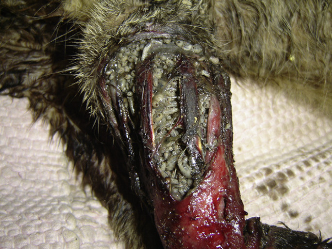
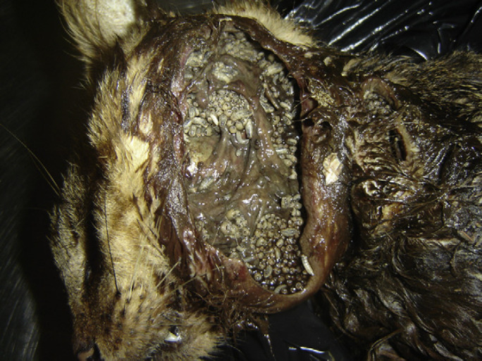
Five cases of intact adult male crossbreed cats with myiasis are presented. Three were stray cats recovered from the streets, presenting with serious wounds admitted in a veterinary clinic in Rio de Janeiro municipality. One of the stray cats with an unknown history had a fracture on the left humerus and with injuries that predisposed it to myiasis infestation (Fig 1). The other two presented with serious lesions in the right front leg and left lateral region of the neck extending to the face, respectively (Fig 2). The remaining two were client-owned animals previously treated with cryosurgery in the Small Animal Hospital of the Universidade Federal Rural do Rio de Janeiro, Municipality of Seropédica. Both client-owned cats were previously treated with cryosurgery; the first one for squamous cell carcinoma causing facial lesions. Thirty-six days after the procedure screwworm infestation was observed in a surgical wound at the level of temporalis muscle. The second cat presented with multiple ulcerative lesions caused by the thermally dimorphic fungus Sporothrix schenckii. Eighteen days following cryosurgery, myiasis was observed in a surgical wound at the level of the left elbow joint. Debridement of necrotic tissue and mechanical removal of accessible larvae were promptly performed as part of the treatment in all animals. For supplementary larval removal, nitempyran (Capstar; Novartis Animal Health) was orally administered following the approximately recommended doses for flea control (1 mg/kg), as this drug has been demonstrated as an efficient larvicidal against screwworm infestations due to C hominivorax on dogs. 9 The animals also received flunixin meglumine (1 mg/kg, SC, 3 days) and enrofloxacin (5 mg/kg, SC, 7 days) as supportive treatment. Larvae were mechanically removed from all animals and fixed in ethanol 70% for identification under stereomicroscope. 1
Fig 1.

Stray cat presenting myiasis by Cochliomyia hominivorax at left front leg.
Fig 2.

Stray cat presenting myiasis by Cochliomyia hominivorax on the left region of the neck extending to the face.
Maggots were classified as third-instar C hominivorax larvae. As demonstrated in previous studies, this infestation is quite common in feline practice in Rio de Janeiro State. 8,10 This suggests that it may occur in other countries throughout neotropical America.
Nitempyran lead to expulsion and larvicidal effects when administered to infested cats as reported on dogs. 9 A few hours after administration larvae actively left wounds in all cats. Larvae that penetrated deeper into the lesion usually die without being expelled, requiring mechanical removal. Another therapeutic option is the use of subcutaneous ivermectin (50 μg/kg) associated to antibiotic therapy, previously reported in cats. 8
The fact that all animals were intact crossbreed males agrees with previous reports in the literature. These are more likely to suffer injury as a result of territorial and reproductive competitive fighting behavior and have a higher incidence of lesions in the front part of the body. Moreover, stray cats are more susceptible to accidents, such as canine attacks and road traffic accidents. 10
In the present study, client-owned cats were successfully treated because the diagnosis was made early and the lesions were not advanced. The cause of the myiasis infestation in both cases was inadequate postoperative wound care. 10
In contrast, the two stray cats with facial and neck lesions died 1 day later; one with a broken front leg died 4 days later. These animals had massive infestation in extensive lesions. Myiasis diagnoses and treatment in stray cats are known to be difficult. Besides avoiding contact with humans because their feral behavior, injured animals often hide in places of limited access, aggravating lesions and making treatment frequently impossible. 8
In comparison to other myiasis previously reported in domestic cats, C hominivorax infestation can be considered more aggressive. Many other infestations such as those caused by Dermatobia hominis, 11 Lucilia eximia, 12 L sericata, 13,14 Chrysomyia erytrocephala, 15 and Cuterebra species, 16 larvae penetrate subcutaneously, are relatively benign and are routinely effectively treated. However, in some cases, Cuterebra infestations can cause serious ophthalmological or neurological disorders if the larvae migrate through atypical host tissues and the outcome can be fatal. 17
Clinical case reports of New World screwworm myiasis are relevant due to its overall importance and frequent occurrence in feline veterinary practice in many countries, especially in outdoor intact male cats. Nitempyran is useful in promoting larval expulsion and should be recommended as treatment for this parasitosis. Small animal practitioners should be aware of this myiasis and be able to appropriately inform owners on its recognition, prevention and treatment.
References
- 1.Guimarães J.H., Papavero N., Prado A.P. As miíases da Região Neotropical, Rev Bras Zool 1, 1983, 239–416. [Google Scholar]
- 2.Grisi L., Massard C.L., Moya G.E., Pereira J. Impacto econômico das principais ectoparasitoses em bovinos no Brasil, Hora Vet 21, 2002, 8–10. [Google Scholar]
- 3.Moya G.E. Erradicação ou manejo integrado das miíases neotropicais das Américas, Pesq Vet Bras 23, 2003, 131–138. [Google Scholar]
- 4.WHO http://www.who.int/zoonoses/outbreaks/glews/en/index.html2007, (accessed December 22, 2008) [Google Scholar]
- 5.Alexander J.L. Screwworms, J Am Vet Med Assoc 228, 2006, 357–367. [DOI] [PubMed] [Google Scholar]
- 6.Soulsby E.J.L. Parasitologia y enfermidades parasitárias en los animales domésticos, 2nd edn., 1987, Interamericana: Mexico. [Google Scholar]
- 7.Scott D.W., Miller W.H., Griffin C.E. Muller & Kirk's Small Animal Dermatology, 6th edn., 2001, W B Saunders: Philadelphia. [Google Scholar]
- 8.Mendes-De-Almeida F., Labarthe N., Guerrero J., et al. Cochliomyia hominivorax myiasis in a colony of stray cats (Felis catus Linnaeus, 1758) in Rio de Janeiro, RJ, Vet Parasitol 146, 2007, 376–378. [DOI] [PubMed] [Google Scholar]
- 9.Machado M.L.S., Rodrigues E.M.P. Emprego do nitenpyram como larvicida em miíases caninas por Cochliomyia hominivorax, Acta Sci Vet 30, 2002, 59–62. [Google Scholar]
- 10.Cramer-Ribeiro B.C., Sanavria A., Oliveira M.Q., Souza F.S., Rocco F.S., Cardoso P.G. Inquérito sobre os casos de miíases por Cochliomyia hominivorax em gatos das zonas norte, sul e oeste e do centro do município do Rio de Janeiro no ano 2000, Braz J Vet Res An Sci 39, 2002, 165–170. [Google Scholar]
- 11.Marcial T., Roman E.M., Pivat I.V. Estúdio retrospectivo de doscientos casos de miíasis presentados en el Hospital de Pequeños Animales ‘Dr Daniel Cabello-Mariani’. Facultad de Ciencias Veterinárias, Universidad Central de Venezuela durante los años de 1996 a 1999, Rev Fac Cs Vet UCV 44, 2003, 87–95. [Google Scholar]
- 12.Madeira N.G., Silveira G.A.R., Pavan C. The occurrence of primary myiasis caused by Phaenicia eximia (Diptera: Calliphoridae), Mem Inst Oswaldo Cruz 84, 1989, 341. [Google Scholar]
- 13.Marluis J.C., Schnack J.A. Cervinazzo, I, Quintana, C. Cochliomyia hominivorax (Coquerel, 1858) and Phaenicia sericata (Meigen, 1826) parasiting domestic animals in Buenos Aires and vicinities (Diptera, Calliphoridae), Mem Inst Oswaldo Cruz 89, 1994, 139. [DOI] [PubMed] [Google Scholar]
- 14.Vignau M.L., Arias D.O. Myiasis cutaneo-ulcerosas en pequeños animales, Parasitol Dia 21, 1997, 36–39. [Google Scholar]
- 15.Rodriguez J.M., Perez M. Cutaneous myiasis in three obese cats, Vet Q 18, 1996, 102–103. [DOI] [PubMed] [Google Scholar]
- 16.Slansky F. Feline Cuterebriasis caused by Lagomorph-Infesting Cuterebra spp. Larva, J Parasitol 93, 2007, 959–961. [DOI] [PubMed] [Google Scholar]
- 17.Wyman M., Starkey R., Weisbrode S., Filko D., Grandstaff R., Ferrebee E. Ophthalmomyiasis (interna posterior) of the posterior segment and central nervous system myiasis: Cuterebra spp in a cat, Vet Ophthalmol 8, 2005, 77–80. [DOI] [PubMed] [Google Scholar]


