Abstract
Introduction
We encountered a colon cancer case with a very rare anomaly of the middle colic artery (MCA) originating from the splenic artery (SA).
Case Presentation
A woman was referred to our hospital for transverse colon cancer. Three-dimensional computed tomography (3D-CT) angiography showed an anomalous MCA originating from the SA rather than from the superior mesenteric artery (SMA) as is typical. Laparoscopic left hemicolectomy with D3 lymph node dissection was performed. The lymph nodes around the SMA were dissected from the caudal view, confirming the absence of a typical MCA. An anomalous SA-originating MCA was identified just below the pancreas, where it was clipped and ligated; subsequently, total mesenteric excision was achieved.
Conclusion
As D3 lymph node dissection for transverse colon cancer is technically difficult, 3D-CT angiography is useful for identifying vascular anomalies preoperatively, thereby avoiding intraoperative injury. This is the first case report of laparoscopic colectomy associated with a SA-originating MCA anomaly.
Keywords: Colon cancer, Middle colic artery, Splenic artery, Anomaly, Laparoscopic surgery
Introduction
Laparoscopic lymph node dissection associated with the middle colic artery (MCA) is considered difficult mainly because the root of the MCA originating from the superior mesenteric artery (SMA) is adjacent to the pancreas. In cases of rare anatomical vascular variations, precise mesenteric excision for transverse colon cancer may be more challenging than it would be otherwise. We herein report a case of successful laparoscopic resection with D3 lymph node dissection for a transverse colon cancer case with an anomalous MCA originating from the splenic artery (SA).
Anomalous MCA originating from SA is reported to be extremely rare [1, 2], and there have been no surgical case reports for colon cancer with this type of vascular anomaly. Using preoperative three-dimensional computed tomography (3D-CT) angiography, the anomalous MCA was preoperatively recognized, and lymph node dissection was safely performed. The CARE Checklist has been completed by the authors for this case report, and attached as supplementary material (for all online suppl. material, see https://doi.org/10.1159/000536672).
Case Presentation
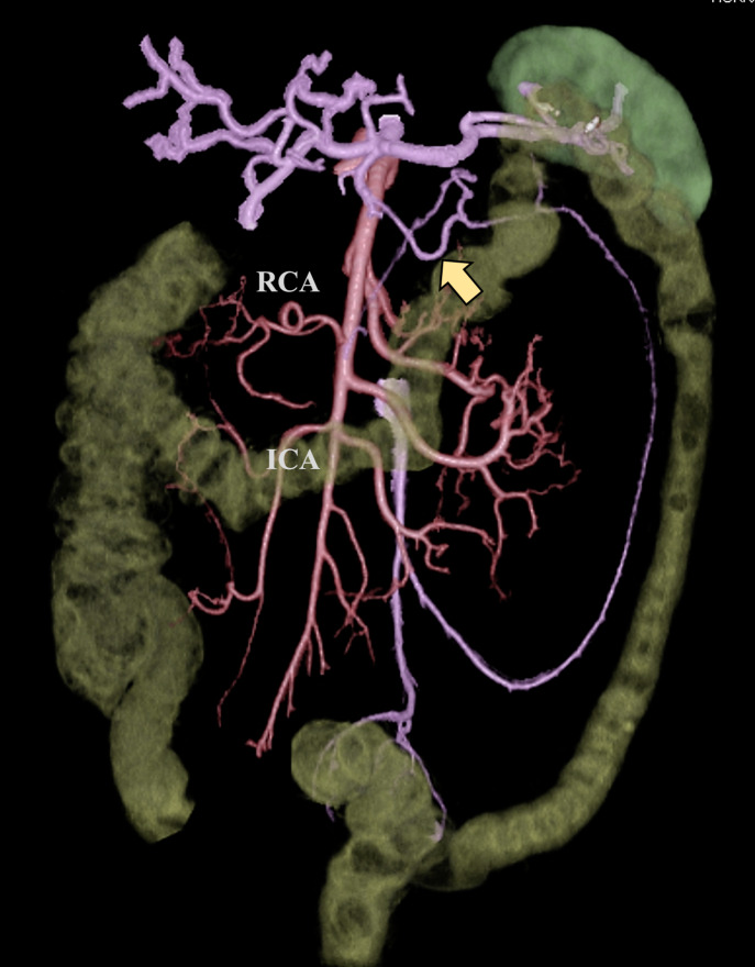
A Japanese woman in her 60s was referred to our hospital for left-side transverse colon cancer. Enhanced CT showed no regional or distant metastasis, so the colon cancer was diagnosed as cT2N0M0 cStage I. 3D-CT angiography showed the absence of a typical MCA originating from the SMA and the presence of an anomalous MCA originating from the celiac artery and the SA (Fig. 1).
Fig. 1.
3D-CT angiography findings. A typical MCA originating from the SMA was absent. An anomalous MCA originating from the SA running below the pancreas body (yellow arrow) was visualized. RCA, right colic artery; ICA, ileocecal artery.
Laparoscopic left hemicolectomy with D3 lymph node dissection was performed with a five-port conventional technique in which the descending and sigmoid colon were mobilized via a medial approach. After entire splenic flexure mobilization, the lymph nodes around the SMA were dissected via the caudal view, and the absence of a typical MCA was confirmed (Fig. 2a). The typical middle colonic vein running parallel to the typical MCA was also absent, with only the marginal artery and vein present. The caudal margin of the pancreas was reached, while the anterior aspect of the SMA was dissected. Total mesenteric dissection of the transverse colon mesentery was performed along the caudal margin of the pancreas. The anomalous MCA originating from the SA was then identified just below the pancreatic body (Fig. 2b), where it was successfully clipped and dissected. The inferior mesenteric vein was present just dorsal to the anomalous MCA and was clipped and dissected. These procedures completed a total mesenteric excision of the tumor area (Fig. 2c). The specimen was removed through a 4-cm incision, and functional end-to-end anastomosis was performed using linear staples. The operative time was 205 min, and the blood loss was 9 mL. There were no immediate or delayed complications. The pathological examination identified 15 lymph nodes harvested in the specimen, with no metastasis.
Fig. 2.
Laparoscopic findings during lymph node dissection. a The lymph nodes around the SMA were dissected via the caudal view, and the absence of a typical MCA was confirmed. b An anomalous MCA originating from the SA was identified below the pancreas body. c Laparoscopic view after completion of total mesenteric excision. Stump of the anomalous MCA (yellow arrow) originating from the SA was observed just below the pancreas body (gray arrow).
Discussion
The MCA supplies blood to the transverse colon and typically (>97% of cases) arises from the SMA [3, 4]. MCA origin anomaly is extremely rare, and only three surgical cases have been reported in the English literature, including 1 case where the MCA originated from the gastroduodenal artery in pancreas tumor surgery [5] and 2 cases where the MCA originated directly from the aorta in open surgery for colon cancer [3, 6].
Anomalous MCA originating from the SA is reported to be extremely rare, and only a few autopsy cases have been reported [1, 2], with no colorectal surgical cases has yet described. Approximately 25–31% of patients have an accessary middle colic artery (acMCA) that originates independently from the MCA and irrigates the area around the splenic flexure [4, 7]. The most common origin for the acMCA is the SMA (88%). Yano et al. [8] reported in their analysis of 3D-CT angiography that 1.4% of acMCAs arose from the celiac artery, although no autopsy or surgical cases have yet been reported. In the present case, a typical MCA was completely absent, so the artery originating from the SA and feeding the left-side transverse colon was defined as the MCA.
Laparoscopic colorectal surgery has been shown to be associated with superior perioperative outcomes to open surgery [9]. Standardization of surgical procedures and advances in surgical instruments have steadily expanded the indications for laparoscopic colorectal surgery. However, in cases involving abnormalities or aberrant vessels, performing laparoscopic procedures can be difficult and sometimes dangerous due to reduced sensation and a limited angle of view. Furthermore, the mesenteric anatomy in the vicinity of the splenic flexure is quite complex, making accurate lymph node dissection challenging [4, 10]. In the last decade, 3D-CT angiography has been reported useful for the preoperative assessment of a patient’s vascular anatomy for laparoscopic colectomy, especially in cases with vascular anomalies, thereby leading to safer surgery [4, 10–12]. In the present patient, the absence of a typical MCA arising from the SMA and the presence of an anomalous MCA originating from the SA were preoperatively recognized based on the information obtained on 3D-CT angiography. 3D-CT angiography was very helpful in this case for determining the location of the anomalous MCA below the pancreas body, and vascular ligation was performed safely, leading to completion of total mesenteric excision for left transverse colon cancer. With advances in 3D imaging technology, preoperative 3D vascular construction is expected to become an essential preoperative evaluation for laparoscopic colorectal cancer surgery in the near future [10]. Preoperative 3D angiography will contribute to safe laparoscopic surgery and reliable lymph node dissection, especially in patients with vascular anomalies.
In conclusion, laparoscopic surgery can be performed safely even for patients with an extremely rare vascular anomaly (e.g., MCA originating from the SA) using information from 3D-CT angiography. To our knowledge, this is the first case report of laparoscopic colectomy associated with an MCA anomaly originating from the SA. We believe this case report will contribute to the development of endoscopic surgery for colorectal cancer.
Statement of Ethics
This study was reviewed and approved by the Ethical Committee of Hokkaido Cancer Center (No. 30-06). Written informed consent was obtained from the patient for publication of the details of her treatment and any accompanying images. The patient’s anonymity was preserved. All authors have been in agreement with the content of the manuscript, in keeping with the latest guidelines of the International Committee of Medical Journal Editors.
Conflict of Interest Statement
The authors have no conflicts of interest to disclose.
Funding Sources
The authors have not received any funding relevant to their study.
Author Contributions
Y.M. is the primary investigator and contributed to conceptualization, data collection, and drafting of the manuscript. N.M., T.K., N.O., T.S., and C.I. were involved in the operation and the management of the patient. A.F. and Y.M. contributed to image processing of 3D-CT. All authors have read and approved the manuscript.
Funding Statement
The authors have not received any funding relevant to their study.
Data Availability Statement
Data sharing is not applicable to this article due to privacy and ethical restrictions. Further inquiries can be directed to the corresponding author.
Supplementary Material
References
- 1. Michels NA, Siddharth P, Kornblith PL, Parke WW. The variant blood supply to the descending colon, rectosigmoid and rectum based on 400 dissections. Its importance in regional resections: a review of medical literature. Dis Colon Rectum. 1965;8:251–78. [DOI] [PubMed] [Google Scholar]
- 2. Sato Y, Takeuchi R, Kawashima T, et al. An anomalous case of a middle colic artery derived from the splenic artery. J Kyorin Med Soc. 1984;15:87–92. [Google Scholar]
- 3. Milnerowicz S, Milnerowicz A, Taboła R. A middle mesenteric artery. Surg Radiol Anat. 2012;34(10):973–5. [DOI] [PMC free article] [PubMed] [Google Scholar]
- 4. Andersen BT, Stimec BV, Kazaryan AM, Rancinger P, Edwin B, Ignjatovic D. Re-interpreting mesenteric vascular anatomy on 3D virtual and/or physical models, part II: anatomy of relevance to surgeons operating splenic flexure cancer. Surg Endosc. 2022;36(12):9136–45. [DOI] [PMC free article] [PubMed] [Google Scholar]
- 5. Kwong MLM, Pelton J. Middle colic artery originating from the gastroduodenal artery discovered during a whipple. Case Rep Surg. 2019;2019:1986084 Published online. Article ID. 1986084. [DOI] [PMC free article] [PubMed] [Google Scholar]
- 6. Dirrigl AM, Zimmermann A, Ockert S, Eckstein HH. Middle mesenteric artery arising from an inflammatory infrarenal aortic aneurysm. J Vasc Surg. 2009;49(2):474–7. [DOI] [PubMed] [Google Scholar]
- 7. Murono K, Nozawa H, Kawai K, Sasaki K, Emoto S, Kishikawa J, et al. Vascular anatomy of the splenic flexure: a review of the literature. Surg Today. 2022;52(5):727–35. [DOI] [PubMed] [Google Scholar]
- 8. Yano M, Okazaki S, Kawamura I, Ito S, Nozu S, Ashitomi Y, et al. A three-dimensional computed tomography angiography study of the anatomy of the accessory middle colic artery and implications for colorectal cancer surgery. Surg Radiol Anat. 2020;42(12):1509–15. [DOI] [PMC free article] [PubMed] [Google Scholar]
- 9. Rossi BWP, Labib P, Ewers E, Leong S, Coleman M, Smolarek S. Long-term results after elective laparoscopic surgery for colorectal cancer in octogenarians. Surg Endosc. 2020;34(1):170–6. [DOI] [PubMed] [Google Scholar]
- 10. Andersen BT, Kazaryan AM, Stimec BV, Edwin B, Rancinger P, Ignjatovic D. Personalized surgery for the splenic flexure cancer: new frontiers. Br J Surg. 2022;109(9):880–1. [DOI] [PMC free article] [PubMed] [Google Scholar]
- 11. Ferrari V, Carbone M, Cappelli C, Boni L, Melfi F, Ferrari M, et al. Value of multidetector computed tomography image segmentation for preoperative planning in general surgery. Surg Endosc. 2012;26(3):616–26. [DOI] [PMC free article] [PubMed] [Google Scholar]
- 12. Maeda Y, Shinohara T, Nagatsu A, Futakawa N, Hamada T. Laparoscopic resection aided by preoperative 3-D CT angiography for rectosigmoid colon cancer associated with a horseshoe kidney: a case report. Asian J Endosc Surg. 2014;7(4):317–9. [DOI] [PubMed] [Google Scholar]
Associated Data
This section collects any data citations, data availability statements, or supplementary materials included in this article.
Supplementary Materials
Data Availability Statement
Data sharing is not applicable to this article due to privacy and ethical restrictions. Further inquiries can be directed to the corresponding author.


