Abstract
The Polycomb group proteins are involved in maintenance of the silenced state of several developmentally regulated genes. These proteins form large aggregates with different subunit compositions. To explore the nature of these complexes and their function, we used the full-length Eed (embryonic ectoderm development) protein, a mammalian homolog of the Drosophila Polycomb group protein Esc, as a bait in the yeast two-hybrid screen. Several strongly interacting cDNA clones were isolated. The cloned cDNAs all encoded the 150- to 200-amino-acid N-terminal fragment of the mammalian homolog of the Drosophila Enhancer of zeste [E(z)] protein, Ezh2. The full-length Ezh2 bound strongly to Eed in vitro, and Eed coimmunoprecipitated with Ezh2 from murine 70Z/3 cell extracts, confirming the interaction between these proteins observed in yeast. Mutations T1031A and T1040C in one of the WD40 repeats of Eed, which account for the hypomorphic and lethal phenotype of eed in mouse development, blocked binding of Ezh2 to Eed in a two-hybrid interaction in yeast and in mammalian cells. These mutations also blocked the interaction between these proteins in vitro. In mammalian cells, the Gal4-Eed fusion protein represses the activity of a promoter bearing Gal4 DNA elements. The N-terminal fragment of the Ezh2 protein abolished the transcriptional repressor activity of Gal4-Eed protein when they were coexpressed in mammalian cells. Eed and Ezh2 were also found to bind RNA in vitro, and RNA altered the interaction between these proteins. These findings suggest that Polycomb group proteins Eed and Ezh2 functionally interact in mammalian cells, an interaction that is mediated by the WD40-containing domain of Eed protein.
Pattern formation or morphogenesis is controlled by a host of parallel mechanisms that regulate gene expression at the level of chromatin structure, transcription, and multiple posttranscriptional steps. Although originally these processes were mostly studied in Drosophila, it has became apparent that the mechanisms and molecules that control development are highly conserved throughout all multicenter organisms. Homeotic genes are key developmental determinants of this kind that have been studied extensively in different organisms. These genes encode transcriptional regulators that mediate the anterior-posterior pattern formation. In turn, homeotic genes expression is under the tight control of other regulators such as gap genes products and Polycomb group (PcG) proteins. While the gap genes products are transcription repressors that initiate spatial restriction of homeotic genes expression early in development, the PcG proteins are required for maintenance and propagation of this repressed state (34, 49, 57, 58). Several models of PcG-mediated repression have been suggested that mostly postulate alterations in chromatin structure induced by PcG proteins (28, 31, 35, 47).
The Drosophila extra sex combs (esc) gene belongs to the PcG gene family (51). Several distinctive features of esc may allow it to serve as a linker between the initial binding of the gap gene product to DNA and the subsequent assembly of PcG protein complexes (48, 52, 53). esc regulates all the homeotic genes that have been tested thus far, and its activity is required only transiently in early Drosophila development; however, in later stages, esc is no longer needed to maintain the homeotic genes in a repressed state. In contrast to esc, once the repression process is initiated, all of the other PcG proteins are required to maintain the silencing of homeotic genes during the later stages of development (34, 50). It has been hypothesized that Esc protein recognizes local, transient changes in chromatin structure induced by the gap proteins and then recruits the rest of PcG complex (15, 43, 48, 49). The protein targets of the action of Esc have not yet been identified.
The esc gene was cloned recently from Drosophila, and its mammalian homolog eed was cloned from the mouse (8, 15, 43–45). Esc and Eed show a high degree of similarity, especially in the C-terminal half of the molecules, which was originally deduced to contain five (45) or six (15) WD40 repeats, but a very recent detailed analysis provided evidence for seven WD40 repeats (29). The N termini are less conserved between these species; for example, mammalian protein contains a domain of 100 amino acids (aa) at the very N terminus which was not found in the Esc. This domain binds the heterogeneous nuclear ribonucleoprotein (hnRNP) K protein (8), the only known molecular partner of Eed. Most of the spontaneous mutations that interfere with esc and eed function were localized in the WD40 domain, suggesting that integrity of this domain is required for its function. Structural analysis of several WD40 repeat-containing proteins revealed that this domain has a propeller-like structure in which each blade corresponds to one WD40 repeat. While in some proteins, like the β subunits of G proteins, the WD40 repeats mediate binding to other proteins (26), in other cases this domain nests metal ion in the middle of propeller and exhibits enzymatic activity, as in galactose oxidase (17). Thus, although the WD40 domains are structurally similar, their functions are seemingly diverse and they may play either structural or catalytic roles.
To gain more insight into the mechanisms of Eed action, we set out to identify the protein(s) that binds the WD40 propeller region of this protein. Using the yeast two-hybrid screen of mammalian cDNA libraries, we isolated cDNA encoding a strongly interacting protein which was identified as Ezh2, a mammalian homolog of the Drosophila Enhancer of zeste [E(z)]. The possible implications of this finding are discussed.
MATERIALS AND METHODS
Cell lines.
Rat glomerular epithelial cells (GEC) (41), mouse pre-B lymphocytes (70Z/3) (30), monkey kidney cells (COS cells) (23) human epidermoid cells (KB cells) (2), and leukemia T cells (Jurkat cells) (37, 41) were grown as described previously.
Reagents.
The bacterial expression vector pGEX-KT was provided by J. Dixon (University of Michigan). The mammalian expression vectors pM1 through pM3 were kindly provided by I. Sadowsky (University of British Columbia, Vancouver, Canada). The luciferase reporter vectors pGL3-Enhancer containing the simian virus 40 (SV40) enhancer and pGL3-Promoter containing the SV40 promoter were purchased from Promega (Madison, Wis.). Glutathione-agarose homopolyribonucleotides covalently bound to agarose and unbound homopolyribonucleotides were obtained from Sigma (St. Louis, Mo.).
RNA extraction and Northern blot analysis.
Total RNA was extracted as previously described (7). Cells or animal tissues were washed with phosphate-buffered saline (PBS). A 2.0-ml volume of solution D (4 M guanidinium thiocyanate, 25 mM sodium citrate, 0.5% Sarkosyl, 0.1 M β-mercaptoethanol) was added to each plate or to 100 mg of animal tissue to lyse the cells. The final RNA was dissolved in water and used for Northern blot analysis. RNA was analyzed essentially as described previously (18). After being denatured in formaldehyde-formamide at 65°C for 15 min, the RNA samples were cooled on ice. A 10-μg portion of total RNA per lane was resolved by electrophoresis in a 1.2% agarose gel containing 2.2 M formaldehyde. The RNA was transferred to a Hybond N+ membrane (Amersham, Little Chalfont, United Kingdom) and “baked” at 80°C for 45 min. The membranes were prehybridized for 2 h at 42°C in prehybridization buffer (50% formamide, 5× Denhardt solution, 5× SSC [1× SSC is 0.15 M NaCl plus 0.015 M sodium citrate], 0.5% sodium dodecyl sulfate [SDS], 0.1 mg of denatured salmon sperm DNA per ml, 0.1 mg of yeast tRNA per ml). After prehybridization, 32P-labeled cDNA probe (2 × 10⁶ cpm/ml) was added and hybridization was carried out overnight at 42°C. After, hybridization, the membranes were washed twice in 2× SSC–0.1% SDS at 22°C for 10 min and then twice in 0.1× SSC–0.1% SDS at 50°C for 20 min and autoradiographed.
Yeast two-hybrid system library screen and DNA sequencing.
The procedure used was based on previously described methods (10, 56). For the screen, the L40 strain of yeast (generated by Stanley Hollenberg) was used. The L40 yeast strain contains both lacZ and HIS3 marker genes under the control of minimal GAL1 promoter fused with multimers of LexA DNA-binding sites.
To construct the bait, the open reading frame of eed was used as a template for PCR. The PCR primers were programmed to contain a BamHI site at the 5′ end and a stop codon and a PstI site at the 3′ end. After digestion, the PCR-generated fragment was subcloned in frame with LexA into the pBTM116 vector, a DNA-binding domain plasmid (pBTM116 was constructed by Paul Bartel and Stan Fields). cDNA libraries prepared from poly(A)+ RNA from mouse embryos 9.5 to 10.5 days postcoitum (generated by Stanley Hollenberg) or Jurkat cell line (Clontech, Palo Alto, Calif.) were used in the screen.
Yeast cells were first transformed with the bait plasmid, and then the L40/LexA-Eed strains were transformed with the cDNA library. Screening for positive clones containing the two plasmids, LexA-Eed fusion protein and the activation domain library plasmids, was done by growing colonies on His− selective media and testing them for β-galactosidase activity (56). The cDNA plasmids were rescued from the primary positive clones and then retransformed back into cells carrying either the wild-type or mutant LexA-Eed plasmid.
Sequencing was performed by the DyeDeoxy Terminator (Perkin-Elmer) cycle-sequencing method.
In vitro transcription and translation.
Partial or complete cDNAs were used as template for in vitro transcription by SP6 or T7 DNA-dependent RNA polymerases to generate mRNAs as previously described (14). In vitro translation in a rabbit reticulocyte cell-free system was performed as specified by the manufacturer (Promega, Madison, Wis.)
In vitro binding studies, immunoprecipitation, and Western blot analysis.
A 2.5-μl volume of the cell-free translational system containing 35S-labeled proteins was added to a suspension of 10 μl of glutathione-agarose beads bearing either the wild-type or mutated Eed protein fused to glutathione S-transferase (GST) in 100 μl of binding buffer (10 mM HEPES-NaOH [pH 7.5], 100 mM KCl, 2.0 mM MgCl2, 0.1% Nonidet P-40). After mixing for 40 min (4°C), the beads were washed three times with 400 μl of binding buffer and were boiled with 30 μl of SDS sample buffer. Released proteins were analyzed by SDS-polyacrylamide gel electrophoresis (PAGE). Binding to agarose beads carrying covalently attached homopolyribonucleotides was performed in the same way.
Western blot analysis with anti-Gal4 monoclonal antibody (0.5 μg/ml; Santa Cruz), anti-LexA rabbit polyclonal antibody (kindly provided by Erica Golemis, The Fox Chase Cancer Center), or anti-Eed rabbit polyclonal antibody was performed as described elsewhere (8). Immunoprecipitation with anti-Enx1 (Ezh2) polyclonal serum (kindly provided by A. Ullrich, Max-Planck-Institute for Biochemistry) was done as described previously (13).
Plasmid constructs.
pM1Eed, pM1EedT1040C, pM1EedT1031A, pM1EedΔBB, pG5GL3enh, and pG5GL3pro were described previously (8). Expression plasmid pMEzh2N was constructed by inserting the BamHI-BglII fragment of the human ezh2 cDNA (kindly provided by Haiming Chen, University of Geneva) into BglII-BamHI-cut pM1 (42). The final construct contained an insert of the ezh2 cDNA fragment encoding the N-terminal 203 aa of the protein under the control of the SV40 promoter. pMEBP1 was constructed by inserting the VP16-cDNA fusion fragment from positive clone 1 (see Fig. 2) into BglII-EcoRI-linearized pM1 vector. The GST-Ezh2 fusion construct was made by inserting the PCR-amplified fragment of the mouse Ezh2 gene encoding the N-terminal aa 1 to 192 of the protein into pGEX-KT vector. GST-Eed was made by cloning PCR-amplified fragment of the human Eed gene corresponding to the aa 56 to 535 piece of the protein into the pGEX-KT vector. Point mutations T1031A and T1040C were introduced into the Eed gene by using the QuikChange site-directed mutagenesis kit from Stratagene. The Eed deletion mutants EedΔN and EedΔC are described elsewhere (8). All constructs were verified by sequencing and cell-free translation.
FIG. 2.
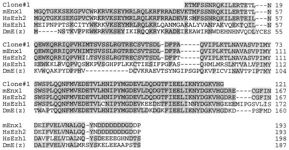
The Eed-interacting clones encode the N-terminal part of Ezh2 protein, a mammalian homolog of the Drosophila E(z) protein. The cDNA inserts from the positive clones were sequenced, and a search for sequence similarity in a database was carried out. All of the clones matched the N-terminal part of the Ezh2 protein. The alignment of the shortest clone with different members of the E(z) protein family is shown. DmE(z), D. melanogaster E(z) protein (20); mEnx1, mouse Enx1 protein (16); HsEzh2, human Ezh2 protein (6); HsEzh1, human Ezh1 protein (1). Only N-terminal parts of the proteins are shown. Identical positions are shaded.
Transient transfection and luciferase activity assay.
COS 7 cells were grown in Dulbecco’s minimal essential medium supplemented with 10% fetal calf serum to approximately 60 to 75% confluency in 100-mm-diameter dishes and were transfected with different plasmids by using the SuperFect reagent (Qiagen Inc., Santa Clarita, Calif.). Briefly, cells were treated with a total of 4 μg of plasmid DNA premixed with 15 μl of the reagent. After 24 to 48 h, transfected cells were washed twice with PBS, scraped with a rubber policeman, and centrifuged in a microcentrifuge. The cell pellet was lysed in 150 μl of ice-cold 1× lysis buffer (Promega). The supernatant was assayed for protein concentration and luciferase activity by the standard Promega method with a luminometer.
Jurkat cells (12) were grown in suspension at 37°C in complete RPMI 1640 medium supplemented with 10% fetal calf serum. They were transfected with SuperFect transfection reagent as specified by the manufacturer (Qiagen Inc.). After 24 to 48 h, transfected cells were centrifuged in a microcentrifuge and washed twice with PBS. The cell pellet was lysed in 150 μl of ice-cold 1× lysis buffer (Promega), and the supernatant was assayed for luciferase activity.
RESULTS
A yeast two-hybrid screen with the Eed protein reveals Ezh2 as a strongly interacting partner.
To gain more insight into the mechanisms that mediate the transcription repressor activity of Eed (8), we used a two-hybrid screening of cDNA libraries in yeast (10). The full-length Eed protein fused to the LexA DNA-binding domain was used as a bait to screen two different libraries. Several strongly interacting cDNA clones were obtained. The cDNA-containing plasmids from the positive clones were isolated and transformed back into yeast strains carrying either the wild-type Eed or the mutant Eed bait construct. Five of six clones showed very strong interaction with the wild-type Eed protein but no interaction at all with the Eed T1040C point mutant (Fig. 1A). Western blot analysis with anti-LexA serum showed similar levels of expression of both baits (Fig. 1B), indicating that the lack of interaction with the mutant bait was not due to its degradation in yeast cells. This mutation, which affects the structure of the third WD40 repeat (45), abrogates the transcription repression activity of Eed in mammalian cells (8), and when homozygous, this mutation is lethal in mice (45). Sequencing of the five cDNA clones revealed that they all encode N-terminal portions of the Ezh2 protein, a mammalian homolog of the Drosophila Enhancer of Zeste protein [E(z)]. The cDNA inserts varied in length but had the same aa 39 to 159 region of Ezh2 protein found in the smallest clone, clone 1 (Fig. 2). These data suggest that the mammalian PcG proteins Eed and Ezh2 interact with each other and indicate that integrity of the WD40 repeat region of Eed is crucial for this interaction.
FIG. 1.
Screening of cDNA libraries using Eed as a bait in yeast two-hybrid system. (A) cDNA libraries from mouse embryos 9.5 to 10.5 days postcoitum or Jurkat cells were screened in yeast against the LexA-Eed bait construct as described in Materials and Methods. The cDNA plasmids from primary positive clones were purified and transformed into yeast strains carrying either LexA-Eed wild-type or LexA-T1040C mutant Eed bait. Three colonies from each transformation were grown on plate with selective media (Leu−, Trp−) and then transferred onto nitrocellulose filter and assayed for β-galactosidase activity. The blue color was developed for 3 h at 30°C. (B) The transformants were also checked for expression of the bait constructs. Cells transformed with pBT116 (Vector), LexA-Eed, and LexA-T1040C plasmids were grown overnight in selective medium, lysed in SDS sample buffer, electrophoresed, transferred onto a polyvinylidene difluoride membrane, and stained with anti-LexA rabbit serum followed by anti-rabbit immunoglobulin G-alkaline phosphatase conjugate. A membrane stained with 5-bromo-4-chloro-3-indolylphosphate/nitroblue tetrazolium phosphatase substrate is shown.
eed and ezh2 genes are expressed in the same mouse tissues and cell lines.
The Ezh2 cDNA was used as a probe to detect the Ezh2 transcript in several mouse tissues and cell lines by Northern blot analysis. The results are shown in Fig. 3A. The 3.3-kb transcript was present in ovaries (lane 2), the mouse pre-B 70Z/3 cell line (lane 6), and human KB cells (lane 5). Another 2-kb transcript, which could be a result of alternative splicing (6), was found in brain and 70Z/3 samples (lanes 1 and 6). Remarkably, Ezh2 mRNA expression in these tissues mirrors that of Eed (8), suggesting that there may be a functional need for these two genes to be coexpressed in the ovaries, brain, and lymphocytes. Since the Eed and Ezh2 transcripts were far more abundant in the pre-B cell line (70Z/3) than in other tissues or cell lines, we tested several other lymphoid cell lines for expression of these two genes. As shown in the Fig. 3B, the Ezh2 and Eed mRNAs were also coexpressed in a wide variety of B- and T-cell lines, providing further evidence that Eed and Ezh2 are functionally related.
FIG. 3.
Northern blot analysis reveals that the ezh2 and eed genes are coexpressed in cell lines and mouse tissues. Portions (10 μg) of total RNA from different sources, as indicated, were run in a 1.2% agarose gel containing 2.2 M formaldehyde, transferred onto a nylon membrane, and probed with either ezh2 (A and B) or eed (B) 32P-labeled probes. After hybridization, the membranes were washed and exposed to X-ray films. Before the hybridization procedure, the membranes were stained for RNA with methylene blue (A, lower panel) as a control for the amounts of RNA. 28S indicates 28S rRNA. The membrane in panel B was boiled in water–0.1% SDS for 10 min after use of the first probe and then rehybridized with the second probe. The positions of RNA size markers are shown on the left. The positions of bands corresponding to ezh2 and eed transcripts are indicated by arrows.
Interaction between Eed and Ezh2 proteins in vitro.
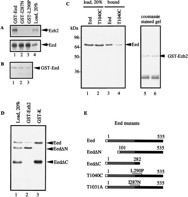
All the Ezh2 clones that we isolated in the two-hybrid system represented only the N-terminal part of the molecule. To test the possibility that the full-length Ezh2 also binds Eed, we used an in vitro binding assay. The glutathione-agarose beads bearing recombinant GST-Eed fusion protein were mixed with 35S-labeled Ezh2 synthesized in a cell-free translational system. As shown in Fig. 4, the full-length Ezh2 protein binds well to the wild-type Eed protein (lane 1) but not at all to either EedI287N (T1031A) (lane 2) or EedL290P (T1040C) (lane 3) mutants. The T1031A mutation affects the same WD40 domain of Eed as does the lethal T1040C mutation but results in nonlethal aberrations in anterior-posterior patterning in the mouse (45). In contrast, 35S-labeled Eed, which can form homodimers (8a), bound to all types of beads with equal affinity. These results show that the full-length Ezh2 binds Eed with high affinity and that the binding is mediated by the propeller domain of Eed. In reciprocal experiments, the 35S-labeled translational products of the wild-type Eed and T1040C mutant were analyzed for binding to GST-Ezh2 (aa 1 to 192) agarose (Fig. 4C). In agreement with the previous experiment, the GST-Ezh2 beads effectively pulled down the wild-type Eed (lane 3) but the T1040C mutant bound the beads less efficiently (lane 4). In the next experiment, we tested two deletion mutants of Eed, EedΔN (aa 101 to 535), and EedΔC (aa 1 to 282) in the binding assay (Fig. 4D). As expected, the EedΔC mutant, which lacks most of the WD40 propeller domain, did not bind GST-Ezh2. Surprisingly, the EedΔN mutant with the deleted N-terminal domain, which was not found in the Drosophila Esc protein (8), bound GST-Ezh2 better than the full-length Eed protein did (lane 2). Binding of these Eed mutants to GST-K (mouse hnRNP-K) had an opposite pattern; i.e., EedΔC but not EedΔN bound the beads (lane 3) (8). The binding of Eed to GST-K rules out the possibility that the EedΔC mutant was nonfunctional (aggregated) or that the EedΔN mutant was “sticky.” Taken together, these results indicate that Ezh2 binds the WD40 propeller domain of Eed.
FIG. 4.
Ezh2 binds Eed in vitro. Ezh2 or Eed mRNAs were translated in a rabbit reticulocyte cell-free system in the presence of [35S]methionine. The translational products were incubated (1 h at 4°C) with glutathione-agarose beads bearing either the wild-type GST-Eed or GST-Eed mutants I287N (T1031A) and L290P (T1040C) (A), GST-Ezh2 (aa 1 to 192) (C and D) or GST-K (D). After binding, the beads were washed and boiled in SDS buffer, and eluted proteins were analyzed by SDS-PAGE and autoradiography. The Coomassie blue-stained gel of the experiment in panel A, lanes 1 to 3, is shown in panel B. (C) Eed and T1040C translational products were bound to GST-Ezh2 beads. Lanes 5 and 6 display the Coomassie blue-stained gel corresponding to lanes 3 and 4. (D) Eed, EedΔN, and EedΔC translational products were mixed and bound to either GST-Ezh2 or GST-K beads. (E) Eed constructs used in the experiments. Open box, GST (A) or His/T7 tag (pET28 vector) (C and D); shaded box, N-terminal part of Eed; solid box, C-terminal propeller domain of Eed.
RNA alters the Eed-Ezh2 interaction in vitro.
Previous studies provided evidence that RNA may be involved in the process of PcG protein assembly and action (8, 32). We therefore tested the effect of RNA on the Ezh2 interaction with Eed by adding different homopolyribonucleotides to the binding reaction (20 μg/ml). As shown in Fig. 5A, binding of 35S-Eed to GST-Ezh2 was blocked by poly(G) (lane 3) but not by the other RNAs (lanes 4 to 6). The binding of 35S-Ezh2 to GST-Eed was also inhibited by poly(G) (data not shown). In the control experiment, poly(G) did not affect the binding of 35S-Eed to GST-K (Fig. 5A, lower panel), showing that the effect of poly(G) was specific to the Eed-Ezh2 interaction. Since this experiment suggested that Eed and/or Ezh2 may bind to RNA, we incubated 35S-Eed, 35S-Ezh2, and 35S-hnRNP K with agarose beads bearing all four homopolyribonucleotides. The beads were washed, and proteins were eluted with SDS and analyzed by gel electrophoresis and autoradiography (Fig. 5B). This experiment revealed that both Eed and Ezh2 bound poly(G) very strongly (most of the added proteins bound RNA) but that they bound poly(U) less strongly and did not bind poly(A) or poly(C). In contrast, 35S-hnRNP K bound poly(C) and poly(U) but not poly(G). Since Eed and Ezh2 were also able to bind RNA when produced and purified from bacteria (data not shown), they most probably bind RNA directly. These series of experiments show that RNA can modulate complex formation between the PcG protein Eed and its partners, hnRNP K and Ezh2.
FIG. 5.
Binding of Eed to Ezh2 in vitro is modulated by RNA. The in vitro binding assay was performed as described in the legend to Fig. 4. (A) The 35S-Eed translational product was bound to either GST-Ezh2 or GST-K beads in the presence of 20 μg of poly(G) (lane 3), poly(C) (lane 4), poly(U) (lane 5), or poly(A) (lane 6) per ml. −, no addition (lane 2). The Coomassie blue-stained gels are shown in the panels below the autoradiograms. Eed, position of 35S-Eed in the gel; GST-Ezh2 and GST-K, positions of the proteins in the Coomassie blue-stained gel. (B) Binding of 35S-Eed, 35S-K, and 35S-Ezh2 to different homopolyribonucleotides. The Eed, hnRNP-K, and Ezh2 translational products were incubated (1 h at 4°C) with agarose beads with covalently attached poly(G) (lane 2), poly(C) (lane 3), poly(U) (lane 4), or poly(A) (lane 5). After binding, the beads were washed and boiled in SDS buffer, and eluted proteins were analyzed by SDS-PAGE and autoradiography.
Interaction between Eed and Ezh2 in vivo.
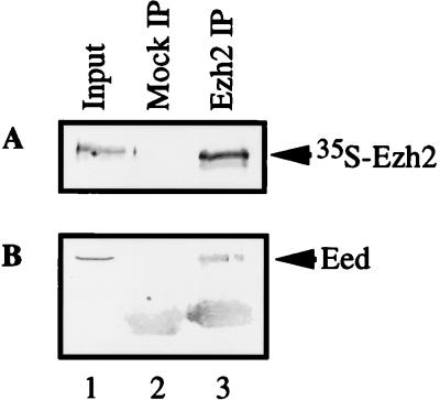
To determine if Eed and Ezh2 exist in a complex in vivo, we carried out a coimmunoprecipitation experiment from the murine 70Z/3 cell extracts that express both Eed (8) and Ezh2 (Fig. 3). Nonimmune or anti-Ezh2 polyclonal serum was incubated with cell extracts. Immunoglobulins were pulled down with protein A-coated beads; after the beads were washed, proteins were eluted with SDS loading buffer. Eluted proteins were analyzed by SDS-PAGE and Western blotting with an anti-Eed polyclonal serum (8). The results showed that the antiserum that specifically immunoprecipitates Ezh2 (Fig. 6A) coprecipitated Eed from 70Z/3 cell extracts, providing evidence that Eed and Ezh2 exist in a complex in vivo.
FIG. 6.
Eed and Ezh2 coprecipitate from 70Z/3 cell extracts. A 5-μl volume of 35S-labeled Ezh2 (A) or 100 μl of 70Z/3 cell extract (B) was incubated with 3 μl of anti-Ezh2 rabbit serum in a final volume of 1 ml of ELB buffer (13). The immunoglobulins were pulled down with protein A-Sepharose, and the beads were washed extensively with the ELB buffer and eluted with SDS loading buffer. The eluted proteins were separated by SDS-PAGE, transferred to a polyvinylidene difluoride membrane, and either exposed to X-ray film (A) or probed with anti-Eed polyclonal serum (B). The position of bands corresponding to Ezh2 and Eed are shown by arrows. IP, immunoprecipitate.
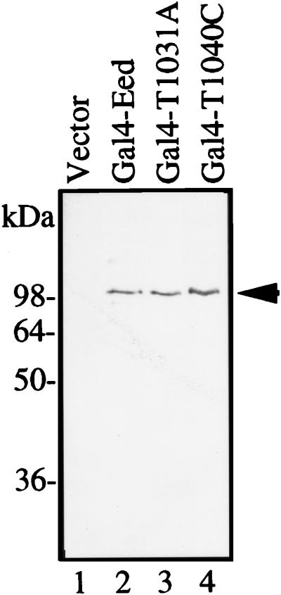
To further test if Eed and Ezh2 interact in mammalian cells, we used two-hybrid interaction system in the human Jurkat and monkey COS cells. Gal4-Eed fusion was used as a DNA-binding bait, and VP16AD-Ezh2 (fragment from aa 39 to 159) fusion was used as the activation domain hybrid. The activity of the firefly luciferase reporter gene driven by the SV40 enhancer and a minimal thymidine kinase promoter containing 5× Gal4 DNA elements was used as a readout. The luciferase activity in cells cotransfected with Gal4-Eed and VP16AD-Ezh2 hybrids was more than 100-fold higher than that generated by any other pair of hybrids including the VP16AD-Ezh2 and Gal4-mutant Eed variants (Table 1). The equal expression of Gal4-Eed fusion proteins in these transfections demonstrates that the observed differences in the two-hybrid interactions were not the result of variations in the levels of bait expression in the transfected cells (Fig. 7, compare lanes 2, 3, and 4).
TABLE 1.
Two-hybrid interaction between Eed and Ezh2 in Jurkat and COS cellsa
| Bait construct | Luciferase activity of VP16AD fusion construct in:
|
|||
|---|---|---|---|---|
| Jurkat cells
|
COS cells
|
|||
| VP16 | VP16-Ezh2 (aa 39 to 159) | VP16 | VP16-Ezh2 (aa 39 to 159) | |
| Gal4 | 1.0 | 1.2 ± 0.2 | 1.0 | 1.5 ± 0.3 |
| Eedb | 0.8 | 1.3 | 0.7 | 0.4 |
| Gal4-Eed | 0.3 ± 0.1 | 147.0 ± 29 | 0.6 ± 0.1 | 313.0 ± 12 |
| Gal4-T1031A | 1.2b | 1.4b | 0.7 ± 0.1 | 0.4 ± 0.2 |
| Gal4-T1040C | 1.0 ± 0.2 | 1.2 ± 0.3 | 0.7 ± 0.2 | 0.5 ± 0.2 |
Cells were transfected with 0.5 μg of the reporter plasmid, bearing the firefly luciferase gene driven by the SV40 enhancer and minimal promoter containing five Gal4-binding elements, and two hybrid constructs (2 μg of each): the Gal4 DNA-binding domain alone (Gal4) or Gal4 fused to the wild-type or mutant Eed (Gal4-Eed, Gal4-T1031A, Gal4-T1040C), or nonfused Eed protein (Eed), and the VP16 activation domain either alone (VP161) or fused to N-terminal fragment of Ezh2 [VP16-Ezh2 (aa 39 to 159)]. Two days after transfection, the cells were analyzed for luciferase activity. Data represent the means ± standard errors calculated from three independent experiments.
The experiment was repeated once.
FIG. 7.
Western blot analysis of Gal4-Eed expressed in COS cells. Mammalian expression plasmid containing either wild-type or mutated Eed fused to Gal4 were transfected into COS cells. After 2 days, total cellular extracts were separated by SDS-PAGE and proteins were transferred from the gel onto a polyvinylidene difluoride membrane and probed with anti-Gal4 monoclonal antibodies (Santa Cruz). The membrane was developed with secondary antibodies conjugated with alkaline phosphatase and 5-bromo-4-chloro-3-indolyl phosphate/nitroblue tetrazolium substrate. The positions of molecular mass markers are shown on the left. Vector, cells were transfected with pM1 plasmid containing no inserts (see Materials and Methods).
Next, we asked if Ezh2 protein could affect the transcription repression activity of Eed protein. As we have recently shown, the Gal4-Eed fusion construct repressed the transcription of the reporter luciferase gene driven by the SV40 promoter and 5×Gal4 DNA elements (8). Here, we coexpressed the reporter plasmid with Gal4-Eed and the N-terminal portion of Ezh2 protein, aa 1 to 203. The results of the luciferase activity measurements are shown in Fig. 8. Gal4-Eed repressed transcription of the reporter gene by 80 to 85% compared to the control. The Gal4-Eed-mediated transcriptional repression was diminished four- to fivefold by coexpression of the Ezh2 fragment (aa 1 to 203). This effect was specific, since the levels of reporter gene expression in the presence of Gal4 were not affected by coexpression of Ezh2. These data strongly support a model where Eed and Ezh2 proteins are close partners acting to control gene expression.
FIG. 8.
Effect of Ezh2 on transcriptional activity of Gal4-Eed. Jurkat cells were transfected with a mixture of reporter and expression plasmids. (A) Plasmids used in the transfection experiments. The reporter plasmid was a luciferase gene pGL3-promoter vector containing an SV40 promoter with five Gal4-binding elements. The expression plasmids were as follows. The mammalian vector, pM1, was used for expression of either the Gal4 DNA-binding domain alone (Gal4) or a fusion of Gal4 DNA-binding domain with the wild-type Eed (Gal4-Eed). Plasmid pM1 expressing the N-terminal fragment of the human Ezh2 protein (N-Ezh2, aa 1 to 203) instead of Gal4 was also used. (B) Result of the transfection experiments. Expression plasmids (1 μg of Gal4 or Gal4-Eed and 3 μg of N-Ezh2) with 0.3 μg of luciferase reporter plasmid were used for cotransfections. The total amount of DNA, 4.3 μg, per transfection was adjusted with pM1. Two days after transfection, cells were analyzed for luciferase activity. The data shown represent the means ± standard errors calculated from three independent experiments.
DISCUSSION
PcG proteins are thought to maintain silenced chromatin states in the homeotic gene loci. Esc (Eed), as a likely linker between the initiation of silencing and assembly of the PcG complex, can exert its action either as a structural constituent of the chromatin or as an enzyme (modifier) that alters the function of the other chromatin components or both. The WD40 domain of the protein is crucial for its function because most of the mutations that affect Eed/Esc functions in vivo were localized in this domain (15, 45). Moreover, the Eed WD40 domain contains transcription repression activity (8). To further explore the mechanisms of Eed action, we set out to identify a protein(s) that binds the C-terminal WD40 propeller region of this protein.
We have shown here that Eed strongly interacts with Ezh2, a mammalian homolog of the Drosophila E(z) protein. This finding was supported by several observations. First, Ezh2 was the strongest interacting clone in the yeast two-hybrid screen with Eed protein as a bait (Fig. 1) and was also found to bind Eed in the mammalian two-hybrid interaction (Table 1). Second, Ezh2 formed a tight complex with Eed in vitro (Fig. 4) and in cell extracts (Fig. 6). Third, the patterns of eed and ezh2 transcripts expression in mouse tissues and several cell lines were very similar, if not identical (Fig. 3). Finally, the interactive domain of Ezh2 diminished the transcription repressor activity of Eed protein in mammalian cells (Fig. 8). Taken together, these observations provide evidence that Eed and Ezh2 act in concert, and it is tempting to speculate that this interaction is required for the function of these proteins.
Alignment of the cloned cDNAs allowed us to localize the Eed-binding domain to the aa 39 to 159 region of Ezh2. This domain was sufficient for binding to Eed in vivo and in vitro (data not shown). The C-terminal half of this fragment (aa 91 to 159 in the human protein) contains an evolutionarily conserved motif (Fig. 2), which can be considered the most likely region mediating binding to Eed. In cross-species expression experiments, some extra copies of the human Ezh2 gene enhanced position effect variegation in Drosophila (24), showing that the chromatin-modifying function of Ezh2 is highly conserved. We have shown that the human protein Ezh2 was able to bind Drosophila GST-Esc fusion protein in vitro (data not shown) indicating that the Ezh2 [E(z)] and Eed (Esc) partnership is likewise evolutionarily conserved.
The observation that either the T1031A or T1040C mutation in the Eed WD40 domain blocks its interaction with Ezh2 suggests that the WD40 domain binds Ezh2 directly. Another possibility is that Ezh2 binds the nonpropeller N-terminal region of the molecule and the failure of Ezh2 to bind to the mutants can be explained by masking of the N terminus by the altered WD40 domain. The latter scenario is less likely because the ability of the Eed N-terminal dimerization domain to homodimerize was not affected by the mutations in the WD40 domain (Fig. 4A) and, moreover, the EedΔC mutant with the WD40 domain deleted failed to bind Ezh2 (Fig. 4D). The postulate that the propeller rather than N-terminal domain of Eed is directly involved in binding to Ezh2 is also supported by the observation that Ezh2 interacts with Esc (data not shown) and that the WD40 domains of Eed and Esc are highly conserved while their N termini are not.
E(z) protein, originally discovered as an enhancer of zeste/white interaction, was later classified as a member of several different groups of proteins, such as PcG, Trithorax (Trx), a group of positive regulators of homeotic genes (21, 22, 25, 33), and, recently, a chromatin modifier with a haplo-suppressor/triplo-enhancer dosage effect on position effect variegation (24). The haplo-suppressor/triplo-enhancer group was originally postulated to encode structural chromatin proteins (27, 40), and recent studies support this hypothesis (9, 39, 54). Thus, E(z) belongs to all of the major systems known to affect chromatin. This indicates that E(z) most probably acts as a basic structural chromatin component and that PcG or other proteins may act to modify its function at specific genomic loci. Consistent with this notion is the observation that in E(z) temperature-sensitive mutants, the immunostaining of several PcG proteins in specific sites on polytene chromosomes was highly reduced at the nonpermissive temperature (5, 36, 38). These mutations have also resulted in the general decondensation of chromatin structure (38), and increased chromosome breakage was observed in flies hemizygous for an E(z) null allele (11).
Because E(z) shares some features with Trx proteins, it is plausible that the E(z)/Ezh2 protein is aggregated, with an open chromatin structure which is easily accessible to the transcriptional machinery. We believe that interaction of Esc/Eed with E(z)/Ezh2 is one of the events in the process of chromatin transformation. Whatever the mechanism of Eed action is, the chromatin modification induced by Esc/Eed early on should be self-perpetuating at the later stages of embryo development after the Eed/Esc protein is no more expressed. This requirement rules out simple models where Esc/Eed contains intrinsic enzymatic activity or recruits other enzymes that modify the function of E(z)/Ezh2, unless there is a mechanism to maintain the modification of E(z) at the silenced loci during or after DNA replication.
We have found that Eed and Ezh2 bind RNA in vitro. The patterns of bound homopolyribonucleotide were quite similar for Eed and Ezh2; both favored poly(G) and poly(U). Remarkably, both Eed and Ezh2 showed a level of affinity for RNA comparable to that seen with the classic RNA-binding protein hnRNP K (4) (Fig. 5B). These data identify Eed and Ezh2 as RNA-binding proteins. We have also shown that poly(G), but not the other polyribonucleotides (used at 20 μg/ml), blocked the interaction between Eed and GST-Ezh2 (Fig. 5A). It is therefore conceivable that there are RNA species involved in the action of Eed and Ezh2 proteins, and it ought to be possible to identify these RNA species by using these proteins as probes. It is also possible that as with hnRNP K (for a review, see reference 3 and citations therein), other nucleic acids, such as single- or double-stranded DNA, are likewise involved in modulating Eed and/or Ezh2 action.
While this work was under review, similar data on the interaction between Esc/Eed and E(z)/Ezh2 were published by others (19, 46, 55), providing independent evidence that these proteins interact in both Drosophila and mammalian cells.
In summary, we have identified an interaction between the mammalian PcG proteins Eed and Ezh2. This interaction is mediated by the Eed WD40 repeat domain, where point mutations abrogate this interaction. Both proteins were found to bind RNA, and RNA altered the Eed-Ezh2 interaction. The above studies provide new information on the mechanisms of initiation and propagation of PcG complexes in the silenced loci.
ACKNOWLEDGMENTS
We thank Haiming Chen for ezh2 cDNA, Erica Golemis for anti-LexA serum, Axel Ullrich for anti-Enx1 serum, and Peter Harte for discussion and valuable suggestions.
This work was supported by grants GM45134 and DK45978 from the NIH, the Northwest Kidney Foundation, and the American Diabetes Association.
REFERENCES
- 1.Abel K J, Brody L C, Vlades J M, Erdos M R, McKinley D R, Castilla L H, Merajver S D, Couch F J, Friedman L S, Ostermeyer E A, Lynch E D, King M-C, Welcsh P L, Osborne-Lawrence S, Spillman M, Bowcock A M, Collins F S, Weber B L. Characterization of EZH1, a human homolog of Drosophila Enhancer of zeste near BRCA1. Genomics. 1996;37:161–171. doi: 10.1006/geno.1996.0537. [DOI] [PubMed] [Google Scholar]
- 2.Bird T A, Schule H, Delaney P B, Sims J E, Thoma B, Dower S K. Evidence that MAP (mitogen-activated protein) kinase activation may be a necessary but not sufficient signal for a restricted subset of responses in IL-1-treated epidermoid cells. Cytokine. 1992;4:429–440. doi: 10.1016/1043-4666(92)90003-a. [DOI] [PubMed] [Google Scholar]
- 3.Bomsztyk K, Van Seuningen I, Suzuki H, Denisenko O, Ostrowski J. Diverse molecular interactions of the hnRNP K protein. FEBS Lett. 1997;403:113–115. doi: 10.1016/s0014-5793(97)00041-0. [DOI] [PubMed] [Google Scholar]
- 4.Burd C G, Dreyfuss G. Conserved structures and diversity of functions of RNA-binding proteins. Science. 1994;265:615–621. doi: 10.1126/science.8036511. [DOI] [PubMed] [Google Scholar]
- 5.Carrington E A, Jones R S. The Drosophila Enhancer of zeste gene encodes a chromatin protein: examination of wild-type and mutant protein distribution. Development. 1996;122:4073–4083. doi: 10.1242/dev.122.12.4073. [DOI] [PubMed] [Google Scholar]
- 6.Chen H, Rossier C, Antonarakis S E. Cloning of a human homolog of the Drosophila Enhancer of zeste gene (EZH2) that maps to the chromosome 21q22.2. Genomics. 1996;38:30–37. doi: 10.1006/geno.1996.0588. [DOI] [PubMed] [Google Scholar]
- 7.Chomczynski P, Sacchi N. Single-step method of RNA isolation by acid guanidinium thiocyanate-phenol-chloroform extraction. Anal Biochem. 1987;162:156–159. doi: 10.1006/abio.1987.9999. [DOI] [PubMed] [Google Scholar]
- 8.Denisenko O N, Bomsztyk K. The product of the murine homolog of the Drosophila extra sex comb gene displays transcriptional repressor activity. Mol Cell Biol. 1997;17:4707–4717. doi: 10.1128/mcb.17.8.4707. [DOI] [PMC free article] [PubMed] [Google Scholar]
- 8a.Denisenko, O. N., and K. Bomsztyk. Unpublished data.
- 9.Eissenberg J C, James T C, Foster-Harnett D M, Harnett T, Ngan T, Elgin S C R. Mutation in a heterochromatin-specific chromosomal protein is associated with suppression of position-effect variegation in Drosophila melanogaster. Proc Natl Acad Sci USA. 1990;87:9923–9927. doi: 10.1073/pnas.87.24.9923. [DOI] [PMC free article] [PubMed] [Google Scholar]
- 10.Fields S, Song O. A novel genetic system to detect protein-protein interactions. Nature. 1989;340:245–246. doi: 10.1038/340245a0. [DOI] [PubMed] [Google Scholar]
- 11.Gatti M, Baker B S. Genes controlling essential cell-cycle functions in Drosophila melanogaster. Genes Dev. 1989;3:438–453. doi: 10.1101/gad.3.4.438. [DOI] [PubMed] [Google Scholar]
- 12.Gillis S, Watson J. Biochemical and biological characterization of lymphocyte regulatory molecules. V. Identification of an interleukin 2-producing human leukemia T cell line. J Exp Med. 1980;152:1709–1719. doi: 10.1084/jem.152.6.1709. [DOI] [PMC free article] [PubMed] [Google Scholar]
- 13.Gunster M, Satijn D, Hamer K, den Blaauwen J, de Bruijn D, Alkema M, van Lohuizen M, van Driel R, Otte A. Identification and characterization of interactions between the vertebrate polycomb-group protein BMI1 and human homologs of polyhomeotic. Mol Cell Biol. 1997;17:2326–2335. doi: 10.1128/mcb.17.4.2326. [DOI] [PMC free article] [PubMed] [Google Scholar]
- 14.Gurevich V V, Pokrovskaya I D, Obukhova T A, Zozulya S A. Preparative in vitro mRNA synthesis using SP6 and T7 RNA polymerase. Anal Biochem. 1991;195:207–213. doi: 10.1016/0003-2697(91)90318-n. [DOI] [PubMed] [Google Scholar]
- 15.Gutjahr T, Frei E, Spicer C, Baumgartner S, White R A H, Noll M. The polycomb-group gene, extra sex combs, encodes a nuclear member of the WD-40 repeat family. EMBO J. 1995;14:4296–4306. doi: 10.1002/j.1460-2075.1995.tb00104.x. [DOI] [PMC free article] [PubMed] [Google Scholar]
- 16.Hobert O, Sures I, Ciossek T, Fuchs M, Ullrich A. Isolation and developmental expression analysis of Enx-1, a novel mouse Polycomb group gene. Mech Dev. 1996;55:171–184. doi: 10.1016/0925-4773(96)00499-6. [DOI] [PubMed] [Google Scholar]
- 17.Ito N, Phillips S E, Stevens C, Ogel Z B, McPherson M J, Keen J N, Yadav K D, Knowles P F. Novel thioether bond revealed by a 1.7 A crystal structure of galactose oxidase. Nature. 1991;350:87–90. doi: 10.1038/350087a0. [DOI] [PubMed] [Google Scholar]
- 18.Johnson R J, Iida H, Alpers C E, Majesky M W, Schwartz S M, Pritzl P, Gordon K, Gwon A M. Expression of smooth muscle cell phenotype by rat mesangial cells in immune complex nephritis: α-smooth muscle actin is a marker of mesangial cell proliferation. J Clin Invest. 1991;87:847–858. doi: 10.1172/JCI115089. [DOI] [PMC free article] [PubMed] [Google Scholar]
- 19.Jones C A, Ng J, Peterson A J, Morgan K, Simon J, Jones R S. The Drosophila esc and E(z) proteins are direct partners in polycomb group-mediated repression. Mol Cell Biol. 1998;18:2825–2834. doi: 10.1128/mcb.18.5.2825. [DOI] [PMC free article] [PubMed] [Google Scholar]
- 20.Jones R S, Gelbart W M. The Drosophila Polycomb-group gene Enhancer of zeste contains a region with sequence similarity to trithorax. Mol Cell Biol. 1993;13:6357–6366. doi: 10.1128/mcb.13.10.6357. [DOI] [PMC free article] [PubMed] [Google Scholar]
- 21.Jones R S, Gelbart W M. Genetic analysis of the enhancer of zeste locus and its role in gene regulation in Drosophila melanogaster. Genetics. 1990;126:185–199. doi: 10.1093/genetics/126.1.185. [DOI] [PMC free article] [PubMed] [Google Scholar]
- 22.Kalisch W E, Rasmuson B. Changes of zeste phenotype induced by autosomal mutations in Drosophila melanogaster. Hereditas. 1974;78:97–104. doi: 10.1111/j.1601-5223.1974.tb01432.x. [DOI] [PubMed] [Google Scholar]
- 23.Kluxen F W, Lubbert H. Maximal expression of recombinant cDNAs in COS cells for use in expression cloning. Anal Biochem. 1993;208:352–356. doi: 10.1006/abio.1993.1060. [DOI] [PubMed] [Google Scholar]
- 24.Laible G, Wolf A, Dorn R, Reuter G, Nislow C, Lebersorger A, Popkin D, Pillus L, Jenuwein T. Mammalian homologues of the Polycomb-group gene Enhancer of zeste mediate gene silencing in Drosophila heterochromatin and at S. cerevisiae telomeres. EMBO J. 1997;16:3219–3232. doi: 10.1093/emboj/16.11.3219. [DOI] [PMC free article] [PubMed] [Google Scholar]
- 25.LaJeunesse D, Shearn A. E(z): a polycomb group gene or a trithorax group gene? Development. 1996;122:2189–2197. doi: 10.1242/dev.122.7.2189. [DOI] [PubMed] [Google Scholar]
- 26.Lambright D G, Sondek J, Bohm A, Skiba N P, Hamm H E, Sigler P B. The 2.0 A crystal structure of a heterotrimeric G protein. Nature. 1996;379:311–319. doi: 10.1038/379311a0. [DOI] [PubMed] [Google Scholar]
- 27.Locke J, Kotarski M A, Tartof K D. Dosage-dependent modofiers of position effect variegation in Drosophila and a mass action model that explains their effect. Genetics. 1988;120:181–198. doi: 10.1093/genetics/120.1.181. [DOI] [PMC free article] [PubMed] [Google Scholar]
- 28.McCall K, Bender W. Probes for chromatin accessibility in the Drosophila bithorax complex respond differently to Polycomb-mediated repression. EMBO J. 1996;15:569–580. [PMC free article] [PubMed] [Google Scholar]
- 29.Ng J, Li R, Morgan K, Simon J. Evolutionary conservation and predicted structure of the Drosophila extra sex combs repressor protein. Mol Cell Biol. 1997;17:6663–6672. doi: 10.1128/mcb.17.11.6663. [DOI] [PMC free article] [PubMed] [Google Scholar]
- 30.Paige C J, Kincaide P W, Ralph P. Murine B cell leukemia line with inducible surface immunoglobulin expression. J Immunol. 1978;121:641–647. [PubMed] [Google Scholar]
- 31.Paro R. Imprinting the determined state into the chromatin of Drosophila melanogaster. Trends Genet. 1990;6:416–421. doi: 10.1016/0168-9525(90)90303-n. [DOI] [PubMed] [Google Scholar]
- 32.Paro R, Messmer S, Moehrle, Orlando V, Zink D. 17th International Congress of Genetics, Birmingham, United Kingdom. 1993. Regulation of stable gene expression at the higher-order chromatin level. Genetics and the Understanding of Life. [Google Scholar]
- 33.Phillips M D, Shearn A. Mutations in polycombeotic, a Drosophila polycomb-group gene, cause a wide range of maternal and zygotic phenotypes. Genetics. 1990;125:91–101. doi: 10.1093/genetics/125.1.91. [DOI] [PMC free article] [PubMed] [Google Scholar]
- 34.Pirrotta V. Chromatin complexes regulating gene expression in Drosophila. Curr Opin Genet Dev. 1995;5:466–472. doi: 10.1016/0959-437x(95)90050-q. [DOI] [PubMed] [Google Scholar]
- 35.Pirrotta V. PcG complexes and chromatin silencing. Curr Opin Genet Dev. 1997;7:249–258. doi: 10.1016/s0959-437x(97)80135-9. [DOI] [PubMed] [Google Scholar]
- 36.Platero J S, Sharp E J, Adler P N, Eissenberg J C. In vivo assay for protein-protein interactions using Drosophila chromosomes. Chromosoma. 1996;104:393–404. doi: 10.1007/BF00352263. [DOI] [PubMed] [Google Scholar]
- 37.Rachie N A, Seger R, Valentine M A, Ostrowski J, Bomsztyk K. Identification of an inducible 85-kDa nuclear protein kinase. J Biol Chem. 1993;268:22143–22149. [PubMed] [Google Scholar]
- 38.Rastelli L, Chen C S, Pirrotta V. Related chromosome binding sites for zeste, suppressors of zeste and Polycomb group proteins in Drosophila and their dependence on Enhancer of zeste function. EMBO J. 1993;12:1513–1522. doi: 10.1002/j.1460-2075.1993.tb05795.x. [DOI] [PMC free article] [PubMed] [Google Scholar]
- 39.Reuter G, Giarre M, Farah J, Gausz J, Spierer A, Spierer P. Dependence of position-effect variegation in Drosophila on dose of a gene encoding an unusual zinc-finger protein. Nature. 1990;344:219–223. doi: 10.1038/344219a0. [DOI] [PubMed] [Google Scholar]
- 40.Reuter G, Spierer P. Position effect variegation and chromatin structure. Bioessays. 1992;14:605–612. doi: 10.1002/bies.950140907. [DOI] [PubMed] [Google Scholar]
- 41.Richardson C A, Gordon K L, Couser W G, Bomsztyk K. IL-1β increases laminin B2 chain mRNA levels and activates NF-κB in rat glomerular epithelial cells. Am J Physiol. 1995;268:F273–F278. doi: 10.1152/ajprenal.1995.268.2.F273. [DOI] [PubMed] [Google Scholar]
- 42.Sadowski I, Ptashne M. A vector for expressing GAL4 (1-147) fusions in mammalian cells. Nucleic Acids Res. 1989;17:7539. doi: 10.1093/nar/17.18.7539. [DOI] [PMC free article] [PubMed] [Google Scholar]
- 43.Sathe S S, Harte P J. The Drosophila extra sex combs protein contains WD motifs essential for its function as a repressor of homeotic genes. Mech Dev. 1995;52:77–87. doi: 10.1016/0925-4773(95)00392-e. [DOI] [PubMed] [Google Scholar]
- 44.Sathe S S, Harte P J. The extra sex combs protein is highly conserved between Drosophila virilis and Drosophila melanogaster. Mech Dev. 1995;52:225–232. doi: 10.1016/0925-4773(95)00403-n. [DOI] [PubMed] [Google Scholar]
- 45.Schumacher A, Faust C, Magnuson T. Positional cloning of a global regulator of anterior-posterior patterning in mice. Nature. 1996;383:250–253. doi: 10.1038/383250a0. [DOI] [PubMed] [Google Scholar]
- 46.Sewalt R G A B, van der Vlag J, Gunster M J, Hamer K M, den Blaauwen J L, Satijn D P E, Hendrix T, van Driel R, Otte A P. Characterization of interaction between the mammalian Polycomb-group proteins Enx1/Ezh2 and Eed suggests the existence of different mammalian Polycomb-group protein complexes. Mol Cell Biol. 1998;18:3586–3595. doi: 10.1128/mcb.18.6.3586. [DOI] [PMC free article] [PubMed] [Google Scholar]
- 47.Simon J. Locking in stable states of gene expression: transcriptional control during Drosophila development. Curr Opin Cell Biol. 1995;7:376–385. doi: 10.1016/0955-0674(95)80093-x. [DOI] [PubMed] [Google Scholar]
- 48.Simon J, Bornemann D, Lunde K, Schwartz C. The extra sex combs product contains WD40 repeats and its time of action implies a role distinct from other Polycomb group products. Mech Dev. 1995;53:197–208. doi: 10.1016/0925-4773(95)00434-3. [DOI] [PubMed] [Google Scholar]
- 49.Simon J, Chiang A, Bender W, Shimell M J, O’Connor M. Elements of the Drosophila bithorax complex that mediate repression by Polycomb group products. Development. 1993;158:131–144. doi: 10.1006/dbio.1993.1174. [DOI] [PubMed] [Google Scholar]
- 50.Singh P B. Molecular mechanisms of cellular determination: their relation to chromatin structure and parental imprinting. J Cell Sci. 1994;107:2653–2668. doi: 10.1242/jcs.107.10.2653. [DOI] [PubMed] [Google Scholar]
- 51.Struhl G. A gene product required for correct initiation of segmental determination in Drosophila. Nature. 1981;293:36–41. doi: 10.1038/293036a0. [DOI] [PubMed] [Google Scholar]
- 52.Struhl G. Role of the esc+ gene product in ensuring the selective expression of segment-specific homeotic genes in Drosophila. J Embryol Exp Morphol. 1983;76:297–331. [PubMed] [Google Scholar]
- 53.Struhl G, Brower D. Early role of the esc+ gene product in the determination of segments in Drosophila. Cell. 1992;31:285–292. doi: 10.1016/0092-8674(82)90428-7. [DOI] [PubMed] [Google Scholar]
- 54.Tschiersch B, Hofmann A, Krauss V, Dorn R, Korge G, Reuter G. The protein encoded by the Drosophila position-effect variegation suppressor gene Su(var)3-9 combines domains of antagonistic regulators of homeotic gene complexes. EMBO J. 1994;13:3822–3831. doi: 10.1002/j.1460-2075.1994.tb06693.x. [DOI] [PMC free article] [PubMed] [Google Scholar]
- 55.van Lohuizen M, Tijms M, Voncken J W, Schumacher A, Magnuson T, Wientjens E. Interaction of mouse Polycomb-group (PcG) proteins Enx1 and Enx2 with Eed: indication for separate PcG complexes. Mol Cell Biol. 1998;18:3572–3579. doi: 10.1128/mcb.18.6.3572. [DOI] [PMC free article] [PubMed] [Google Scholar]
- 56.Vojtek A B, Hollenberg S M, Cooper J A. Mammalian Ras interacts directly with the serine/threonine kinase Raf. Cell. 1993;74:205–214. doi: 10.1016/0092-8674(93)90307-c. [DOI] [PubMed] [Google Scholar]
- 57.White R A H, Lehmann R. A gap gene, hunchback, regulates the spatial expression of Ultrabithorax. Cell. 1986;47:311–321. doi: 10.1016/0092-8674(86)90453-8. [DOI] [PubMed] [Google Scholar]
- 58.Zhang C-C, Bienz M. Segmental determination in Drosophila conferred by hunchback (hb), a repressor of the homeotic gene Ultrabothorax (Ubx) Proc Natl Acad Sci USA. 1992;89:7511–7515. doi: 10.1073/pnas.89.16.7511. [DOI] [PMC free article] [PubMed] [Google Scholar]


