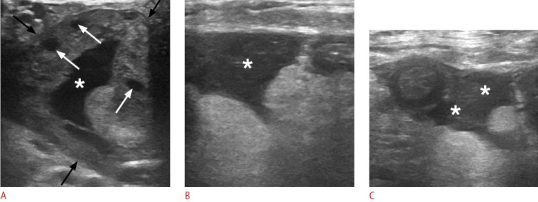
Fig. 18. Ruptured hemorrhagic ovarian cyst in a 35-year-old woman presenting with acute left lower quadrant pain.
A. Sonographic image of the left lower abdomen reveals an enlarged left ovary (black arrows) with a well-defined cystic lesion (asterisk) featuring an irregular, collapsed wall. Note the presence of normal follicles (white arrows). B, C. The concomitant presence of intraperitoneal fluid with low-level echoes encircling small bowel loops (asterisks) suggests a hemoperitoneum resulting from cyst rupture.

