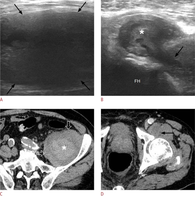
Fig. 22. Hemorrhagic iliopsoas bursitis in a 63-year-old man.
A. Sonographic image at the level of the left lower quadrant shows a well-defined fluid-filled collection (black arrows) with internal lowlevel echoes, indicative of a distended iliopsoas bursa. B. Sonographic image at a more distal level than image A again demonstrates the bursa situated anterior to the femoral head (FH), containing hyperechoic material (asterisk). Note the communication between the bursa and the left hip joint (black arrow). C. Corresponding axial contrast-enhanced computed tomography image reveals the distended bursa (open arrows) with internal high-attenuating hemorrhagic material (asterisk). The hyperdense content exhibited high attenuation on the scan prior to intravenous contrast administration (not shown). D. The bursa is depicted at its expected anatomical location anterior to the left hip, in communication with the joint (black arrow).

