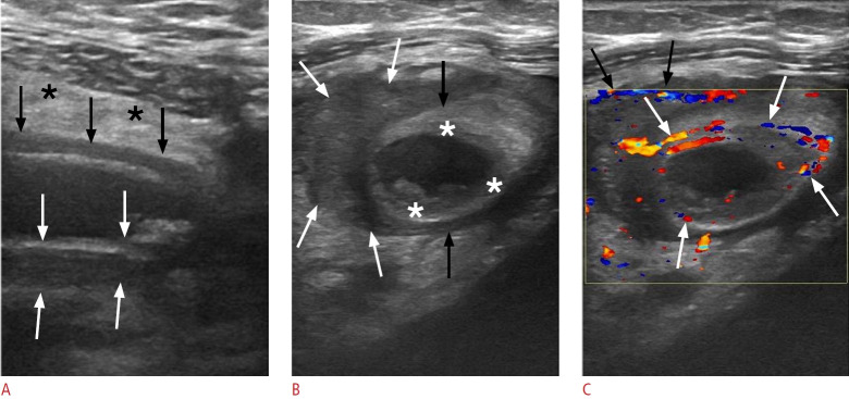
Fig. 7. Acute appendicitis in a 33-year-old woman.
A. Longitudinal sonogram of the inflamed appendix shows a noncompressible, blind-ending tubular structure with an outer diameter of 12.3 mm (black arrows), accompanied by marked mural thickening (between the white arrows). The surrounding mesentery appears hyperechoic (black asterisks). B. Sonographic image captured across the short axis of the appendix (black arrows) reveals hypoechogenic areas within the submucosal layer, indicative of edema (white asterisks). A heterogeneous, hypoechoic collection (white arrows) surrounds the appendix, which is suggestive of perforation. C. Transverse color Doppler sonography of the inflamed appendix reveals mural hyperemia (white arrows) and hypervascularity along the periphery of the periappendiceal fluid collection (black arrows).

