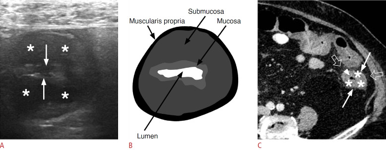
Fig. 9. Ischemic colitis in a 78-year-old woman.
A. Sonographic image through the short axis of the descending colon reveals mural thickening characterized by a hypoechoic pattern and loss of stratification, indicative of submucosal edema (asterisks) affecting a 20-cm segment. The image also shows a narrowed bowel lumen (white arrows). B. A schematic representation of image A illustrates submucosal edema within the mural colonic layers. C. Axial contrastenhanced computed tomography image demonstrates circumferential wall thickening of the descending colon, extending from the splenic flexure to the rectosigmoid junction. A low-density ring of submucosal edema (asterisks) is visible between the enhancing mucosa (arrowhead) and serosa (white arrows), accompanied by pericolic fat stranding (open arrows).

