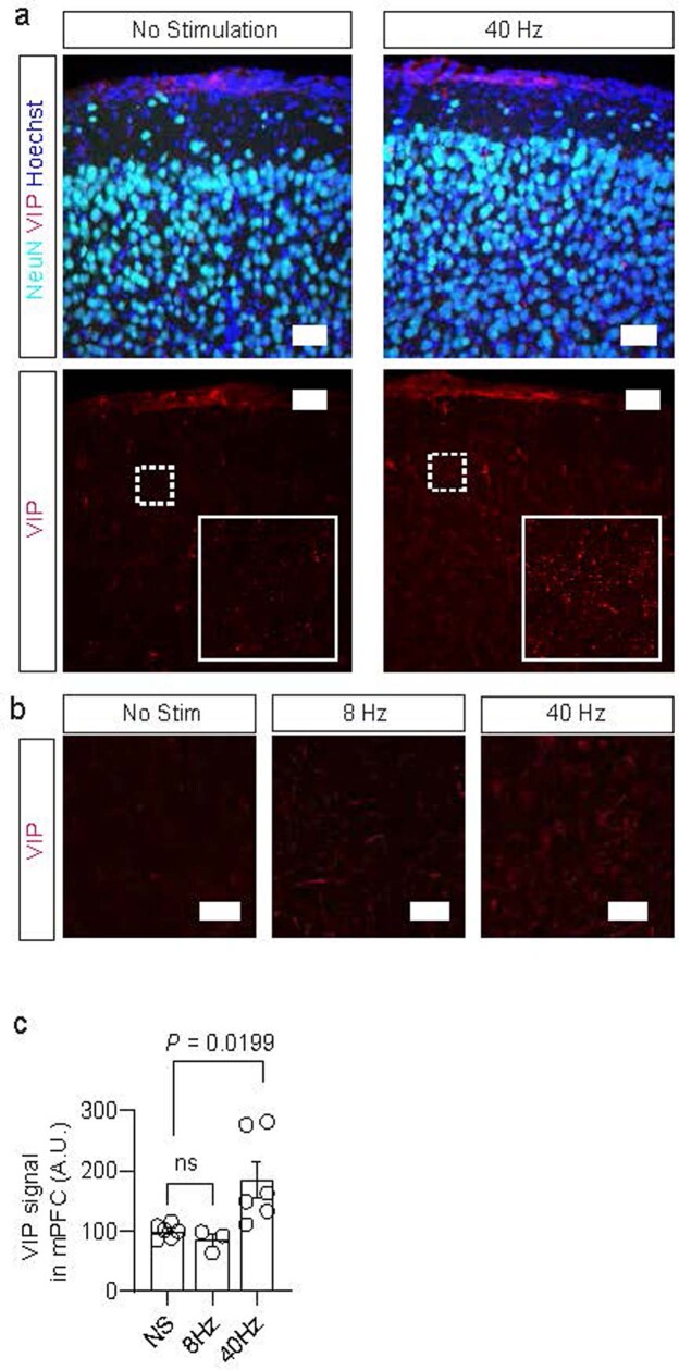
Extended Data Fig. 10. Immunohistochemistry for vasoactive intestinal peptide (VIP) following gamma stimulation.
a. Example images of VIP (red) and NeuN (cyan) immunohistochemistry in 6-month-old 5XFAD mouse prefrontal cortex following gamma stimulation or no-stimulation control. Insets reveal increased VIP immunofluorescence in gamma-treated mice. Scale bar, 50 μm. b. Example images of VIP immunohistochemistry in 6-month-old 5XFAD mouse cortex following gamma stimulation or control (red, VIP). Scale bar, 20 μm. c. Quantification of VIP signal intensity (n = 5 mice for no stimulation, 3 mice for 8 Hz stimulation, and mice for 40 Hz stimulation; each data point represents the mean from 3 z-stacks of the prefrontal cortex; data is presented as the mean ± s.e.m.; *P was calculated by one-way ANOVA followed by Dunnett’s multiple comparisons test).

