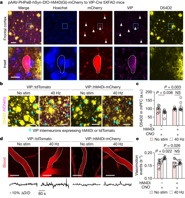
Fig. 4. VIP neurons mediate gamma-mediated glymphatic clearance.
a, Top row, example image of frontal cortex of 6-month-old VIP-Cre 5XFAD mice after receiving PHP.eB.AAV.Syn.DIO-hM4Di-mCherry. Bottom row, magnified view of indicated region in the top row. This experiment was repeated twice. Scale bar: 50 μm (top row) and 10 μm (bottom row). b, Example confocal z-stack maximum intensity images of medial prefrontal cortex (mPFC) of 6-month-old VIP-Cre 5XFAD mice injected with PHP.eB.AAV.Syn.DIO-hM4Di-mCherry (VIP::hM4Di-mCherry) or control (VIP::tdTomato) and labelled with D54D2 and mCherry (indicated with cyan arrowheads) receiving no stimulation or 40 Hz stimulation. Scale bars, 100 μm. c, Quantification of amyloid in experiment represented in b (n = 7 (no stimulation + tdTomato), 8 (40 Hz + tdTomato), 7 (no stimulation + hM4Di) and 7 (40 Hz + hM4Di) VIP-Cre 5XFAD mice; data are mean ± s.e.m.; P values by two-way ANOVA followed by Fisher’s LSD test). d, Example images and time series of arterial pulsatility after multisensory stimulation. Scale bars, 50 μm. e, Quantification of arterial pulsatility (n = 5 VIP-Cre 5XFAD per group; data are mean ± s.e.m.; P values by two-way ANOVA followed by Fisher’s LSD test).

