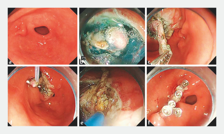
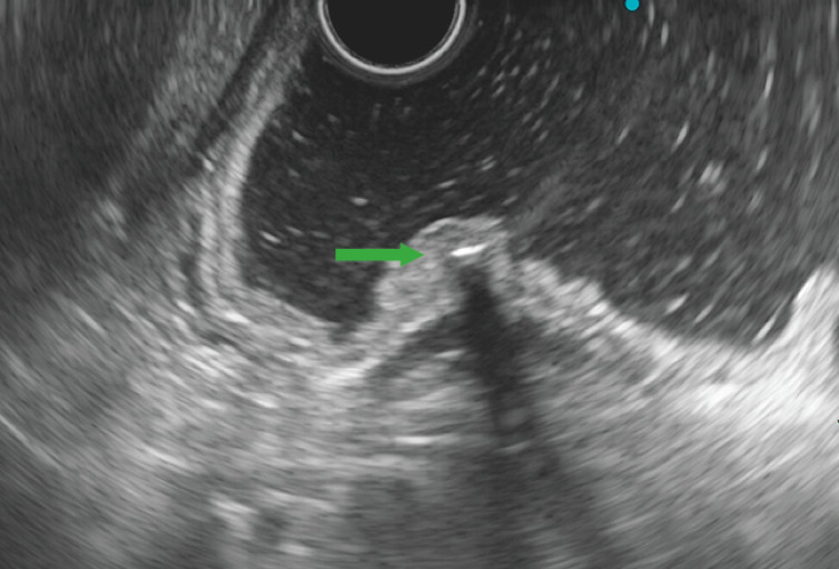
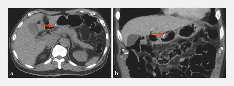
A 65-year-old man was referred to our hospital with a half-year history of upper abdominal pain. Endoscopy showed a submucosal eminence on the anterior wall of the gastric antrum ( Fig. 1 a ). Endoscopic ultrasonography (EUS) revealed a hyperechoic lesion in the gastric submucosa ( Fig. 2 ). A computed tomography (CT) scan showed a long, high density shadow in the gastric antrum, locally protruding into the serosal cavity ( Fig. 3 ). Emergency endoscopy was performed with the patient under general anesthesia and with endotracheal intubation ( Video 1 ).
Fig. 1.
Endoscopic images showing: a a submucosal eminence on the anterior wall of the gastric antrum; b partial exposure of the fishbone; c attempts to extract the fishbone using foreign body forceps; d snare traction being employed; e endoscopic full-thickness resection being performed; f closure of the perforation with metal clips.
Fig. 2.
Endoscopic ultrasonography image showing a hyperechoic lesion in the gastric submucosa.
Fig. 3.
Computed tomography images showing the location and depth of the fishbone (red arrow) on: a transverse plane; b coronal plane.
Removal of an embedded gastric fishbone by traction-assisted endoscopic full-thickness resection.
Video 1
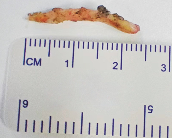
The mucosa of the gastric antrum was circumferentially incised, exposing one side of the fishbone ( Fig. 1 b ). Attempts to extract it using foreign body forceps were unsuccessful, indicating significant adhesion with the surrounding tissues ( Fig. 1 c ). Snare traction was then employed ( Fig. 1 d ). Subsequently, we performed traction-assisted endoscopic full-thickness resection (EFTR), revealing that the base of the fishbone was enveloped within the omentum ( Fig. 1 e ). After the adhesions had been dissected, a 2.5-cm long fishbone was successfully extracted ( Fig. 4 ) and the perforation was immediately closed with several metal clips ( Fig. 1 f ). The operative and postoperative periods were uneventful, without any complications.
Fig. 4.
Photograph of the extracted fishbone.
A fishbone invading the intrinsic muscularis and serosa of the gastric wall is rare 1 . Removal is often more challenging when there has been prolonged penetration of the gastric wall, and the risk of complications increases 2 3 . We performed traction using a snare combined with endoclips to assist in ETFR to successfully remove the fishbone. In this case, laparoscopic and open surgery were avoided.
Endoscopy_UCTN_Code_CCL_1AB_2AF
Funding Statement
Guangzhou Traditional Chinese Medicine and Traditional Chinese-Western Medicine Integration Science and Technology Project
Footnotes
Conflict of Interest The authors declare that they have no conflict of interest.
Endoscopy E-Videos https://eref.thieme.de/e-videos .
E-Videos is an open access online section of the journal Endoscopy , reporting on interesting cases and new techniques in gastroenterological endoscopy. All papers include a high-quality video and are published with a Creative Commons CC-BY license. Endoscopy E-Videos qualify for HINARI discounts and waivers and eligibility is automatically checked during the submission process. We grant 100% waivers to articles whose corresponding authors are based in Group A countries and 50% waivers to those who are based in Group B countries as classified by Research4Life (see: https://www.research4life.org/access/eligibility/ ). This section has its own submission website at https://mc.manuscriptcentral.com/e-videos .
References
- 1.Chiu YH, Hou SK, Chen SC et al. Diagnosis and endoscopic management of upper gastrointestinal foreign bodies. Am J Med Sci. 2012;343:192–195. doi: 10.1097/MAJ.0b013e3182263035. [DOI] [PubMed] [Google Scholar]
- 2.Birk M, Bauerfeind P, Deprez PH et al. Removal of foreign bodies in the upper gastrointestinal tract in adults: European Society of Gastrointestinal Endoscopy (ESGE) Clinical Guideline. Endoscopy. 2016;48:489–496. doi: 10.1055/s-0042-100456. [DOI] [PubMed] [Google Scholar]
- 3.Fan T, Wang CQ, Song YJ et al. Granulomatous inflammation of greater omentum caused by a migrating fishbone. J Coll Physicians Surg Pak. 2022;32:S124–S126. doi: 10.29271/jcpsp.2022.Supp2.S124. [DOI] [PubMed] [Google Scholar]


