Abstract
Purpose of Review
This article seeks to elucidate the mechanisms underlying the bidirectional relationship between the gut and the heart, focusing on the pathophysiology of heart failure. We have previously demonstrated that Heart failure (HF) has significant effects on splanchnic vasculature and leads to key alterations in the gut microbiome, portending greater comorbidity with HF.
Recent Findings
A growing field of research is focused on the effects of a “leaky gut” in the development of disease across organ systems. The leaky gut hypothesis centers on intestinal epithelial barrier dysfunction causing increased permeability of the gut and subsequent alterations to gut composition by endotoxins and microbial metabolites. Changes in the quantities of metabolites including short-chain fatty acids, trimethylamine N-oxide and other amino acid metabolites, and various bile acid species have been shown to result in gut dysbiosis and worsening HF.
Summary
The gut plays a highly significant role in HF prognosis and requires greater attention for future therapeutic interventions. Treatments targeting gut composition could have very beneficial effects on HF prognosis.
Keywords: Heart failure, Gut microbiome, Microbial metabolites, Splanchnic vasculature
Introduction
Heart failure (HF) currently affects over 6.7 million adults in the USA alone, and the prevalence is expected to rise to 8.5 million by 2030 [1]. It is a complex clinical syndrome with signs and symptoms that result from a structural or functional impairment of ventricular filling or ejection [2]. Typical signs of HF include elevated jugular venous pressure, pulmonary crackles, and peripheral/abdominal edema, with typical symptoms including breathlessness and fatigue [2]. Additional non-cardiac complications of HF arise from the downstream effects on other systems and organs, particularly the gastrointestinal system [3]. Patients with HF often complain of abdominal symptoms, including early satiety, abdominal discomfort and swelling, and sometimes even abnormal bowel habits. However, asking about these symptoms has not been emphasized in the traditional bedside evaluation. This connection between the gut and HF has yet to be clearly defined. However, with the increasing prevalence of HF, there is a need to develop new comprehensive therapeutic approaches, and novel insights into the role of the gut in the pathogenesis and pathophysiology of HF may allow this connection to be exploited for more effective HF treatment [4]. As previously mentioned, the current understanding of the heart-gut axis is incomplete, and there remains a lack of understanding of how gut dysbiosis and other related metabolic disturbances in gut microbiota contribute to both HF disease progression and the promotion of risk factors such as atherosclerosis and hypertension [5]. Several gut microbial metabolic pathways, including the production of trimethylamine N-oxide (TMAO), phenylacetylglutamine (PAGln), and short-chain fatty acids (SCFAs), have been shown to play an important role in the progression of HF [4, 5]. In addition, the abdominal compartment (more specifically the splanchnic vasculature) may further contribute to hemodynamic derangements [6]. For example, increased ventricular filling pressures, a hallmark of HF, can co-occur with increased splanchnic congestion. Thus, there is important evidence that the abdominal region can serve an important role in the prognosis of HF. In this review, we aim to not only underline the physiologic consequences of HF on the gut but also the role the gut microbiome plays in HF pathophysiology.
Physiologic Consequences of Impaired Hemodynamics on the Gut
Heart failure has been traditionally defined by impaired forward flow exemplified by a low cardiac output. From a hemodynamic standpoint, the problem has always been attributed to a forward failure caused by the heart’s inability to pump enough blood to the vital organs [7]. However, increasing evidence has shown a concurrent significant backward failure causing edema, leakiness, and engorgement of veins among other symptoms [6]. The inability of the ventricles to effectively pump out the blood entering them also increases ventricular filling pressures. This backward failure may produce a disproportionate arterial-venous blood distribution in the vasculature, leading to systemic venous congestion that impedes blood exiting the abdominal organs including the intestines, liver, and kidneys [8].
Importance of Splanchnic Vasculature
Systemic congestion typically involves abdominal congestion, as the splanchnic vasculature holds up to 65% of the total blood volume in a typical circulatory system [9]. Three midline branches of the abdominal aorta supply blood to the abdominal organs: the celiac artery supplying blood to the foregut (stomach, spleen, and pancreas), the superior mesenteric artery to the midgut (pancreas, small intestines, colon), and the inferior mesenteric artery to the hindgut (colon). About 25% of cardiac output flows to the splanchnic system through these three main arteries. The hepatic portal vein also plays an important role in the splanchnic vasculature as 75% of splanchnic arterial flow reaches the liver only after moving through abdominal organs and entering the portal vein [10]. The remaining 25% of blood delivered to the liver gets there directly via the hepatic artery. Vascular channels (sinusoids) within the liver funnel both venous and arterial blood to the hepatic central veins, which then drain into the inferior vena cava and flow back to the heart. Volume recruitment from the splanchnic compartment to the systemic circulation is a key physiological response to stressors like physical activity and blood loss.
Within the abdominal vasculature, α1, α2, and β2 adrenergic receptors play an important role in congestive HF. When activated, α-adrenergic receptors cause vasoconstriction by constricting the hepatic arterial smooth muscle. β-adrenergic receptors cause vasodilation by decreasing vascular resistance and increasing blood flow through the hepatic artery. The α-adrenergic receptors are largely concentrated in the splanchnic arteries. The increased sympathetic stimulation results in catecholamine release causing vasoconstriction as the pressure in capacitance vessels rises [6]. With this progressive volume overload from splanchnic venous congestion, systemic congestion with increased intra-abdominal pressure (IAP) occurs [6]. In one study, 60% of patients with heart failure also had elevated IAP [11]. That study also importantly noted that persistently increased IAP despite diuresis has been associated with impaired renal function in congestive HF patients. Recent evidence suggests that modulating the greater splanchnic nerve may impact excess fluid redistribution, thereby potentially improving cardiac filling pressures and exercise capacity [12•].
The spleen is another organ that acts as an important part of the splanchnic vasculature, receiving about 5% of cardiac output [13]. Blood flow through the spleen modulates splanchnic congestion as the splenic vein joins with the superior mesenteric vein to form the portal vein [13]. Splenic contraction may provide an incremental increase in systemic blood volume during exertion especially in the setting of severe cardiac impairment. With increasing splanchnic congestion in HF, the microvascular pressure within the spleen tends to increase, and fluid flows out of the spleen, thereby overloading the lymphatic system and leading to interstitial edema [6, 13].
Inflammatory Hypothesis of Heart Failure
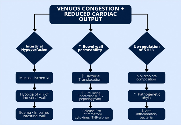
Hemodynamic derangements in HF can directly disrupt intestinal mucosa structure, with impaired cardiac output associated with intestinal edema, ischemia, and inflammation (Fig. 1) [4]. Specifically, venous congestion and splanchnic ischemia in HF may induce bowel hypoperfusion, leading to hypoxia of the villa of the bowel wall [14]. Disruption of this intestinal mucosal barrier leads to intestinal permeability and resultant translocation of gut bacteria and microbial products into the circulatory system (so called “leaky” gut) [14]. The diffusion of bacterial endotoxins and inflammatory cytokines into systemic circulation can activate the inflammatory response that is characteristic of HF [14]. Besides inducing systemic proinflammatory cytokines, endotoxins can also lead to an intracardiac inflammatory response that directly damages cardiomyocytes. For example, lipopolysaccharide (LPS) is a well-known endotoxin that diminishes ventricular contractility by binding to toll-like receptor 4 (TLR4) on cardiomyocytes.
Fig. 1.
Pathophysiological mechanisms behind the “gut hypothesis” of HF, particularly the association of splanchnic hemodynamics with disruption of the intestinal wall and gut dysbiosis
Abnormal sodium and fluid handling in the gut can co-occur with gut dysfunction caused by hypoperfusion. Sodium-hydrogen exchanger 3 (NHE3) is a heavily regulated electrolyte channel within the gut involved in regulating the exchange of sodium and protons. NHE3 is crucial for maintaining the balance between sodium secretion into, and absorption from, the gut. Proper NHE3 functioning is essential to maintaining salt, volume, and acid-base homeostasis. In congestive HF, the function of the NHE3 channel is upregulated because of hypoxia from venous congestion [15]. As a result, reabsorption of Na+ increases, and the subsequent increase in fluid absorption generates an unusual salt and water load on the cardiovascular system, worsening heart dysfunction. Therefore, NHE3 inhibition in the intestine can result in luminal sodium and water retention, leading to reduction in intestinal sodium and phosphate absorption and increased stool excretion, and even lower blood pressure [16]. Indeed, novel NHE3 inhibitors such as tenapanor have been used to treat hyperphosphatasemia in chronic kidney disease, while inhibitory properties of sodium glucose cotransporter-2 (SGLT2) inhibitors on NHE3 may explain some of their well-established benefits in the heart failure population [17].
Gut Microbial Contributions to Metabolic Aspects of Heart Failure
The gut luminal hypoxia that can occur in HF patients can significantly affect the composition of the commensal gut microbiota. A common theme in the gut microbiome of patients with HF is a shift to pathogenetic phyla and a diminished number of bacteria with anti-inflammatory properties. A study by Pasini et al. indicates that, in comparison to healthy controls, HF patients had significantly increased quantities of pathogenic bacteria such as Candida [18]. Additionally, it was found that the extent of colonization by pathogenic bacteria like Clostridium difficile correlates with HF severity. These observed patterns of gut microbial dysbiosis in HF have also been noted in other cardiovascular diseases like coronary artery disease and in chronic systemic diseases like type 2 diabetes. Importantly, a common symptom of these conditions is a state of inflammation, which is often associated with a disrupted gut microbiome.
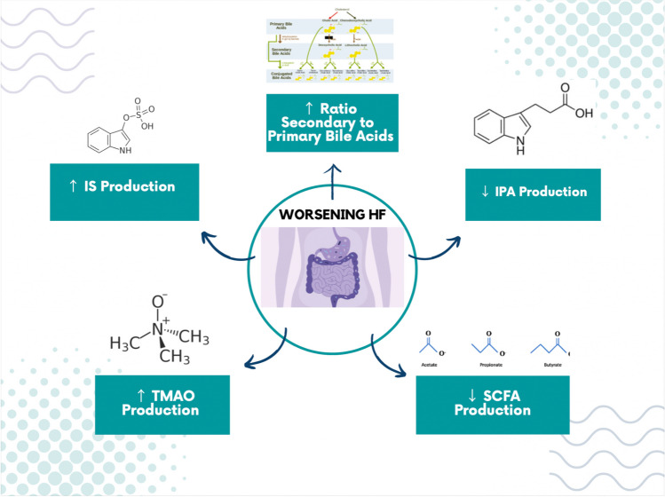
The gut microbiome’s role as a contributor to HF pathogenesis and adverse outcomes has increasingly been suspected, but delineation of precise pathways and participants involved is unclear. The gut microbiome produces metabolites that can be readily absorbed into the host bloodstream, which likely play a key role in mediating the inflammatory state characteristic of HF (Fig. 2). Approximately 10% of circulating small metabolites in mammalian blood is gut microbiome-related, implicating the gut as a significant environmental factor in human diseases [19]. Using metabolic approaches to better characterize the nature of these gut metabolites during HF can provide significant findings to better guide future diagnosis and therapy.
Fig. 2.
Gut microbial metabolites with active roles in HF pathophysiology. The metabolites fill important roles in susceptibility to HF and have been linked to adverse outcomes over time. The metabolites represent biomarkers and potential avenues for future therapeutics in treating HF
Short-Chain Fatty Acids
Short-chain fatty acids (SCFAs) are the result of fermentation of indigestible nutrients like fiber and complex carbohydrates by the gut microbes. They act on several different G-protein coupled receptors, allowing them to play an essential role in regulating fluid and electrolyte homeostasis along with maintenance of the epithelial barrier. Common SCFAs like acetate, butyrate, and propionate act as protectors of intestinal mucosa [20, 21]. Butyrate and propionate specifically aid in regulating blood pressure and exhibit anti-inflammatory effects through T-cell activation. Butyrate also plays an essential role in regulating the integrity of the intestinal barrier and suppressing intestinal and extra-intestinal inflammation. Not surprisingly, it has been demonstrated that depletion of these SCFAs, and known SCFA producers, is associated with disruption of the intestinal barrier and dysbiosis of gut microbiota in HF patients. Using 16S ribosomal RNA sequencing in fecal samples, Kamo et al. demonstrated that patients with HF have an increasingly depleted number of SCFA-producing gut microbes [22].
Not only are SCFAs needed for intestinal barrier function, but they also have a cardioprotective role. Kaye et al. demonstrated that mice fed a diet lacking in prebiotic fiber were prone to hypertension and cardiac hypertrophy, but when SCFAs were introduced to these mice, with no other alterations to their diet, these phenotypes were no longer displayed [23]. However, despite the demonstrated beneficial role of SCFAs, there are still debates regarding the therapeutic use of SCFA supplementation. Some studies have shown that neither acetate nor fiber could change cardiac remodeling and override the development of HF [24, 25•].
Trimethylamine N-oxide
Trimethylamine N-oxide (TMAO) is a gut microbiome-mediated metabolite that has been identified as a biomarker for cardiovascular disease and provides a well-characterized example of gut-host interactions with downstream effects in HF [26]. Dietary nutrients containing a trimethylamine (TMA) moiety, including choline, phosphatidylcholine, and carnitine, can be metabolized by the gut microbial enzyme TMA lyase, which cleaves the TMA from the parent nutrient. Once cleaved, the free TMA in the intestinal lumen can be absorbed into hepatic circulation where host enzymes, particularly hepatic flavin monooxygenase 3 (FMO3), convert TMA into TMAO [26].
TMAO has a multitude of demonstrated negative effects on the cardiovascular system. For example, high TMAO levels have been linked to atherosclerotic heart disease and overall major adverse cardiovascular events (MACE) [27, 28]. Murine studies have demonstrated that mice treated with supplemental TMAO or choline before HF development saw worsening pulmonary edema and increased systolic dysfunction. However, these effects were reversed with dietary changes or inhibition of microbial enzymes generating TMA/TMAO [29, 30]. However, the exact TMAO host receptor through which TMAO promotes HF is still unknown. Elevated TMAO levels are common in HF patients and correlate with diastolic dysfunction, which may mean that venous congestion plays an important role in altering the gut microbiome to increase TMAO production. Such a finding would be of immense significance considering that elevated baseline TMAO levels have been implicated as a predictor of mortality in patients with HF [31].
Amino Acid Metabolites
Gut bacteria play an essential role in fermenting and metabolizing specific dietary amino acids (AA) such as phenylalanine, tryptophan, and tyrosine. Tryptophan is metabolized into indole which can then be converted into either indole-3-propionate (IPA) or indoxyl sulfate (IS). Indoles like IPA play an important role in maintaining the integrity of the intestinal wall, reducing both permeability and the entering of circulating inflammatory cytokines [32]. It has been postulated that IPA acts as a mitochondrial modulator in cardiomyocytes and has been shown to alter cardiac function in an ex vivo mouse model [33]. As is true for most microbial metabolites, only certain bacteria in the gut microbiome can metabolize amino acids to make IPA. Hence, changes in the composition of the gut microbiota could easily result in decreased IPA levels. A result of this shift in composition is increased permeability of the intestinal wall and the mechanisms that describe the “gut hypothesis” of HF.
Although microbial metabolism of dietary AAs can produce beneficial downstream effects, other effects can be harmful. As previously mentioned, tryptophan can be converted into IS instead of IPA, and tyrosine and phenylalanine can be metabolized into p-cresyl sulfate (pCS). Both IS and pCS are uremic toxins that exhibit biological toxicity on the kidney and cardiovascular system [34]. Furthermore, IS specifically has demonstrated adverse cardiac effects in cells. In a human monocytic cell line, treatment with IS increased expression of TNF-ɑ and IL-1β, two pro-inflammatory cytokines that stimulate adverse cardiac remodeling including hypertrophy and collagen fibrosis [35]. Another study demonstrated that high IS levels were associated with cardiac dysfunction, particularly diastolic dysfunction, and a heightened risk of cardiac events in patients with dilated cardiomyopathy [36]. Therefore, a therapeutic benefit may be obtained by targeting the gut microbial enzymes that generate pCS and IS to reduce or inhibit the pCS/IS-mediated pro-thrombotic phenotype [37•]. Removal of uremic toxins from the GI tract, before they are systemically absorbed, also has the potential to be an effective treatment. Previous studies have demonstrated that oral uremic toxin adsorbents can reverse HF by stopping myocardial apoptosis [38].
Phenylacetylglutamine (PAGln), a meta-organismal metabolite derived from microbial fermentation of phenylalanine to phenylacetic acid followed by glutamine conjugation by host liver enzymes, has also been linked to adverse cardiac events in patients with chronic kidney disease [39]. PAGln fosters heightened thrombotic risks in animal model studies and in human platelet studies, which likely accounts for the observed association between elevated PAGln levels and clinical thrombotic event risks [40]. Moreover, the effects of PAGln, which possesses structural similarity to catecholamines, were shown to be mediated via adrenergic receptors [40]. In addition, our group recently reported a striking association between PAGln levels and prevalent HF, as well as the ability of PAGln to foster a negative inotropic effect on both epinephrine-stimulated isolated cardiomyocytes and stimulation of the gene responsible for cardiomyocyte natriuretic peptide expression in vitro and in vivo [41•]. In patients with HF, plasma PAGln levels served as an independent predictor of an increased risk of adverse cardiovascular events in HF that was complementary to NT-proBNP levels [42•, 43].
Bile Acids
Primary bile acids (BA) are synthesized in the liver through the oxidation of cholesterol. BAs are actively secreted into the bile along with cholesterol and phospholipids and play an essential role as emulsifiers through the absorption of lipids and fat-soluble vitamins. Primary BAs are predominantly recycled and reabsorbed in the ileum via the hepatic portal vein in the enterohepatic cycle. Those that are not recirculated to the liver end up in the colon where the gut microbiota use bile salt hydrolases to convert them into secondary BAs [44].
BA studies have shown contradicting effects on cardiac structure and function. In the past, it was thought that primary BAs caused only negative inotropic and chronotropic effects [45, 46]. Recent studies have shown that bile acids can have varying effects depending on which BA receptor is expressed on the cardiomyocyte. For example, one study demonstrated that BAs binding to Takeda G protein-coupled receptor 5 (TGR5) improved myocardial survival following physiologic and hemodynamic stress in mice [47]. However, another investigation demonstrated that BAs binding to a different receptor, the farnesoid X receptor, resulted in cardiac apoptosis and contributed to myocardial ischemic injury [48].
The types of BAs present in one’s body differ between healthy individuals and individuals with chronic HF, in part because of gut dysbiosis. Most notably, Mayerhofer et al. found a higher ratio of secondary BAs to primary BAs in patients with chronic HF [49]. Although this may lead to an assumption that primary BAs are more beneficial for individuals, a separate study indicated that secondary BAs can also have positive effects in reversing HF [50]. This study, where patients with HF were treated with the secondary BA ursodeoxycholic acid, found that the BA exerted anti-inflammatory properties and improved peripheral blood flow by trapping LPS, an inflammatory cytokine, in micelles. These contrasting findings suggest that since individual BAs have varying effects on HF patients, the development of HF may be associated more with an imbalance of bile acids rather than a specific, or group of specific, BAs.
Conclusions and Future Direction
Heart failure remains an immense health burden in the USA despite the plethora of treatment regimens now available as many of those regimens have proven ineffective. Recent studies have demonstrated the impact of hemodynamic derangements on gut physiology and the important subsequent contribution to the development and prognosis of HF. Future studies analyzing host cardio-abdominal-renal interactions are warranted, especially in the context of continuous congestion. Understanding more about splanchnic blood vessels and microcirculation may also offer alternative diagnostic routes for HF, and current clinical trials are investigating promising new drug (e.g., NHE3 inhibitors) or device (e.g., splanchnic nerve ablation or renal denervation) therapies for relieving splanchnic congestion [51].
Importantly, a growing body of evidence demonstrates that alterations in the gut microbiome may serve as a mediator in furthering the pathogenesis of many of the complications that arise from HF. Specifically, intestinal barrier breakdown, microbial translocation, and an altered composition of the gut microbiome have all been shown to have an immense impact on HF pathogenesis. Furthermore, with increased investigation of microbial metabolites like SCFAs, TMAO, BAs, and amino acid metabolites, we have been able to increase our understanding of the intricate host-microbe interactions in HF. These metabolites also serve as promising therapeutic targets and, as we move into an era of personalized medicine, measuring an individual’s specific metabolite levels could help direct personalized pharmacologic or dietary interventions.
Author contributions
Mr. Mahenthiran wrote the initial draft of the manuscript, and Ms Wilcox and Dr. Tang provided critical input and revisions for the manuscript.
Funding
This work was supported in part by grants from the National Institutes of Health (R01HL126827).
Declarations
Conflict of Interest
Dr. Tang has received consultancy fees from Sequana Medical, Cardiol Therapeutics, Genomics, Zehna Therapeutics, Boston Scientific, WhiteSwell, and Kiniksa Pharmaceuticals, CardiaTec Biosciences, Intellia Therapeutics, Bristol Myers Squibb, Alleviant Medical, and has received honorarium from Springer Nature, Belvoir Media Group, and American Board of Internal Medicine. Mr. Mahenthiran and Ms Wilcox have no relationships to disclose.
Human and Animal Rights and Informed Consent
This article does not contain any studies with human or animal subjects performed by any of the authors.
Footnotes
Publisher’s Note
Springer Nature remains neutral with regard to jurisdictional claims in published maps and institutional affiliations.
References
Papers of particular interest, published recently, have been highlighted as: • Of importance
- 1.Bozkurt B, Ahmad T, Alexander KM, Baker WL, Bosak K, Breathett K, et al. Heart failure epidemiology and outcomes statistics: a report of the Heart Failure Society of America. J Card Fail. 2023;29(10):1412–1451. doi: 10.1016/j.cardfail.2023.07.006. [DOI] [PMC free article] [PubMed] [Google Scholar]
- 2.Heidenreich PA, Bozkurt B, Aguilar D, Allen LA, Byun JJ, Colvin MM, et al. 2022 AHA/ACC/HFSA guideline for the management of heart failure: executive summary: a report of the American College of Cardiology/American Heart Association Joint Committee on Clinical Practice guidelines. J Am Coll Cardiol. 2022;79(17):1757–1780. doi: 10.1016/j.jacc.2021.12.011. [DOI] [PubMed] [Google Scholar]
- 3.Rogler G, Rosano G. The heart and the gut. Eur Heart J. 2014;35(7):426–430. doi: 10.1093/eurheartj/eht271. [DOI] [PubMed] [Google Scholar]
- 4.Tang WHW, Li DY, Hazen SL. Dietary metabolism, the gut microbiome, and heart failure. Nat Rev Cardiol. 2019;16(3):137–154. doi: 10.1038/s41569-018-0108-7. [DOI] [PMC free article] [PubMed] [Google Scholar]
- 5.Mamic P, Snyder M, Tang WHW. Gut microbiome-based management of patients with heart failure: JACC review topic of the week. J Am Coll Cardiol. 2023;81(17):1729–1739. doi: 10.1016/j.jacc.2023.02.045. [DOI] [PubMed] [Google Scholar]
- 6.Verbrugge FH, Dupont M, Steels P, Grieten L, Malbrain M, Tang WHW, et al. Abdominal contributions to cardiorenal dysfunction in congestive heart failure. J Am Coll Cardiol. 2013;62(6):485–495. doi: 10.1016/j.jacc.2013.04.070. [DOI] [PubMed] [Google Scholar]
- 7.Schrier RW, Abraham WT. Hormones and hemodynamics in heart failure. N Engl J Med. 1999;341(8):577–585. doi: 10.1056/NEJM199908193410806. [DOI] [PubMed] [Google Scholar]
- 8.Mullens W, Abrahams Z, Francis GS, Sokos G, Taylor DO, Starling RC, et al. Importance of venous congestion for worsening of renal function in advanced decompensated heart failure. J Am Coll Cardiol. 2009;53(7):589–596. doi: 10.1016/j.jacc.2008.05.068. [DOI] [PMC free article] [PubMed] [Google Scholar]
- 9.Greenway CV. Role of splanchnic venous system in overall cardiovascular homeostasis. Fed Proc. 1983;42(6):1678–1684. [PubMed] [Google Scholar]
- 10.Gelman S, Mushlin PS. Catecholamine-induced changes in the splanchnic circulation affecting systemic hemodynamics. Anesthesiology. 2004;100(2):434–439. doi: 10.1097/00000542-200402000-00036. [DOI] [PubMed] [Google Scholar]
- 11.Mullens W, Abrahams Z, Skouri HN, Francis GS, Taylor DO, Starling RC, et al. Elevated intra-abdominal pressure in acute decompensated heart failure: a potential contributor to worsening renal function? J Am Coll Cardiol. 2008;51(3):300–306. doi: 10.1016/j.jacc.2007.09.043. [DOI] [PubMed] [Google Scholar]
- 12.•.Fudim M, Ponikowski PP, Burkhoff D, Dunlap ME, Sobotka PA, Molinger J, et al. Splanchnic nerve modulation in heart failure: mechanistic overview, initial clinical experience, and safety considerations. Eur J Heart Fail. 2021;23(7):1076–1084. doi: 10.1002/ejhf.2196. [DOI] [PMC free article] [PubMed] [Google Scholar]
- 13.Kaufman S, Deng Y. Splenic control of intravascular volume in the rat. J Physiol. 1993;468:557–565. doi: 10.1113/jphysiol.1993.sp019788. [DOI] [PMC free article] [PubMed] [Google Scholar]
- 14.Gallo A, Macerola N, Favuzzi AM, Nicolazzi MA, Gasbarrini A, Montalto M. The gut in heart failure: current knowledge and novel frontiers. Med Princ Pract. 2022;31(3):203–214. doi: 10.1159/000522284. [DOI] [PMC free article] [PubMed] [Google Scholar]
- 15.Inoue BH, dos Santos L, Pessoa TD, Antonio EL, Pacheco BP, Savignano FA, et al. Increased NHE3 abundance and transport activity in renal proximal tubule of rats with heart failure. Am J Physiol Regul Integr Comp Physiol. 2012;302(1):R166–R174. doi: 10.1152/ajpregu.00127.2011. [DOI] [PubMed] [Google Scholar]
- 16.Spencer AG, Labonte ED, Rosenbaum DP, Plato CF, Carreras CW, Leadbetter MR, et al. Intestinal inhibition of the Na+/H+ exchanger 3 prevents cardiorenal damage in rats and inhibits Na+ uptake in humans. Sci Transl Med. 2014;6(227):227ra36 [DOI] [PubMed]
- 17.Packer M. Activation and inhibition of sodium-hydrogen exchanger is a mechanism that links the pathophysiology and treatment of diabetes mellitus with that of heart failure. Circulation. 2017;136(16):1548–1559. doi: 10.1161/CIRCULATIONAHA.117.030418. [DOI] [PubMed] [Google Scholar]
- 18.Pasini E, Aquilani R, Testa C, Baiardi P, Angioletti S, Boschi F, et al. Pathogenic gut flora in patients with chronic heart failure. JACC Heart Fail. 2016;4(3):220–227. doi: 10.1016/j.jchf.2015.10.009. [DOI] [PubMed] [Google Scholar]
- 19.Wikoff WR, Anfora AT, Liu J, Schultz PG, Lesley SA, Peters EC, Siuzdak G. Metabolomics analysis reveals large effects of gut microflora on mammalian blood metabolites. Proc Natl Acad Sci USA. 2009;106(10):3698–3703. doi: 10.1073/pnas.0812874106. [DOI] [PMC free article] [PubMed] [Google Scholar]
- 20.Topping DL, Clifton PM. Short-chain fatty acids and human colonic function: roles of resistant starch and nonstarch polysaccharides. Physiol Rev. 2001;81(3):1031–1064. doi: 10.1152/physrev.2001.81.3.1031. [DOI] [PubMed] [Google Scholar]
- 21.Peng L, Li ZR, Green RS, Holzman IR, Lin J. Butyrate enhances the intestinal barrier by facilitating tight junction assembly via activation of AMP-activated protein kinase in Caco-2 cell monolayers. J Nutr. 2009;139(9):1619–1625. doi: 10.3945/jn.109.104638. [DOI] [PMC free article] [PubMed] [Google Scholar]
- 22.Kamo T, Akazawa H, Suda W, Saga-Kamo A, Shimizu Y, Yagi H, et al. Dysbiosis and compositional alterations with aging in the gut microbiota of patients with heart failure. PloS One. 2017;12(3):e0174099. doi: 10.1371/journal.pone.0174099. [DOI] [PMC free article] [PubMed] [Google Scholar]
- 23.Kaye DM, Shihata WA, Jama HA, Tsyganov K, Ziemann M, Kiriazis H, et al. Deficiency of prebiotic fiber and insufficient signaling through gut metabolite-sensing receptors leads to cardiovascular disease. Circulation. 2020;141(17):1393–1403. doi: 10.1161/CIRCULATIONAHA.119.043081. [DOI] [PubMed] [Google Scholar]
- 24.Jama HA, Fiedler A, Tsyganov K, Nelson E, Horlock D, Nakai ME, et al. Manipulation of the gut microbiota by the use of prebiotic fibre does not override a genetic predisposition to heart failure. Sci Rep. 2020;10(1):17919. doi: 10.1038/s41598-020-73614-y. [DOI] [PMC free article] [PubMed] [Google Scholar]
- 25.•.Awoyemi A, Mayerhofer C, Felix AS, Hov JR, Moscavitch SD, Lappegård KT, et al. Rifaximin or Saccharomyces boulardii in heart failure with reduced ejection fraction: results from the randomized GutHeart trial. EBioMedicine. 2021;70:103511. doi: 10.1016/j.ebiom.2021.103511. [DOI] [PMC free article] [PubMed] [Google Scholar]
- 26.Tang WH, Kitai T, Hazen SL. Gut microbiota in cardiovascular health and disease. Circ Res. 2017;120(7):1183–1196. doi: 10.1161/CIRCRESAHA.117.309715. [DOI] [PMC free article] [PubMed] [Google Scholar]
- 27.Koeth RA, Wang Z, Levison BS, Buffa JA, Org E, Sheehy BT, et al. Intestinal microbiota metabolism of L-carnitine, a nutrient in red meat, promotes atherosclerosis. Nat Med. 2013;19(5):576–585. doi: 10.1038/nm.3145. [DOI] [PMC free article] [PubMed] [Google Scholar]
- 28.Tang WH, Wang Z, Levison BS, Koeth RA, Britt EB, Fu X, et al. Intestinal microbial metabolism of phosphatidylcholine and cardiovascular risk. N Engl J Med. 2013;368(17):1575–1584. doi: 10.1056/NEJMoa1109400. [DOI] [PMC free article] [PubMed] [Google Scholar]
- 29.Organ CL, Li Z, Sharp TE, 3rd, Polhemus DJ, Gupta N, Goodchild TT, et al. Nonlethal inhibition of gut microbial trimethylamine n-oxide production improves cardiac function and remodeling in a murine model of heart failure. J Am Heart Assoc. 2020;9(10):e016223. doi: 10.1161/JAHA.119.016223. [DOI] [PMC free article] [PubMed] [Google Scholar]
- 30.Organ CL, Otsuka H, Bhushan S, Wang Z, Bradley J, Trivedi R, et al. Choline diet and its gut microbe-derived metabolite, trimethylamine n-oxide, exacerbate pressure overload-induCED heart failure. Circ Heart Fail. 2016;9(1):e002314. doi: 10.1161/CIRCHEARTFAILURE.115.002314. [DOI] [PMC free article] [PubMed] [Google Scholar]
- 31.Tang WH, Wang Z, Shrestha K, Borowski AG, Wu Y, Troughton RW, et al. Intestinal microbiota-dependent phosphatidylcholine metabolites, diastolic dysfunction, and adverse clinical outcomes in chronic systolic heart failure. J Card Fail. 2015;21(2):91–96. doi: 10.1016/j.cardfail.2014.11.006. [DOI] [PMC free article] [PubMed] [Google Scholar]
- 32.Zhao ZH, Xin FZ, Xue Y, Hu Z, Han Y, Ma F, et al. Indole-3-propionic acid inhibits gut dysbiosis and endotoxin leakage to attenuate steatohepatitis in rats. Exp Mol Med. 2019;51(9):1–14. doi: 10.1038/s12276-019-0304-5. [DOI] [PMC free article] [PubMed] [Google Scholar]
- 33.Gesper M, Nonnast ABH, Kumowski N, Stoehr R, Schuett K, Marx N, et al. Gut-derived metabolite indole-3-propionic acid modulates mitochondrial function in cardiomyocytes and alters cardiac function. Front Med (Lausanne) 2021;8:648259. doi: 10.3389/fmed.2021.648259. [DOI] [PMC free article] [PubMed] [Google Scholar]
- 34.Lekawanvijit S. Role of Gut-derived protein-bound uremic toxins in cardiorenal syndrome and potential treatment modalities. Circ J. 2015;79(10):2088–2097. doi: 10.1253/circj.CJ-15-0749. [DOI] [PubMed] [Google Scholar]
- 35.Lekawanvijit S, Adrahtas A, Kelly DJ, Kompa AR, Wang BH, Krum H. Does indoxyl sulfate, a uraemic toxin, have direct effects on cardiac fibroblasts and myocytes? Eur Heart J. 2010;31(14):1771–1779. doi: 10.1093/eurheartj/ehp574. [DOI] [PubMed] [Google Scholar]
- 36.Shimazu S, Hirashiki A, Okumura T, Yamada T, Okamoto R, Shinoda N, et al. Association between indoxyl sulfate and cardiac dysfunction and prognosis in patients with dilated cardiomyopathy. Circ J. 2013;77(2):390–396. doi: 10.1253/circj.CJ-12-0715. [DOI] [PubMed] [Google Scholar]
- 37.•.Nemet I, Funabashi M, Li XS, Dwidar M, Sangwan N, Skye SM, et al. Microbe-derived uremic solutes enhance thrombosis potential in the host. mBio. 2023:e0133123. New study examining tryptophan metabolites from gut microbiome—p-cresyl sulfate and indolyl sulfate—contributing to a prothrombotic phenotype. [DOI] [PMC free article] [PubMed]
- 38.Asanuma H, Chung H, Ito S, Min KD, Ihara M, Takahama H, et al. AST-120, an adsorbent of uremic toxins, improves the pathophysiology of heart failure in conscious dogs. Cardiovasc Drugs Ther. 2019;33(3):277–286. doi: 10.1007/s10557-019-06875-z. [DOI] [PubMed] [Google Scholar]
- 39.Poesen R, Claes K, Evenepoel P, de Loor H, Augustijns P, Kuypers D, et al. Microbiota-derived phenylacetylglutamine associates with overall mortality and cardiovascular disease in patients with CKD. J Am Soc Nephrol. 2016;27(11):3479–3487. doi: 10.1681/ASN.2015121302. [DOI] [PMC free article] [PubMed] [Google Scholar]
- 40.Nemet I, Saha PP, Gupta N, Zhu W, Romano KA, Skye SM, et al. A Cardiovascular disease-linked gut microbial metabolite acts via adrenergic receptors. Cell. 2020;180(5):862–877.e22. doi: 10.1016/j.cell.2020.02.016. [DOI] [PMC free article] [PubMed] [Google Scholar]
- 41.•.Romano KA, Nemet I, Prasad Saha P, Haghikia A, Li XS, Mohan ML, et al. Gut microbiota-generated phenylacetylglutamine and heart failure. Circ Heart Fail. 2023;16(1):e009972. doi: 10.1161/CIRCHEARTFAILURE.122.009972. [DOI] [PMC free article] [PubMed] [Google Scholar]
- 42.•.WHW T, Nemet I, Li XS, Wu Y, Haghikia A, Witkowski M, Koeth RA, Demuth I, König M, Steinhagen-Thiessen E, Bäckhed F, Fischbach MA, Deb A, Landmesser U, Hazen SL. Prognostic value of gut microbe-generated metabolite phenylacetylglutamine in patients with heart failure. Eur J Heart Fail. 2023; 10.1002/ejhf.3111. Longitudinal follow-up of two independent heart failure cohorts demonstrated association between elevated levels of phenylacetylglutamine and long-term adverse outcomes independent of traditional risk factors, cardio-renal indices, and TMAO.
- 43.Wei H, Wu J, Wang H, Huang J, Li C, Zhang Y, et al. Increased circulating phenylacetylglutamine concentration elevates the predictive value of cardiovascular event risk in heart failure patients. J Intern Med. 2023;294(4):515–530. doi: 10.1111/joim.13653. [DOI] [PubMed] [Google Scholar]
- 44.Foley MH, O'Flaherty S, Barrangou R, Theriot CM. Bile salt hydrolases: gatekeepers of bile acid metabolism and host-microbiome crosstalk in the gastrointestinal tract. PLoS Pathog. 2019;15(3):e1007581. doi: 10.1371/journal.ppat.1007581. [DOI] [PMC free article] [PubMed] [Google Scholar]
- 45.Binah O, Rubinstein I, Bomzon A, Better OS. Effects of bile acids on ventricular muscle contraction and electrophysiological properties: studies in rat papillary muscle and isolated ventricular myocytes. Naunyn Schmiedebergs Arch Pharmacol. 1987;335(2):160–165. doi: 10.1007/BF00177718. [DOI] [PubMed] [Google Scholar]
- 46.Joubert P. An in vivo investigation of the negative chronotropic effect of cholic acid in the rat. Clin Exp Pharmacol Physiol. 1978;5(1):1–8. doi: 10.1111/j.1440-1681.1978.tb00645.x. [DOI] [PubMed] [Google Scholar]
- 47.Eblimit Z, Thevananther S, Karpen SJ, Taegtmeyer H, Moore DD, Adorini L, et al. TGR5 activation induces cytoprotective changes in the heart and improves myocardial adaptability to physiologic, inotropic, and pressure-induced stress in mice. Cardiovasc Ther. 2018;36(5):e12462. doi: 10.1111/1755-5922.12462. [DOI] [PMC free article] [PubMed] [Google Scholar]
- 48.Pu J, Yuan A, Shan P, Gao E, Wang X, Wang Y, et al. Cardiomyocyte-expressed farnesoid-X-receptor is a novel apoptosis mediator and contributes to myocardial ischaemia/reperfusion injury. Eur Heart J. 2013;34(24):1834–1845. doi: 10.1093/eurheartj/ehs011. [DOI] [PMC free article] [PubMed] [Google Scholar]
- 49.Mayerhofer CCK, Ueland T, Broch K, Vincent RP, Cross GF, Dahl CP, et al. Increased secondary/primary bile acid ratio in chronic heart failure. J Card Fail. 2017;23(9):666–671. doi: 10.1016/j.cardfail.2017.06.007. [DOI] [PubMed] [Google Scholar]
- 50.von Haehling S, Schefold JC, Jankowska EA, Springer J, Vazir A, Kalra PR, et al. Ursodeoxycholic acid in patients with chronic heart failure: a double-blind, randomized, placebo-controlled, crossover trial. J Am Coll Cardiol. 2012;59(6):585–592. doi: 10.1016/j.jacc.2011.10.880. [DOI] [PubMed] [Google Scholar]
- 51.Kittipibul V, Ganesh A, Coburn A, Coyne BJ, Gray JM, Molinger J, et al. Splanchnic nerve modulation effects on surrogate measures of venous capacitance. J Am Heart Assoc. 2023;12(14):e028780. doi: 10.1161/JAHA.122.028780. [DOI] [PMC free article] [PubMed] [Google Scholar]


