Summary
Given the growing interest in the role of zinc in the onset and progression of diseases, there is a crucial demand for reliable methods to modulate zinc homeostasis. Using a dietary approach, we provide validated strategies to alter whole-body zinc in mice, applicable across species. For confirmation of zinc status, animal growth rates as well as plasma and urine zinc levels were evaluated. The accessible and cost-effective methodology outlined will increase scientific rigor, ensuring reproducibility in studies exploring the impact of zinc deficiency and repletion on the onset and progression of diseases.
Keywords: Zinc Deficiency, Zinc Repletion, Mouse Model, Dietary approach
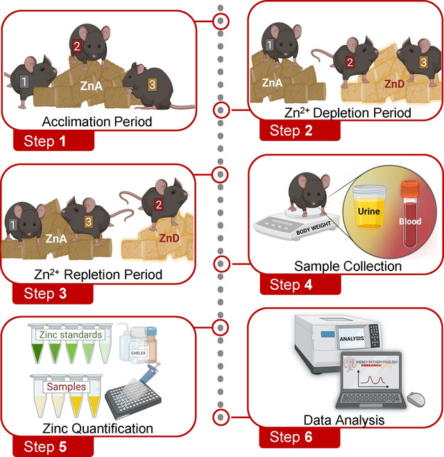
Graphical Abstract

Introduction
Zn2+ is the second most abundant trace metal in the human body, constituting 2 to 3 grams1. Skeletal muscle and bone are major Zn2+ reservoirs, accounting for approximately 50% and 30% of total body Zn2+, respectively2–5. Lower Zn2+ fractions are distributed across various tissues, including the kidney, prostate, liver, gastrointestinal tract, skin, lung, brain, heart, and pancreas5–9. Within these organs, intracellular Zn2+ serves as an essential cofactor for the catalytic activity and structural integrity of over 300 proteins, including those involved in synthesis of macromolecules and cell division10–13.
Since its initial recognition in 196314, Zn2+ deficiency has been increasingly implicated as a hidden culprit in multiple health conditions and chronic diseases such as diabetes, kidney diseases, cancers, neurodegenerative diseases, gastrointestinal disorders, respiratory infections, and skin conditions5,15–17. For more than 60 years, epidemiological, clinical, and experimental studies have focused on determining the role of Zn2+ deficiency in the development of these conditions and diseases14,17–20. Given the continued interest in the role of zinc in the onset and progression of diseases, more research is needed to identify the impact of this important micronutrient. The knowledge gained from these valuable studies will advocate for effective approaches that integrate Zn2+ supplementation into existing therapeutic strategies that address a plethora of conditions and diseases.
To directly study the impact of this important micronutrient on health and disease, standardized methods are required to modulate zinc homeostasis. Using a dietary approach, we provide validated strategies to alter whole-body zinc in mice, applicable across species. We also provide qualitative and quantitative methods to ensure the zinc status of experimental animals. The outlined accessible and cost-effective protocol will elevate scientific rigor, ensuring reproducibility in studies exploring the impact of zinc deficiency and repletion on the onset and progression of a multitude of health conditions and diseases.
Materials and Equipment
4% Chelex Solution
Suspend Chelex 100 Chelating Resin (2 g) in double distilled H20 (50 mL).
On a rocker, shake Chelex Solution for 2 – 3 minutes.
Centrifuge Chelex Solution at 6,000rpm for 1.5 – 2 minutes.
Collect clear portion of supernatant and aliquot 10 mL into multiple clear conical tubes.
Store 4% Chelex Solution aliquots at −80° C until use.
Tris Buffer
In an aliquot of 4% Chelex Solution (10 mL), suspend a Tris Buffered Saline Tablet.
Using a rocker, gently agitate the buffer until the tablet is dissolved.
Aliquot Tris Buffer (5 mL) into black conical tubes.
Store Tris Buffer aliquots at 4°C until use.
Zn Detector Solution
Into an aliquot of Tris Buffer (5 mL), dispense Zn Detector (25 μl).
Note: The Zn Detector is light sensitive.
Vortex for 10 seconds.
Immediately use Zn Detector Solution for zinc quantification.
ZnCl2 Standards (Table 1)
Table 1.
| ZnCl2 Standards | Tris Buffer | ZnCl2 Concentration | ||
|---|---|---|---|---|
| 100 μL of 1 mM | + | 900 μL | = | 100 μM |
| 500 μL of 100 μM | + | 500 μL | = | 50 μM |
| 500 μL of 50 μM | + | 500 μL | = | 25 μM |
| 500 μL of 25 μM | + | 500 μL | = | 12.5 μM |
| 500 μL of 12.5 μM | + | 500 μL | = | 6.25 μM |
| 500 μL of 6.25 μM | + | 500 μL | = | 3.125 μM |
| 500 μL of 3.125 μM | + | 500 μL | = | 1.56 μM |
| 500 μL of 1.56 μM | + | 500 μL | = | 0.78 μM |
| 500 μL of 0.78 μM | + | 500 μL | = | 0.39 μM |
| 500 μL of 0.39 μM | + | 500 μL | = | 0.195 μM |
| 500 μL of 0.195 μM | + | 500 μL | = | 0.097 μM |
| ------- | + | 1000 μL | = | 0 μM |
Using reagents provided in the Zinc Quantification Kit, prepare a 1 mM ZnCl2 Standard by suspending 100 mM ZnCl2 Standard (10 μL) into Assay Buffer (990 μL).
To prepare a 100 μM ZnCl2 Standard, suspend 1 mM ZnCl2 (100 μL) into Tris Buffer (900 μL).
To prepare the designed concentrations of ZnCl2 Standards, begin with the 100 μM ZnCl2 Standard and perform a serial dilution in Tris Buffer.
-
Store ZnCl2 Standards at 4°C until use.
Note: Discard ZnCl2 Standards after two uses.
Before you Begin
Institutional permissions
All animal experiments performed were in accordance with the Animal Care and Use Committee of Wright State University and were conducted in accordance with the National Institutes of Health Guide for the Care and Use of Laboratory Animals. The appropriate licenses and permission for animal experiments should be obtained before proceeding with this protocol.
Step-by-Step Method Details
I. Acclimation Period
Timing: [2 weeks]
Acclimation of Mice to Zn2+-Adequate Diet
- CRITICAL: Group house mice at 27°C in a 12:12-hour light-dark cycled room.
- Until 6 weeks of age, maintain C57BL/6 mice on a standard chow diet.
-
At 6 weeks old, acclimate mice to a Zn2+-adequate (ZnA) diet for 2 weeks.
- Provide mice with ad libitum access to food and purified drinking H2O.
NOTE: Since the quality of tap H2O may vary between facilities, use commercially available purified drinking H2O. - Each week, weigh individual mice to monitor growth rate.
- At the end of the acclimation period, proceed to “Step-by-Step Method Details” section.
II. Zn2+ Depletion Period
Timing: 10 weeks
- CRITICAL: Since mice are social animals, group housing provides a less stressful environment than individually housing mice. If mice must be housed individually, ensure that all experimental groups are housed similarly. To ensure that mice consume similar nutritional content, pair-feed mice by providing the ZnA group with the amount consumed by ZnD mice. Avoid using glass H2O bottles, as glass may be a source of Zn2+. To avoid consumption of fecal Zn2+, weekly mouse cage changes are required.
-
aGroup 1 (Zn2+ Adequate): For the control group, maintain a subset of the ZnA mice on the ZnA diet for up to 10 weeks. Continue to provide ZnA mice with commercially available purified drinking H2O.
-
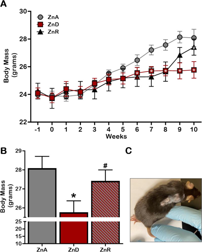
bGroup 2 (Zn2+ Deficient): Switch a subset of ZnA mice to an ingredient-matched, ZnD diet for 10 weeks. To maintain a Zn2+-free environment, provide ZnD mice deionized, distilled drinking H2O in plastic containers.CRITICAL: Visual signs of chronic Zn2+ deficiency include growth retardation (Figure 1B), hair loss (Figure 1C) and loose stool. However, the absence of these visual signs should not disqualify mice from studies. Zn2+ levels must be quantified to verify changes in Zn2+ levels (see “Zn2+ Quantification” Step). NOTE: While Zn2+ deficiency was confirmed at week-10 (Figure 2), this status may occur at an earlier timepoint. As such, plasma and urine Zn2+ levels should be quantified to identify timepoint of changes in Zn2+ levels (see “Zn2+ Quantification” Step).
-
a
Figure 1: Zn2+ homeostasis plays a critical role in growth and development.
To investigate the impact of dietary Zn restriction on mouse health, mice were fed a Zn2+-adequate diet (ZnA, grey) or Zn2+-deficient diet (ZnD, red) for 10 weeks. Effects on weekly weight gain (Figure 1A), body mass at week-10 (Figure 1B), and skin health (Figure 1C) were examined. *p < 0.05 vs ZnA, #p < 0.05 vs ZnD.
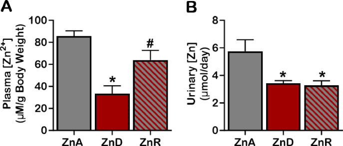
Figure 2: Impact of dietary Zn2+ restriction and repletion on circulating Zn2+ levels.
To probe the influence of dietary Zn restriction on Zn2+ levels, mice were fed a Zn2+-adequate diet (ZnA, grey) or Zn2+-deficient diet (ZnD, red) for 10 weeks. Effects on plasma (Figure 1A), and urine (Figure 1A) Zn2+ levels were assessed. *p < 0.05 vs ZnA, #p < 0.05 vs ZnD.
III. Zn2+ Repletion Period
Timing: 2 weeks
-
c
Group 3 (Zn2+ Repletion): For Zn2+ repletion studies, after 8 weeks on a ZnD-diet (as described for Group 2), return mice to the ZnA-diet and purified drinking H2O (as described for Group 1). For the remaining 2 weeks of the study, maintain this subset of mice (ZnR) on the ZnA-protocol.
IV. Sample Collection
- Plasma Samples
-
From the submandibular vein, collect blood samples from mice.NOTE: Collection method should be perfected to avoid hemolysis.
- Centrifuge collected blood samples at 1500 × g at 4 °C for 15 minutes.
- Collect and aliquot plasma (20 μL) into clear microcentrifuge tubes.
- Proceed to “Zn2+ Quantification” Step.
- Optional Step: Store plasma samples at −80°C for up to 12 months.
-
-
Urine Samples
To collect 24-hour urine samples, house mice in metabolic cages.
Centrifuge collected urine samples at 2000 × g at 4 °C for 10 minutes.
Collect and aliquot debris-free urine (100 μL) into black microcentrifuge tubes.
- Proceed to “Zn2+ Quantification” Step.
- Optional Step: Store urine samples at −80°C for up to 12 months.
V. Zinc Quantification
Timing: [30 mins]
NOTE: Perform all steps in a dark area.
Dispense Tris Buffer (30 μl) into appropriate wells of a black, clear bottom 96-well plate.
In duplicate, pipet Zn2+ Standards (10 μl) and unknown samples (10 μl) into the appropriate wells of the 96-well plate.
To protect Zn2+ Standards and unknown samples from light, cover 96-well plate with aluminum foil.
Using a rocker, gently shake 96-well plate at room temperature for 15 minutes to ensure mixing of Zn2+ Standards and unknown samples with Tris Buffer.
With a fluorescence plate reader, measure background fluorescence (Ex/Em = 485/525) of Zn2+ Standards and unknown samples.
Dispense Zn Detector (30 μl) into the appropriate wells of the 96-well plate.
After covering the 96-well plate with aluminum foil, gently shake on a rocker for 10 seconds.
Incubate covered 96-well plate at room temperature for 15 minutes and then measure Zn Detector fluorescence (Ex/Em = 485/525).
VI. Data Analysis
To obtain net fluorescence, subtract background fluorescence from Zn Detector fluorescence.
Using the net fluorescence, calculate Zn2+ concentration of unknown samples by extrapolating values from the Zn2+ standard curve.
Expected Outcomes
Impact of Dietary Zn2+ Restriction and Repletion on Growth and Development.
To assess the impact of Zn2+ restriction on nutritional status, body mass was measured in mice placed on a ZnA or ZnD-diet for 10 weeks (Figure 1). Until week-5, mice on a ZnD diet had similar body masses as mice maintained on the ZnA diet (Figure 1A). However, starting at week 6, ZnD mice experienced slower weight gain. Furthermore, between 6 – 8 weeks, some ZnD mice experienced alopecia (Figure 1C). While average food consumption was not significantly different (14.74 g ± 0.21 vs 15.21 g ± 0.54), at week-10, mice on the ZnD diet had significantly less body mass (25.769 g ± 0.601) compared to ZnA mice (28.08 g ± 0.626) (Figure 1B).
To assess the impact of Zn2+ repletion (ZnR) on nutritional status, at week 8, ZnD mice were returned to a ZnA diet. Zn2+ repleted mice experienced an accelerated growth rate (Figure 1A). After 2 weeks, body mass (27.42 g ± 0.583) and food consumption (15.78 g ± 0.39) of ZnR mice (Figure 1B) was comparable to mice maintained on the ZnA diet. These results indicate that chronic dietary Zn2+ restriction impairs growth, and acute dietary Zn2+ repletion improves development.
Impact of Dietary Zn2+ Restriction and Repletion on Plasma and Urinary Zn2+ Levels.
Using the Zn2+ Quantification protocol, plasma Zn2+ levels were quantified and normalized to mouse body mass (Figure 2A). At week-10, mice maintained on the ZnA-diet had plasma Zn2+ levels of 3.19 μM/g Body Mass. In contrast, plasma Zn2+ levels were 40% lower in mice placed on a ZnD diet (1.29 μM/g Body Mass). For ZnD mice returned to the ZnA-diet (ZnR) for 2 weeks, plasma Zn2+ levels (2.44 μM/g Body Mass) increased and were comparable to mice maintained on the ZnA diet.
The impact of dietary Zn2+ restriction and repletion on urinary Zn2+ excretion was also examined (Figure 2B). Similar to plasma Zn2+, urinary Zn2+ levels were reduced in ZnD mice (3.43 μmol/day ± 0.19 vs 5.76 μmol/day ± 0.84). However, after 2 weeks of Zn2+ repletion, urinary Zn2+ levels remained low (3.27μmol/day ± 0.34) and were comparable to mice maintained on the ZnD diet. Collectively, these findings demonstrate that chronic dietary Zn2+ restriction is effective at inducing Zn2+ deficiency. Furthermore, while acute dietary Zn2+ repletion improves circulating Zn2+ levels, urinary Zn2+excretion is not yet restored.
Limitations
This protocol was developed and optimized in adult, male, wild-type mice on a C57Bl/6 background. Various factors influence the handling of Zn2+, including genetic composition, age, and sex. Given the potential diversity in how Zn2+ is handled, the impact of dietary Zn2+ depletion and repletion may vary between experimental animals within a study. Consequently, the intricate interplay of genetic factors, age-related dynamics, and sex-specific differences can contribute to distinct responses to Zn2+ manipulation. This important consideration underscores the significance of establishing baseline Zn2+ levels. By obtaining a reference point, Zn2+ changes can be assessed not only between different experimental groups but also longitudinally, with each animal also serving as its own control. This rigorous approach provides a comprehensive assessment of the impact of Zn2+ dynamics on physiological and pathophysiological processes.
Troubleshooting
Problem 1: No changes in plasma and/or urinary Zn2+ levels after Zn2+ depletion Potential solution: Extend duration of Zn2+ Depletion Period
To maintain whole-body Zn2+ homeostasis, Zn2+ is released from Zn2+ stores into the circulation. As such, a longer depletion period may be required to deplete Zn2+ stores and subsequently reduce serum and urine Zn2+ levels.
Problem 2: Reduced plasma and urinary Zn2+ levels after Zn2+ repletion
Potential Solution: Extend duration of Zn2+ Repletion Period
While ZnR mice (2.44 μM/g Body Mass) have increased plasma Zn2+ levels compared to ZnD mice (1.29 μM/g Body Mass), these levels are not yet comparable to ZnA mice (3.19 μM/g Body Mass). Furthermore, urinary Zn2+ levels are still depleted (3.27 μmol/day ± 0.34 vs 5.76 μmol/day ± 0.84). Repletion of Zn2+ stores within organs proceed repletion of serum and urinary Zn2+ levels. As such, a longer repletion period may be required when using serum and urine Zn2+ as markers of Zn2+ status.
Resource availability
Lead contact
Further information and requests for resources and reagents should be directed to and will be fulfilled by the lead contact, Clintoria R. Williams, PhD, FAHA (clintoria.williams@wright.edu).
Materials availability
There are no newly generated materials associated with this protocol.
Data and code availability
This study did not generate/analyze [datasets/code].
Table of Key Resources.
| REAGENT or RESOURCE | SOURCE | IDENTIFIER |
|---|---|---|
| Chemicals | ||
| Chelex 100 Chelating Resin | Bio-Rad, Hercules, CA | 1421253 |
| Tris Buffer Saline Table | Millipore-Sigma, MO, USA | T5030-100TAB |
| Protease Inhibitor Cocktail | Millipore-Sigma, MO, USA | P8340-1ML |
| Critical Commercial Assays | ||
| Zinc Quantification Kit | Abcam, Boston, MA | ab176725 |
| Experimental Models | ||
| C57BL/6J Male Mice | The Jackson Laboratory | JAX:000664 |
| Other | ||
| Standard Chow Diet | Teklad, Inotiv, IN, USA | Teklad 2018 |
| Zn2+ Adequate Diet (50 ppm) | Teklad, Inotiv, IN, USA | TD 85420 |
| Zn2+ Deficient Diet (1 ppm) | Teklad, Inotiv, IN, USA | TD 85419 |
| Deionized Distilled H2O | Millipore-Sigma, MO, USA | 6442-88 |
| Purified Drinking H2O | Aquafina, Sam’s Club | 988708 |
| Black, Clear Bottom 96-well Plate | Thermo Fisher Scientific, Rochester, NY | 265301 |
| 15 ml Conical Tubes (Clear) | Celltreat, Pepperell, MA | 230411-R |
| 15ml Conical Tubes (Black) | Celltreat, Pepperell, MA | 229431 |
| 50 ml conical tube (Clear) | Thermo Fisher Scientific, Rochester, NY | 14-432-22 |
| Microcentrifuge Tubes (Black) | Celltreat, Pepperell, MA | 229437 |
| Microcentrifuge Tubes (Clear) | Electron Microscopy Sciences, Hatfield, PA | 63027-02 |
New and Noteworthy:
This methods paper details 1) dietary approaches to alter zinc homeostasis in rodents, and 2) qualitative and quantitative methods to ensure the zinc status of experimental animals. The outlined accessible and cost-effective protocol will elevate scientific rigor, ensuring reproducibility in studies exploring the impact of zinc deficiency and repletion on the onset and progression of a multitude of health conditions and diseases.
Acknowledgements
This work was supported by the National Institute of Diabetes and Digestive and Kidney Diseases Grants: R21 DK119879 (to C.R.W.), and R01 DK-133698 (to C.R.W.); by the American Heart Association Grant 16SDG27080009 (to C. R.W.); and by the American Society of Nephrology KidneyCure Transition to Independence Grant (to C.R.W.).
Footnotes
Declaration of interests
The authors declare no competing interests.
References
- 1.Plum L. M., Rink L. & Hajo H. The essential toxin: impact of zinc on human health. Int J Environ Res Public Health 7, 1342–1365 (2010). [DOI] [PMC free article] [PubMed] [Google Scholar]
- 2.Frontera W. R. & Ochala J. Skeletal Muscle: A Brief Review of Structure and Function. Behavior Genetics vol. 45 183–195 Preprint at 10.1007/s00223-014-9915-y (2015). [DOI] [PubMed] [Google Scholar]
- 3.Molenda M. & Kolmas J. The Role of Zinc in Bone Tissue Health and Regeneration—a Review. Biological Trace Element Research vol. 201 5640–5651 Preprint at 10.1007/s12011-023-03631-1 (2023). [DOI] [PMC free article] [PubMed] [Google Scholar]
- 4.Skiba G. et al. Influence of the Zinc and Fibre Addition in the Diet on Biomechanical Bone Properties in Weaned Piglets. Animals 12, (2022). [DOI] [PMC free article] [PubMed] [Google Scholar]
- 5.Ume A. C., Wenegieme T. Y., Adams D. N., Adesina S. E. & Williams C. R. Zinc Deficiency: A Potential Hidden Driver of the Detrimental Cycle of Chronic Kidney Disease and Hypertension. Kidney360 4, 398–404 (2023). [DOI] [PMC free article] [PubMed] [Google Scholar]
- 6.To P. K., Do M. H., Cho J. H. & Jung C. Growth modulatory role of zinc in prostate cancer and application to cancer therapeutics. Int J Mol Sci 21, (2020). [DOI] [PMC free article] [PubMed] [Google Scholar]
- 7.Molenda M. & Kolmas J. The Role of Zinc in Bone Tissue Health and Regeneration—a Review. Biological Trace Element Research vol. 201 5640–5651 Preprint at 10.1007/s12011-023-03631-1 (2023). [DOI] [PMC free article] [PubMed] [Google Scholar]
- 8.Chasapis C. T., Spiliopoulou C. A., Loutsidou A. C. & Stefanidou M. E. Zinc and human health: An update. Archives of Toxicology vol. 86 521–534 Preprint at 10.1007/s00204-011-0775-1 (2012). [DOI] [PubMed] [Google Scholar]
- 9.Roohani N., Hurrell R., Kelishadi R. & Schulin R. Zinc and its importance for human health: An integrative review. Journal of Research in Medical Sciences (2013). [PMC free article] [PubMed] [Google Scholar]
- 10.Briassoulis G., Briassoulis P., Ilia S., Miliaraki M. & Briassouli E. The Anti-Oxidative, Anti-Inflammatory, Anti-Apoptotic, and Anti-Necroptotic Role of Zinc in COVID-19 and Sepsis. Antioxidants vol. 12 Preprint at 10.3390/antiox12111942 (2023). [DOI] [PMC free article] [PubMed] [Google Scholar]
- 11.Berger M. M. et al. ESPEN micronutrient guideline. Clinical Nutrition 41, 1357–1424 (2022). [DOI] [PubMed] [Google Scholar]
- 12.Beyersmann D. & Haase H. Functions of zinc in signaling, proliferation and differentiation of mammalian cells. BioMetals vol. 14 (2001). [DOI] [PubMed] [Google Scholar]
- 13.Dresen E. et al. Overview of oxidative stress and the role of micronutrients in critical illness. Journal of Parenteral and Enteral Nutrition vol. 47 S38–S49 Preprint at 10.1002/jpen.2421 (2023). [DOI] [PubMed] [Google Scholar]
- 14.Prasad A. S. Discovery of human zinc deficiency: Its impact on human health and disease. Advances in Nutrition vol. 4 176–190 Preprint at 10.3945/an.112.003210 (2013). [DOI] [PMC free article] [PubMed] [Google Scholar]
- 15.Maywald M. & Rink L. Zinc in Human Health and Infectious Diseases. Biomolecules vol. 12 Preprint at 10.3390/biom12121748 (2022). [DOI] [PMC free article] [PubMed] [Google Scholar]
- 16.Rink L. & Gabriel P. Zinc and the immune system. Proc Nutr Soc 59, 541–52 (2000). [DOI] [PubMed] [Google Scholar]
- 17.Ume A. C., Wenegieme T. Y., Adams D. N., Adesina S. E. & Williams C. R. Zinc Deficiency: A Potential Hidden Driver of the Detrimental Cycle of Chronic Kidney Disease and Hypertension. Kidney360 4, 398–404 (2023). [DOI] [PMC free article] [PubMed] [Google Scholar]
- 18.Maywald M. & Rink L. Zinc in Human Health and Infectious Diseases. Biomolecules vol. 12 Preprint at 10.3390/biom12121748 (2022). [DOI] [PMC free article] [PubMed] [Google Scholar]
- 19.Plum L. M., Rink L. & Hajo H. The essential toxin: Impact of zinc on human health. International Journal of Environmental Research and Public Health vol. 7 1342–1365 Preprint at 10.3390/ijerph7041342 (2010). [DOI] [PMC free article] [PubMed] [Google Scholar]
- 20.Williams C. R. et al. Zinc deficiency induces hypertension by promoting renal Na+ reabsorption. Am J Physiol Renal Physiol 316, F646–F653 (2019). [DOI] [PMC free article] [PubMed] [Google Scholar]
Associated Data
This section collects any data citations, data availability statements, or supplementary materials included in this article.
Data Availability Statement
This study did not generate/analyze [datasets/code].


