

Abstract
Introduction and importance
Glomus tumors are benign soft tissue tumors of the glomus body, most regularly found in the sublingual region of the digits, palms, and soles. Extra digital lesions are uncommon and might be difficult to diagnose.
Case presentation
We report a rare case of a 38-year-old man who presented with a painful nodule on his right upper arm. A definite diagnosis was made by histopathological study. A complete surgical excision was performed to avoid recurrence.
Clinical discussion
Glomus tumors form less than 2 % of all soft tissue tumors. The tumor was first reported by Wood in 1812. It typically appears like a small blue-red solitary papule in the hand especially the digits, which are the most prevalent location for glomus tumors with an incidence rate of up to 75 %. The histopathology findings of glomus tumor, are three components: glomus cells, vasculature, and smooth muscle cells. The preferred method of treatment is total excision to prevent a recurrence.
Conclusion
Eventually, the glomus tumor is fairly a rare benign tumor that physicians should keep in mind as a deferential diagnosis when facing a subcutaneous nodule and don't rule out when the tumor is extradigital.
Keywords: Glomus tumor, Benign, Extradigital, Nodule, Surgery, Upper arm
Highlights
-
•
Glomus tumors are benign soft tissue tumors of the glomus body, most regularly found in the sublingual region of the digits.
-
•
Extra digital lesions are uncommon and might be difficult to diagnose.
-
•
We described a case presented with a painful nodule on his right upper arm.
-
•
A histopathological study is the only method to confirm the diagnosis.
-
•
The treatment of choice is complete surgical excision to prevent a recurrence.
1. Introduction
Glomus tumors (GT), also known as paragangliomas, are uncommon benign vascular lesions emerging from the glomus body, representing roughly 1 % of all hand tumors [1,2]. A glomus body functions to regulate blood flow through dermal capillaries to control blood pressure and temperature [2]. The most usual place for the GT to arise is in the hands, especially the digits [3]. It generally presents with the symptoms of cold hypersensitivity, tenderness, pain, and nail deformation. The exact trigger for this tumor remains unclear [4]. In this report, we present a case of an extra digital glomus tumor unusually located on the upper arm. This case report has been reported in line with SCARE Criteria [5].
2. Case presentation
In July 2022, a 38-year-old man came to the Dermatology department in our hospital complaining of a painful nodule on the lateral inferior aspect of his right upper arm. There was no specific history of cold sensitivity or prior trauma. The mass was previously excised but it appeared again about a year ago. The patient did not have any allergies or diseases in his past medical history and did not take any medications. There was only a familial history of diabetes type 2 in both parents. Physical examination showed a purplish, tender nodule, with regular round edges measuring 1 cm × 1 cm. The nodule was rough and immobile [Fig. 1]. The upper limb showed no signs of neurological or vascular damage. A surgical biopsy specimen was taken under local anesthetic and sent to the pathology department. The hematoxylin-eosin stain of the biopsy showed a subcutaneous well-circumscribed tumor. The tumor was composed of sheets of uniform cells with scant eosinophilic cytoplasm, indistinct borders, and round-to-ovoid central nuclei with inconspicuous nucleoli. There were branching vessels lined by endothelial cells and surrounded by uniform cells [Fig. 2]. No cellular atypia is identified, and the Ki-67 rate is very low. The diagnosis of the glomus tumor was made and the treatment was complete surgical excision to avoid recurrence. The patient was discharged a few hours later, without any complications. As of the latest follow-up in October 2023, the patient has not reported any significant pain, issues at the wound site, or complications associated with the surgical procedure.
Fig. 1.
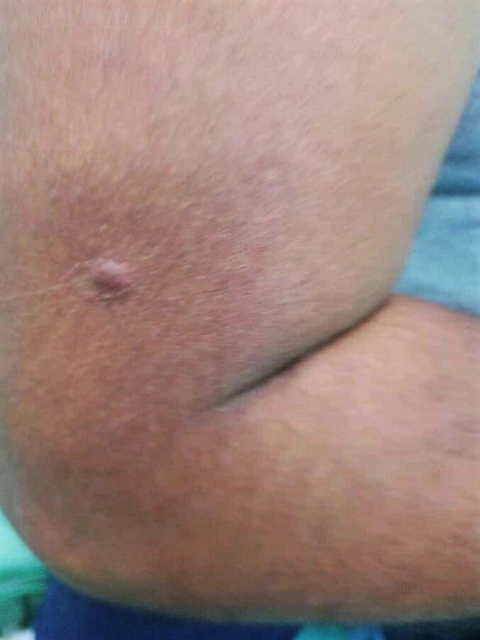
Clinical image shows a purplish, tender nodule, with regular round edges measuring 1 cm × 1 cm. The nodule was rough and immobile.
Fig. 2.

Hematoxylin and eosin-stain (A-D). Microscopic images of the nodule. (A and B) The low-power magnification shows branching vessels lined by endothelial cells and surrounded by uniform cells (40× and 100×). (C and D) The high-power magnification reveals sheets of uniform cells with scant eosinophilic cytoplasm, indistinct borders, and round-to-ovoid central nuclei with inconspicuous nucleoli (200× and 400×).
3. Discussion
Glomus tumor, also known as Barré–Mason syndrome or tumor of Popoff, is a rare benign neoplasm made up of the arterial part of a contractile neuromyoarterial structure called a glomus body [2]. Glomus tumors form less than 2 % of all soft tissue tumors [6]. The tumor was first reported by Wood in 1812. Then, Barré and Mason described the histological aspects of the tumor in 1924 [7,8]. It typically appears like a small blue-red solitary papule in the hand especially the digits, which are the most prevalent location for glomus tumors with an incidence rate of up to 75 % [9,10]. That is because the glomus body is more concentrated in the digits, especially the subungual area. The classical triad of glomus tumor symptoms includes severe pain, pinpoint tenderness, and cold sensitivity [2]. In a retrospective chart review of 50 patients who were diagnosed with glomus tumors from 1989 to 2009, 66 % of tumors were located in the digits and only five patients with extradigital lesions were located in the upper extremities. Also, we found that both digital and extradigital tumors are more common in females than males, with a mean age of 42.3 years [11]. Our patient is a 38-year-old man with a glomus tumor on the lateral inferior aspect of his upper arm, which makes it an unusual case. Also, the classical symptoms of a glomus tumor are absent. Furthermore, while our case report highlights the unusual presentation of an extradigital glomus tumor in the upper arm, we recognize the importance of contributing to the existing literature by providing a comprehensive review of published case reports and case series on glomus tumors in this atypical anatomical location. To bring additional insights and contribute meaningfully to the literature, we will include a table summarizing key details from relevant studies, such as patient demographics, clinical presentations, diagnostic modalities, and treatment outcomes [Table 1]. We believe that in extradigital glomus tumors, it is not necessary to present this typical trio to reach a diagnosis [17]. Several imaging techniques, including plain film radiography, ultrasound, computed tomography, single-photon emission computed tomography, and magnetic resonance imaging (MRI) scans, have been employed to enhance diagnostic accuracy, with MRI recognized as the most dependable for detecting glomus tumors [3]. In our patient, given the initial suspicion of dermatofibroma rather than a glomus tumor, we opted for a biopsy as the primary investigation. The glomus tumor should be differentiated from other benign lesions such as sebaceous cysts, angiomas, angiolipoma, neuromas, dermatofibroma, and leiomyoma [8,9]. The tumor is clinically identified by pain, tenderness, and sensitivity to temperature. However, an accurate initial diagnosis is rarely achieved, leading to prolonged suffering for patients who are frequently subjected to inappropriate treatments over several years [18]. The histopathology findings of glomus tumor, are three components: glomus cells, vasculature, and smooth muscle cells [4]. The preferred method of treatment is total excision to prevent a recurrence [8].
Table 1.
Comparison of cases of glomus tumor in the arm in the literature: patient demographics, clinical presentation, diagnostic modalities, and treatment outcomes.
| Study | Patient demographics | Clinical presentation | Diagnostic modalities | Treatment outcome |
|---|---|---|---|---|
| Takei TR, et al. 1995 [12] | 57-year-old woman | Pain, tenderness, cold sensitivity | Biopsy | Complete surgical excision, the symptoms had fully resolved |
| Yilmaz Tomak et al. 2003 [13] |
56-year-old | Pain and cold sensitivity | Biopsy | Complete surgical excision, No recurrence |
| Gökhan Temiz et al. 2016 [14] |
Five cases One woman only Mean age 35-year-old |
Pain and tenderness | Ultrasound Biopsy |
Complete surgical excision, No recurrence |
| Eman A. Tawfik et al. 2021 [15] |
64-year-old man | Pain, tenderness, cold sensitivity | Ultrasound Biopsy |
Complete surgical excision, the symptoms had fully resolved |
| Elmas, L et al. 2022 [16] |
59-year-old man | Pain and discomfort | Dermoscopic Biopsy |
Punch excision no need to surgical excision |
| Our case report 2022 |
38-year-old man | Painless, no cold sensitivity | Biopsy | Complete surgical excision, No recurrence |
4. Conclusion
Eventually, the glomus tumor is a fairly rare benign tumor that should be considered in the differential diagnosis of subcutaneous nodules, even when presenting in extradigital locations. A histopathological study is the only method to confirm the diagnosis. The treatment of choice is complete surgical excision to prevent a recurrence.
Provenance and peer review
Not commissioned, externally peer-reviewed.
Consent
Written informed consent was obtained from the patient for the publication of this case report and accompanying images. A copy of the written consent is available for review by the Editor-in-Chief of this journal.
Ethical approval
No ethical approval was needed for this case report.
Funding
This research did not receive any specific grant from funding agencies in the public, commercial, or not-for-profit sectors.
Author contribution
Moatasem Hussein Al-janabi: study design, data collections, data analysis, and writing.
Ghina Abdallah: study design, and writing.
Hasan Deeb: study design, and writing.
Firas Melhem: performed surgery.
Rabab Salloum: in reviewing the manuscript.
Guarantor
Rabab Salloum.
Registration of research studies
Not applicable.
Conflict of interest statement
The authors have no conflicts of interest to declare.
References
- 1.Hrubý J., Novotný R., Spaček M., Mitáš P., Hlubocký J., Janák D., Povýšil C., Lindner J. Surgical extirpation of glomus tumor from rare localization on the upper extremity. Case Rep. Vasc. Med. 2013;2013 doi: 10.1155/2013/570945. (Epub 2013 Sep 25. PMID: 24187644; PMCID: PMC3800625) [DOI] [PMC free article] [PubMed] [Google Scholar]
- 2.Morey V.M., Garg B., Kotwal P.P. Glomus tumours of the hand: review of literature. J. Clin. Orthop. Trauma. Oct-Dec 2016;7(4):286–291. doi: 10.1016/j.jcot.2016.04.006. (Epub 2016 Sep 1. PMID: 27857505; PMCID: PMC5106475). [DOI] [PMC free article] [PubMed] [Google Scholar]
- 3.Anley C., Vrettos B., Roche S., Solomons M. A glomus tumour of the elbow: a case report and review of the literature. Should. Elb. Jan 2014;6(1):60–62. doi: 10.1111/sae.12041. (Epub 2013 Oct 12. PMID: 27582912; PMCID: PMC4986653) [DOI] [PMC free article] [PubMed] [Google Scholar]
- 4.El Jouari O., Gallouj S., Elloudi S., Senhaji G., Rimani M., Mernissi F.Z. A painless glomus tumor: a case report. J. Med. Case Rep. Oct 18 2018;12(1):302. doi: 10.1186/s13256-018-1837-2. (PMID: 30333057; PMCID: PMC6193307) [DOI] [PMC free article] [PubMed] [Google Scholar]
- 5.Sohrabi C., Mathew G., Maria N., Kerwan A., Franchi T., Agha R.A. The SCARE 2023 guideline: updating consensus Surgical CAse REport (SCARE) guidelines. Int. J. Surg. Lond. Engl. 2023;109(5):1136. doi: 10.1097/JS9.0000000000000373. [DOI] [PMC free article] [PubMed] [Google Scholar]
- 6.Gombos Z., Zhang P.J. Glomus tumor. Arch. Pathol. Lab. Med. 2008;132(9):1448–1452. doi: 10.5858/2008-132-1448-GT. [DOI] [PubMed] [Google Scholar]
- 7.Wood W. On painful subcutaneous tubercle. Edinb. Med. Surg. J. 1812;8(31):283–291. [PMC free article] [PubMed] [Google Scholar]
- 8.Senhaji G., Gallouj S., El Jouari O., Lamouaffaq A., Rimani M., Mernissi F.Z. Rare tumor in unusual location - glomus tumor of the finger pulp (clinical and dermoscopic features): a case report. J. Med. Case Rep. 2018;12(1):196. doi: 10.1186/s13256-018-1721-0. [DOI] [PMC free article] [PubMed] [Google Scholar]
- 9.Glazebrook K., Laundre B., Schiefer T., Inwards C. Imaging features of glomus tumors. Skelet. Radiol. 2011;40:855–862. doi: 10.1007/s00256-010-1067-1. [DOI] [PubMed] [Google Scholar]
- 10.Schiefer T.K., Parker W.L., Anakwenze O.A., Amadio P.C., Inwards C.Y., Spinner R.J. Extradigital glomus tumors: a 20-year experience. Mayo Clin. Proc. 2006;81:1337–1344. doi: 10.4065/81.10.1337. [DOI] [PubMed] [Google Scholar]
- 11.Chou Tingmao, Pan Shin Chen, Shieh Shyh Jou, Lee Jin Wei, Chiu Haw Yen, Ho Chien Liang. Glomus tumor. Ann. Plast. Surg. 2016;76:S35–S40. doi: 10.1097/SAP.0000000000000684. [DOI] [PubMed] [Google Scholar]
- 12.Takei T.R., Nalebuff E.A. Extradigital glomus tumour. J. Hand Surg. 1995;20(3):409–412. doi: 10.1016/s0266-7681(05)80105-6. [DOI] [PubMed] [Google Scholar]
- 13.Tomak Y., Dabak N., Ozcan H. Extradigital glomus tumor of the triceps tendon as a cause of elbow pain: a case report. J. Shoulder Elb. Surg. 2003;12(4):401–402. doi: 10.1016/s1058-2746(02)00029-0. [DOI] [PubMed] [Google Scholar]
- 14.Temiz G., Şirinoğlu H., Demirel H., Yeşiloğlu N., Sarıcı M., Filinte G.T. Extradigital glomus tumor revisited: painful subcutaneous nodules located in various parts of the body. Indian J. Dermatol. 2016;61(1):118. doi: 10.4103/0019-5154.174080. [DOI] [PMC free article] [PubMed] [Google Scholar]
- 15.Tawfik E.A., Gad A., Taeimah M., Gadallah N. Extradigital Glomus tumor of the forearm identified during neuromuscular ultrasound: a case report. J. Diagn. Med. Sonography. 2021;37(6):592–597. doi: 10.1177/87564793211033240. [DOI] [Google Scholar]
- 16.Elmas L., Akdoğan N., Gököz Ö. Extradigital glomus tumor of the arm. Dermatol. Surg. 2022;48(10):1119–1120. doi: 10.1097/dss.0000000000003571. [DOI] [PubMed] [Google Scholar]
- 17.AlNuaim B., Binsulaiman N., Alkohlani A., Al-Ghannam A., AlMohsen Z., Al-Saati M. Diagnosis of glomus tumor of the elbow: a case report. Int. J. Surg. Case Rep. 2022;90 doi: 10.1016/j.ijscr.2021.106709. [DOI] [PMC free article] [PubMed] [Google Scholar]
- 18.Van Geertruyden J., Lorea P., Goldschmidt D., de Fontaine S., Schuind F., Kinnen L., Ledoux P., Moermans J.P. Glomus tumours of the hand. A retrospective study of 51 cases. J. Hand Surg. (Br.) Apr 1996;21(2):257–260. doi: 10.1016/s0266-7681(96)80110-0. (PMID: 8732413) [DOI] [PubMed] [Google Scholar]