



Abstract
Background
Frontal lobe signs in progressive supranuclear palsy (PSP) are prevalent and occur early in the disease. Although they are recognized in clinical practice, studies are needed to systematically investigate them for an in‐depth understanding of the neurological substrate and their potential prognostic implications in the disease.
Objectives
To study the predictive role of frontal lobe signs in PSP, as well as to describe their neuropsychological and anatomical correlations.
Methods
Nine recognized signs of frontal lobe dysfunction were assessed in 61 patients with PSP. Those signs able to predict PSP Rating Scale (PSPRS) score at baseline were selected, a survival analysis was performed and associations with neuropsychological tests and cortical thickness parameters in brain MRI were studied.
Results
Grasping, anosognosia and orobuccal apraxia predicted the PSPRS score independently of age, gender, clinical subtype and disease duration. The occurrence of groping in the first 4 years could be a predictor of survival. Grasping and anosognosia were associated with frontal cognitive dysfunction, whereas orobuccal apraxia and groping were related to a more widespread cognitive impairment, involving temporal–parietal areas. Presence of groping showed an extensive cortical atrophy, with predominant prefrontal, temporal and superior parietal cortical thinning.
Conclusions
Grasping, groping, anosognosia and orobuccal apraxia are easily evaluable bedside clinical signs that reflect distinct stages of disease progression. Grasping, anosognosia and orobuccal apraxia predict disease disability in patients with PSP, and early onset groping could be a survival predictor.
Keywords: progressive supranuclear palsy, frontal‐lobe signs, disability in PSP, prognostic in PSP
Progressive supranuclear palsy (PSP) is a neurodegenerative disease with a heterogeneous clinical spectrum that results in a variety of motor and non‐motor symptoms. 1 The historically most studied phenotype has been the Richardson's Syndrome (RS), and examination of these patients has mainly focused on motor and oculomotor aspects. However, the definition of diverse phenotypes involving various cortical structures has underscored the significance of evaluating signs and symptoms arising from dysfunction within frontal, temporal and parietal areas. 1 Although frequently recognized in clinical practice, 2 signs of cortical dysfunction have been rarely evaluated systematically in patients with PSP, their neural correlates are uncertain, and their usefulness as potential prognostic markers of the disease is unknown.
The applause sign has been the most widely studied frontal lobe sign in PSP. 3 , 4 , 5 , 6 , 7 It is considered a sign of perseveration due to frontal lobe dysfunction, which correlates with phonetic verbal fluency, and potentially present in any brain pathology involving medial prefrontal areas. 5 It has also been associated with a deficit of inhibitory control secondary to dysfunction of the pallidum and subthalamic nucleus. 6 Although from a diagnostic point of view the applause sign is nonspecific of PSP, 5 it has been linked to disease severity. 6 However, when compared with specific PSP functional scales such as PSP Rating Scale (PSPRS), no correlation with this sign has been found. 7 In addition to the applause sign, other frontal lobe signs described in patients with PSP include utilization behavior, defined as a compulsive tendency to grasp and manipulate any object within reach in a purposeful manner; grasping, which is the reflex of grabbing an object placed in the patient's hand; groping, referring to the involuntary non‐purposeful manual or visual attraction to an object; echolalia, defined as the involuntary repetition of words or phrases uttered by another person; echopraxia, referring to the imitation of movements made by another person; greed for food, which is the act of eating eagerly and excessively regardless of the amount of food; emotional incontinence, involving sudden, involuntary, and disproportionate or incongruent laughter and/or crying relative to the emotional state; anosognosia, defined as the lack of perception of symptoms in the subject; and orobuccal or buccofacial apraxia, which refers to the inability to imitate or follow commands to perform purposeful movements, despite the preservation of automatic movements of these muscles. 8 , 9 , 10 , 11 , 12 , 13 , 14
Frontal lobe signs are highly prevalent and are often present from the early stages of PSP, 8 , 15 , 16 serving as potential markers of cortical involvement. A systematized bedside assessment of these signs could be an efficient strategy to detect patients at greater risk of functional decline. Our aim was to study their association with the PSPRS, a specific and validated scale in PSP patients that captures a broad spectrum of clinical manifestations and predicts disease survival. 17 , 18 , 19 , 20 Moreover, the systematic study of these signs and their correlation with clinical, neuropsychological and neuroimaging findings may lead to a deeper understanding of their clinical implications and neurological correlates.
Methods
Patient Selection
We assessed 61 consecutive patients attending the Movement Disorders Clinic at Hospital de la Santa Creu i Sant Pau, Barcelona. All patients met the clinical diagnosis of progressive supranuclear palsy (PSP) based on the NINDS‐SPS and MDS‐PSP diagnostic criteria for the categories of “probable PSP” and “possible PSP”, during the period from 2015 to 2022. 1 , 21 All patients were assessed at baseline by a neurologist and a neuropsychologist expert in movement disorders. Retrospectively, the clinical subtype of patients evaluated between 2015 and 2017 was recoded based on MDS‐PSP criteria, according to the predominant phenotype at the onset of the disease. Exclusion criteria were brain abnormalities detected in imaging studies, a history of major psychiatric disorders prior to the PSP diagnosis, cerebrovascular disease, and inability to perform cognitive assessments. We also obtained mortality records during patient follow‐up and data were censored on December 15, 2022.
The study was approved by the Sant Pau Hospital Ethical Committee (IIBSP‐PSP‐2014‐34), and written informed consent was obtained from all patients.
Patient Assessment
The presence of the following clinical signs was systematically evaluated in all patients: utilization behavior, grasping, groping, orobuccal apraxia, echolalia, echopraxia, anosognosia, greed for food, and emotional incontinence. These items were evaluated following the available literature recommendations, 10 , 11 , 22 , 23 and their procedure is summarized in the (Table S1.). PSPRS scale was administered to all patients. In addition, a neuropsychological evaluation was performed using the following tests: Frontal Assessment Battery (FAB), Direct and Inverse Digit Span tests, phonemic and animal semantic fluency, Trail Making Test (TMT) part A and B, Symbol Digit Modalities Test (SDMT), Stroop test, Frontal Behavioral Inventory (FBI), Neuropsychiatric Inventory (NPI), Lille Apathy Rating Scale (LARS), Starkstein Apathy Scale, Free and Cued Selective Reminding Test (FCSRT), Consortium to Establish a Registry for Alzheimer's Disease (CERAD) visual memory test, Visual Object and Space Perception Battery (VOSP), Boston Naming Test, Rey Complex Figure (ROCFT), Poppelreuter test, Clock Drawing Test and Geriatric Depression Scale (GDS). Social cognition was estimated from Structured Interview of Reported Symptoms (SIRS) and mini Social Cognition and Emotional Assessment (miniSEA) items.
The association between these frontal lobe signs with disease severity and disability, as measured by the PSPRS scale was studied. For the statistical analysis, the PSPRS score was adjusted by removing the items “grasping/imitation/utilization behavior” and “emotional incontinence” from the “mentation” section, since they were included among the signs being evaluated. We also examined the relation between evaluated clinical signs and the different PSP clinical subtypes. Additionally, we studied the association between the most relevant signs related with functional impairment and scores on neuropsychological tests was studied to identify their cognitive correlates. Cortical thickness analysis was conducted in a sub‐sample of 27 patients who had available cerebral MRI. Finally, a survival analysis was conducted based on most relevant clinical signs.
MRI Acquisition and Pre‐Processing
Structural MRI was collected from a subgroup of participants at baseline. T1‐weighted scans were acquired in a 3 Tesla Philips Achieva station (Philips Medical System, Best, the Netherlands), using a dedicated axial T13D‐magnetization‐prepared rapid gradient‐echo (T13D‐MP‐RAGE) MRI: Repetition time/Echo time (TR/TE) 500/50 milliseconds, flip‐angle = 8°, field of view (FOV) = 23 cm, an in‐plane resolution of 256 × 256 and 1‐mm slice thickness.
Structural image processing was performed using the Computational Anatomy Toolbox (Cat12) 24 and its standard processing pipeline, which includes skull stripping, bias correction and spatial normalization, followed by the estimation of cortical thickness using projection‐based thickness (PBT). 25 The pre‐processing steps were revised using quality‐control metrics built withing the CAT 12 toolbox and manually inspected for errors.
Statistical Analysis
Categorical demographic and clinical variables were expressed as percentages, while quantitative variables were expressed as mean and standard deviation. The difference in means between groups for quantitative variables was tested using independent two‐tailed t‐test, Wilcoxon rank‐sum test, ANOVA and Kruskal‐Wallis test with Dunn correction. Chi‐square test, Monte Carlo test and proportion comparison using confidence intervals were used for the categorical variables. Significant variables in the univariate analysis and clinically relevant variables were included in a multiple linear regression model. Kaplan–Meier representation was used for the survival analysis, and statistical significance and hazard ratios (HR) were obtained using the log‐rank test, univariate and multivariate Cox regressions. The level of statistical significance was set at p < 0.05. Data analysis was performed using R software version 4.3.0. Statistical analyses for structural imaging were carried out using the CAT toolbox. Multiple regression analyses were used to evaluate the presence or absence of clinical signs, such as grasping or groping, adjusted by age, gender and disease evolution as covariates of no interest. The resulting statistical maps were submitted to permutation testing using threshold‐free cluster enhancement (TFCE); results were considered significant using a probability (p) threshold of P < 0.001.
Data Sharing
The datasets used and/or analyzed during the current study are available from the corresponding author on reasonable request.
Results
Sample
The mean age of patients at visit was 73.5 years, 54.1% were female, and the mean duration of disease until the clinical assessment was 3.14 years. The most frequent clinical subtype of PSP was RS (55.74%), followed by parkinsonism (13.11%), progressive gait freezing (11.47%), speech and language (8.19%), corticobasal syndrome (6.56%), and frontal subtype (4.92%). There were no differences in age, gender, or years of education between PSP subtypes.
Clinical Assessments
When examining nine different signs related to frontal lobe dysfunction, 78.7% of patients showed at least two frontal lobe signs at the time of evaluation and 97% of patients had one frontal lobe sign in the first 4 years of the disease. Notably, grasping was the most prevalent sign (63.93%), followed by greed for food (60.65%) and echopraxia (50.82%) (Table 1). Our findings revealed that grasping, groping, anosognosia, and orobuccal apraxia, were significantly associated with disease severity as assessed by the adjusted PSPRS scale (See Fig. 1 and Table S2 in the Supporting Information for the prevalence of frontal lobe signs and their associations with disease severity). We also found a moderate correlation with disease duration for adjusted PSPRS (r = 0.32, P = 0.01), as well as differences in adjusted PSPRS scores between clinical subtypes of PSP (P < 0.05). Furthermore, our multiple linear regression analysis unveiled that grasping, orobuccal apraxia, and anosognosia were predictive of the PSPRS score (regression coefficients of 8.56, 10.07, and 9.12, respectively; P < 0.05), independently of age, gender, years of education, disease duration, and clinical subtype (Table 2). An additional model was performed by grouping the subtypes into cortical, RS, and subcortical, and statistical significance was maintained for grasping, orobuccal apraxia, RS subtype, and disease duration (P = 0.02, P < 0.01, P < 0.01, and P < 0.01, respectively).
TABLE 1.
Demographics and main clinical data
| Mean (SD) or N (%) | |
|---|---|
| Age | 73.5 (7.18) |
| Gender | 33 W (54.1) |
| Education years | 10.4 (3.83) |
| Disease duration (Y) | 3.14 (1.46) |
| Subtypes | |
| PSP‐RS | 34 (55.74) |
| PSP‐P | 8 (13.11) |
| PSP‐PGF | 7 (11.47) |
| PSP‐SL | 5 (8.19) |
| PSP‐CBS | 4 (6.56) |
| PSP‐F | 3 (4.92) |
| Adjusted PSPRS score | 34.2 (15.3) |
| Grasping | 39 (63.93) |
| Groping | 20 (32.79) |
| Anosognosia | 14 (22.95) |
| Orobuccal apraxia | 7 (11.47) |
| Greed for food | 37 (60.65) |
| Echolalia | 14 (22.95) |
| Echopraxia | 31 (50.82) |
| Utilization behavior | 6 (9.84) |
| Emotional incontinence | 30 (49.18) |
Figure 1.
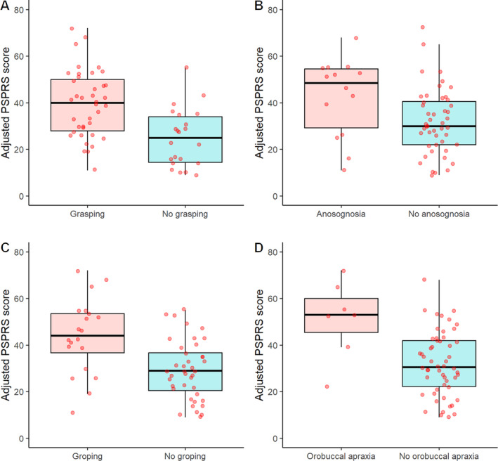
Adjusted PSPRS score for patients with grasping (A), anosognosia (B), groping (C) and orobuccal apraxia (D).
TABLE 2.
Multivariate predictive model of the adjusted PSPRS score
| RC | SE | T value | P value | |
|---|---|---|---|---|
| Age | 0.04 | 0.21 | 0.18 | 0.86 |
| Gender (female) | −5.29 | 2.79 | −1.89 | 0.06 |
| Education | −0.1 | 0.38 | −0.25 | 0.8 |
| Disease duration | 2.4 | 1 | 2.38 | 0.02 |
| Grasping | 8.56 | 3.14 | 2.73 | <0.01 |
| Groping | 0.96 | 3.6 | 0.27 | 0.79 |
| Anosognosia | 9.12 | 4.06 | 2.25 | 0.03 |
| Orobuccal apraxia | 10.07 | 4.89 | 2.06 | 0.04 |
| PSP‐RS subtype | 11.68 | 4.15 | 2.81 | <0.01 |
| PSP‐P subtype | 5.34 | 5.9 | 0.9 | 0.37 |
| PSP‐PGF subtype | −1 | 5.63 | −0.18 | 0.86 |
| PSP‐SL subtype | −5.34 | 5.9 | −0.9 | 0.37 |
Note: The model's goodness‐of‐fit was assessed using the residual standard error and the R‐squared values. Multiple R‐squared: 0.71. Adjusted R‐squared: 0.63. F‐statistic: 8.65. P value <0.001. Adjusted R‐squared value showed that the model's goodness‐of‐fit was reliable even when considering the number of covariates included. The overall model was found to be statistically significant. In the final analysis, frontal and CBS subtypes were excluded due to an insufficient number of observations; however, it was verified that including them in the model maintained statistical significance for the same variables.
Abbreviations: RC, regression coeficient; SE, standard error.
Clinical Signs among PSP Subtypes
Regarding to the different PSP subtypes, proportion comparison using confidence intervals showed a significantly higher prevalence of anosognosia in frontal subtype, of orobuccal apraxia in SL and CBS subtypes, groping in RS, frontal and SL subtypes, utilization behavior in frontal and CBS subtypes, of echolalia in RS, PGF and SL subtypes, of echopraxia in CBS subtype, grasping was less prevalent in PSP‐P and SL subtypes, greed for food was more frequent in PGF, frontal and CBS subtypes, and emotional incontinence in SL subtype (Table S3 in Supporting Information). Figure 2 summarizes the frequency of each explored sign according to the PSP clinical subtype. After grouping the subtypes into cortical, RS, and subcortical, and generating a frontal index reflecting the burden of frontal signs in each patient, significant differences between groups were observed (F = 5.11, P < 0.01), specifically between cortical and subcortical subtypes (mean 3.8 and 2, respectively, P = 0.03) and RS and subcortical subtypes (mean 3.53 and 2, respectively, P = 0.02).
Figure 2.
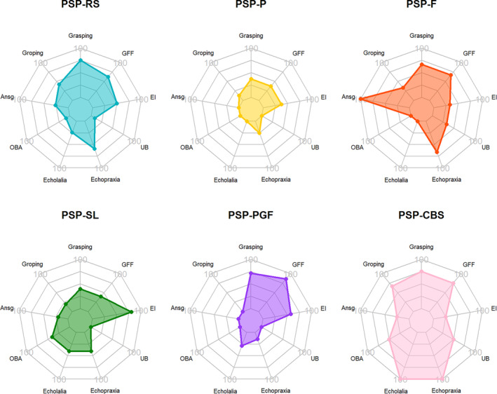
Frequency of frontal lobe signs among PSP subtypes.
Neuropsychological Assessments
Patients with grasping obtained lower scores primarily on frontal domain tests such as FBI, LARS, clock drawing test, and GDS. Patients with anosognosia similarly exhibited poorer performance on FBI, GDS, LARS, and Starkstein apathy scale, highlighting frontal domain involvement. In contrast, patients with groping and orobuccal apraxia displayed posterior domain impairments, reflected in lower scores in semantic fluency, Poppelreuter test, clock copy tests, CERAD recall test, and VOSP number location test. Social cognition impairments were also common among most patients. For detailed results, please refer to Table S4 and Figure S1. in the Supporting Information.
Neuroimaging Analysis
27 patients had T1‐weighted images suitable for pre‐processing (demographics in Table S5 of Supporting Information). After the pre‐processing steps, one subject was excluded due to movement artifacts. The analysis was covaried by age, gender and disease duration. Patients with grasping showed reduced cortical thickness in the superior right frontal cortex but did not reach the significance threshold. Patients with groping showed reduced cortical thickness in the bilateral middle frontal cortex, right superior frontal cortex, right middle temporal gyrus and right precuneus (Fig. 3).
Figure 3.
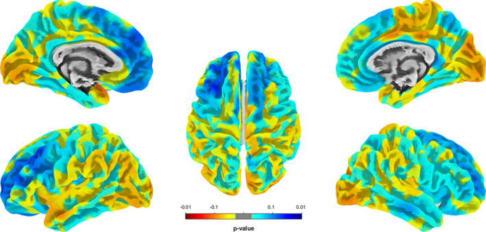
Cortical areas in patients with groping showing significant thinning in bilateral middle frontal cortex, right superior frontal cortex, right middle temporal gyrus and right precuneus.
Survival Analysis
For the censoring date, 15 patients had died (24.59%), 38 patients were alive (62.29%), and 8 patients were lost to follow‐up (13.11%). In the latter group, observation was censored at the date of the last visit. Survival from symptom onset was represented using the Kaplan–Meier curve. The median survival was not reached (Fig. 4A). The survival rate at 7.69 years was 63.2% (95% CI 0.49–0.82). When analyzing survival according to the presence of grasping, groping, anosognosia, and orobuccal apraxia, no statistically significant differences were found. However, when evaluated for the early presence of any of these signs in the first 4 years of the disease, log‐rank test and univariate Cox analysis showed the presence of groping as a potential risk factor for mortality. The median survival in the Kaplan–Meier representation of this group was 6.76 years, with a survival rate of 41.5% compared to 76.6% in those without early onset groping (HR 3.47; 95% CI 1.17–10.25, P = 0.03) (Fig. 4B). After controlling by age, gender, education years, disease duration and PSP subtypes in a multivariate Cox models, a statistically significant association between early onset groping and survival was also found (Table S6 of Supporting Information). A survival model was also conducted according to the cortical, RS, and subcortical subtypes of PSP, revealing higher mortality for the cortical subtype compared to the subcortical (HR 10.65, P = 0.03) (additional information in Table S7 and Fig. S2 of the Supporting Information).
Figure 4.
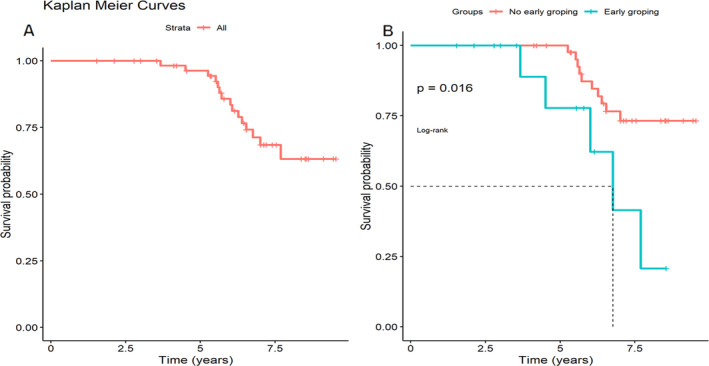
Survival curve of patients with PSP in our sample (A) and survival analysis according to early onset of groping in the first 4 years (B).
Discussion
Among the known clinical predictors of disability in PSP is the RS subtype, frequently associated with a higher rate of falls, cognitive impairment, shorter interval to wheelchair and functional dependence, and lower survival rate. 26 , 27 , 28 Additionally, other indicators of poor prognosis identified in literature are early falls, dementia, early dysphagia and urinary incontinence. Male gender, a short interval to first milestone and older age have also been found as independent predictors. 26 In line with the evidence, in our sample RS subtype also appeared as an independent predictor of disability using the PSPRS scale, and there was a trend for lower disability in the female gender. To the best of our knowledge, predictive value of disability and prognostic role of the frontal bedside clinical signs analyzed in this study has never been explored before. These signs were frequent and of early onset in our sample. We identified grasping, groping, anosognosia and orobuccal apraxia as being associated with disability measured by PSPRS. Furthermore, grasping, anosognosia and orobuccal apraxia were predictors of disability independently of age, gender, years of education, disease duration and clinical subtype of PSP. The early presence of groping emerged as a potential mortality factor in our study. The lower prevalence of these signs in our patients with PSP‐P subtype, often related to better prognosis and higher survival rate compared to RS and cortical subtypes, reinforces these findings. 29 On the other hand, a relation has been described between baseline cognitive and affective status and the rate of disease progression; in fact, shorter disease duration, more severe depression and lower performance on executive function tests at baseline have been associated with greater disability evaluated by PSPRS. 30 In this line, we found in our study that patients with grasping, anosognosia, orobuccal apraxia and groping had greater impairment of frontal functions such as executive tasks, impulse control, apathy and social cognition. In addition, we found additional impairment of visuospatial functions in patients with groping and orobuccal apraxia. While these alterations were first attributed to executive deficits in PSP, 31 it was later found that up to one third of patients had prominent memory and visuospatial dysfunction, unrelated to duration but rather to disease stage. 32 From a topographical perspective, these findings agree with the fronto‐parietal involvement suggested in patients with orobuccal apraxia 14 and in normal groping behavior. 33 , 34 Cortical thickness analysis showed greater atrophy in prefrontal cortex in patients with grasping and groping, independently of age, gender and disease duration, although it only remained significant for groping after corrections. Patients with groping also showed greater atrophy in middle temporal gyrus and right precuneus. These areas are linked to spatial relations for body movement control, called motor imagery, besides attentional processes or mental imagery, 35 as well as to visual and proprioceptive reaching behavior. 36 Since these regions are part of the fronto‐parietal network, future studies with functional MRI are required to search for network disruption at this level. In summary, clinical, neuropsychological and neuroimaging findings in patients with groping suggest that this survival‐related sign is linked to an impairment not only in frontal but also in posterior areas, which are characteristically associated with more advanced neuropathological stages of neuronal and astrogial tau deposition. 37
Terminological confusion between different clinical signs of frontal lobe dysfunction is common in the literature. In particular, the terms “grasping,” “groping” and “utilization behavior” are often used interchangeably. Nevertheless, this study shows the importance of correctly defining and exploring each sign, both for the topographical implications and for its differential association to disease disability. In the initial version of the PSPRS, grasping/imitative/utilizing behavior and emotional incontinence were included as “mentation” items. 18 However, in the later version of the modified PSPRS scale (mPSPRS), grasping/imitative/utilizing behavior items did not reach the severe milestone limit and was therefore removed from this section. 19 Although this study didn't assess the disability attributable per se to these clinical signs, the results indicate that their assessment could be important to quickly and easily detect patients with disease‐related disability.
Limitations
The sample size in this study was limited, since PSP is considered a rare disease. This may influence the generalization of results, and a larger sample size could show a more accurate picture of the associations found. Confirmatory studies involving a larger patient cohort and applying multiple comparison corrections are also warranted to validate the relationship between frontal signs and neuropsychological outcomes. Due to limitations in the availability of brain MRI in some patients, neuroimaging analysis could only be performed in patients with grasping and groping. This limitation could affect the global and complete understanding of the neuroanatomical correlates of the remaining clinical signs. For these subtle clinical signs, structural and functional connectivity, along with diffusivity, would be of interest in future studies. Finally, regarding survival analysis, no increased mortality was observed in the PSP‐RS subtype in our sample. Our classification of PSP subtypes based on phenotype at onset may not capture the full spectrum of disease progression, as many patients transitioned to the RS subtype during their illness. Other studies classify subtypes based on the final phenotype or at the time of evaluation. Also, the study's limited sample size and low event count may have reduced statistical power and hindered our ability to detect significant differences in mortality. This makes the results should be interpreted with caution and requires future larger studies to confirm these trends.
Conclusions
Grasping, anosognosia, orobuccal apraxia and groping constitute easily assessable bedside clinical signs in patients with PSP. While grasping, anosognosia and orobuccal apraxia may predict disease disability, early onset groping could be a survival predictor in clinical practice. These signs reflect distinct stages within the disease spectrum. While patients with grasping and anosognosia predominantly exhibit dysfunction in frontal areas, orobuccal apraxia and groping demonstrate an additional more diffuse involvement of posterior regions. If these findings are validated in confirmatory cohorts, the assessment of frontal lobe signs could be a useful screening tool to identify PSP patients with increased disability in clinical practice. This will allow to better target the patient's resources and needs.
Author Roles
(1) Research project: A. Conception, B. Organization, C. Execution; (2) Statistical Analysis: A. Design, B. Execution, C. Review and Critique; (3) Manuscript Preparation: A. Writing of the first draft, B. Review and Critique.
I.R.B.: 1A, 2A, 2B, 3A
A.H.B.: 1C, 2C, 3B
I.A.B.: 2B, 2C, 3B
S.M.H.: 2C, 3B
J.K.: 1B, 3B
J.P.M.: 1A, 1B, 1C, 2C, 3B
Dislosures
Ethical Compliance Statement: The study was approved by the Sant Pau Hospital Ethical Committee (IIBSP‐PSP‐2014‐34), and written informed consent was obtained from all patients. We confirm that we have read the Journal's position on issues involved in ethical publication and affirm that this work is consistent with those guidelines.
Funding Sources and Conflicts of Interest: The authors declare no conflicts of interest related to this work. This research did not receive specific grants from any funding agency in the public, commercial, or not‐for‐profit sectors.
Financial Disclosures For The Previous 12 Months: I.R.B. is supported by a grant of Instituto de Salud Carlos III (exp. number CM22/00072). He has received speaker honoraria from Zambon. A.H.B has no conflicts of interest to declare. S.M.‐H. has received honoraria for lecturing from Teva, Zambon, UCB, and Roche, and reports grants from the Huntington's Disease Society of America (Human Biology Project) and from Fondo de Investigaciones Sanitarias from Instituto de Salud Carlos III. I.A.B. is supported by a grant of Instituto de Saluds Carlos III (exp. number JR22/00059). He has received speaker honoraria from Medtronic. J.K. has served on advisory or speakers’ boards and received honoraria from UCB, TEVA, Zambon, and Bial. J.P. has served on advisory/speaker boards and received honoraria from Zambon, AbbVie, Allergan, Esteve, Lundbeck, Ipsen and Bial.
Supporting information
Table S1. Clinical assessment of frontal lobe signs.
Table S2. Associations between clinical signs and PSPRS score.
Table S3. Interval confidence analysis of different clinical signs prevalence between PSP subtypes.
Table S4. Comparison of means for significant neuropsychological tests in patients with grasping, anosognosia, orobuccal apraxia and groping.
Table S5. Comparison of populations for brain MRI cortical thickness analysis.
Table S6. Multivariate survival models adjusted for a covariate.
Table S7. Univariate survival Cox model according to cortical, Richardson's syndrome and subcortical PSP subtypes.
Figure S1. Relative frequency of significant differences in cognitive domains in patients with grasping, anosognosia, orobuccal apraxia and groping.
Figure S2. Survival curve and Log‐rank test according to cortical, Richardson's syndrome and subcortical subtypes of PSP.
Contributor Information
Jaime Kulisevsky, Email: jkulisevsky@santpau.cat.
Javier Pagonabarraga, Email: jpagonabarraga@santpau.cat.
References
- 1. Höglinger GU, Respondek G, Stamelou M, et al. Clinical diagnosis of progressive supranuclear palsy: the movement disorder society criteria. Mov Disord Off J Mov Disord Soc 2017;32(6):853–864. [DOI] [PMC free article] [PubMed] [Google Scholar]
- 2. Olfati N, Shoeibi A, Litvan I. Clinical spectrum of tauopathies. Front Neurol 2022;13:944806. [DOI] [PMC free article] [PubMed] [Google Scholar]
- 3. Chatterjee K, Choudhury S, Trivedi S, Basu P, Kumar H. Clinical significance of applause sign in patients with progressive Supranuclear palsy. Can J Neurol Sci J Can Sci Neurol 2022;49(6):809–812. [DOI] [PubMed] [Google Scholar]
- 4. Dubois B, Slachevsky A, Pillon B, Beato R, Villalponda JM, Litvan I. “Applause sign” helps to discriminate PSP from FTD and PD. Neurology 2005;64(12):2132–2133. [DOI] [PubMed] [Google Scholar]
- 5. Luzzi S, Fabi K, Pesallaccia M, Silvestrini M, Provinciali L. Applause sign: is it really specific for parkinsonian disorders? Evidence from cortical dementias. J Neurol Neurosurg Psychiatry 2011;82(8):830–833. [DOI] [PubMed] [Google Scholar]
- 6. Schönecker S, Hell F, Bötzel K, Wlasich E, Ackl N, Süßmair C, et al. The applause sign in frontotemporal lobar degeneration and related conditions. J Neurol 2019;266(2):330–338. [DOI] [PubMed] [Google Scholar]
- 7. Somme J, Gómez‐Esteban JC, Tijero B, Berganzo K, Lezcano E, Zarranz JJ. The applause sign and neuropsychological profile in progressive supranuclear palsy and Parkinson's disease. Clin Neurol Neurosurg 2013;115(8):1230–1233. [DOI] [PubMed] [Google Scholar]
- 8. Horta‐Barba A, Pagonabarraga J, Martínez‐Horta S, et al. Cognitive and behavioral profile of progressive supranuclear palsy and its phenotypes. J Neurol 2021;268(9):3400–3408. [DOI] [PubMed] [Google Scholar]
- 9. Ghika J, Tennis M, Growdon J, Hoffman E, Johnson K. Environment‐driven responses in progressive supranuclear palsy. J Neurol Sci 1995;130(1):104–111. [DOI] [PubMed] [Google Scholar]
- 10. Iaccarino L, Chieffi S, Iavarone A. Utilization behavior: what is known and what has to Be known? Behav Neurol 2014;2014:e297128. [DOI] [PMC free article] [PubMed] [Google Scholar]
- 11. Erro R, Barone P, Moccia M, Amboni M, Vitale C. Abnormal eating behaviors in progressive supranuclear palsy. Eur J Neurol 2013;20(3):e47–e48. [DOI] [PubMed] [Google Scholar]
- 12. Hakimi M, Maurer CW. Pseudobulbar affect in parkinsonian disorders: a review. J Mov Disord 2019;12(1):14–21. [DOI] [PMC free article] [PubMed] [Google Scholar]
- 13. Santos‐Santos MA, Mandelli ML, Binney RJ, et al. Features of patients with nonfluent/Agrammatic primary progressive aphasia with underlying progressive Supranuclear palsy pathology or Corticobasal degeneration. JAMA Neurol 2016;73(6):733–742. [DOI] [PMC free article] [PubMed] [Google Scholar]
- 14. Ozsancak C, Auzou P, Dujardin K, Quinn N, Destée A. Orofacial apraxia in corticobasal degeneration, progressive supranuclear palsy, multiple system atrophy and Parkinson's disease. J Neurol 2004;251(11):1317–1323. [DOI] [PubMed] [Google Scholar]
- 15. Pellicano C, Assogna F, Cellupica N, et al. Neuropsychiatric and cognitive profile of early Richardson's syndrome, progressive Supranuclear palsy‐parkinsonism and Parkinson's disease. Parkinsonism Relat Disord 2017;45:50–56. [DOI] [PubMed] [Google Scholar]
- 16. Gerstenecker A, Duff K, Mast B, Litvan I. Behavioral abnormalities in progressive supranuclear palsy. Psychiatry Res 2013;210(3):1205–1210. [DOI] [PMC free article] [PubMed] [Google Scholar]
- 17. Hall DA, Forjaz MJ, Golbe LI, et al. Scales to assess clinical features of progressive Supranuclear palsy: MDS task force report. Mov Disord Clin Pract 2015;2(2):127–134. [DOI] [PMC free article] [PubMed] [Google Scholar]
- 18. Golbe LI, Ohman‐Strickland PA. A clinical rating scale for progressive supranuclear palsy. Brain J Neurol. 2007;130(Pt 6):1552–1565. [DOI] [PubMed] [Google Scholar]
- 19. Grötsch MT, Respondek G, Colosimo C, et al. A modified progressive Supranuclear palsy rating scale. Mov Disord 2021;36(5):1203–1215. [DOI] [PubMed] [Google Scholar]
- 20. Wills AM, Pantelyat A, Espay A, et al. A modified progressive Supranuclear palsy rating scale for virtual assessments. Mov Disord Off J Mov Disord Soc 2022;37(6):1265–1271. [DOI] [PMC free article] [PubMed] [Google Scholar]
- 21. Litvan I, Agid Y, Calne D, et al. Clinical research criteria for the diagnosis of progressive supranuclear palsy (Steele‐Richardson‐Olszewski syndrome): report of the NINDS‐SPSP international workshop. Neurology 1996;47(1):1–9. [DOI] [PubMed] [Google Scholar]
- 22. Leiguarda RC, Pramstaller PP, Merello M, Starkstein S, Lees AJ, Marsden CD. Apraxia in Parkinson's disease, progressive supranuclear palsy, multiple system atrophy and neuroleptic‐induced parkinsonism. Brain J Neurol 1997;120(Pt 1):75–90. [DOI] [PubMed] [Google Scholar]
- 23. Tranulis C, Corin E, Kirmayer LJ. Insight and psychosis: comparing the perspectives of patient, entourage and clinician. Int J Soc Psychiatry 2008;54(3):225–241. [DOI] [PubMed] [Google Scholar]
- 24. Gaser C, Dahnke R, Thompson PM, Kurth F, Luders E, Initiative ADN. CAT – A Computational Anatomy Toolbox for the Analysis of Structural MRI Data [Internet]; bioRxiv; 2022 [cited 2023 Jul 30]. p. 2022.06.11.495736. Available from: 10.1101/2022.06.11.495736v1. [DOI]
- 25. Dahnke R, Yotter RA, Gaser C. Cortical thickness and central surface estimation. Neuroimage 2013. Jan;15(65):336–348. [DOI] [PubMed] [Google Scholar]
- 26. O'Sullivan SS, Massey LA, Williams DR, et al. Clinical outcomes of progressive supranuclear palsy and multiple system atrophy. Brain J Neurol 2008;131(Pt 5):1362–1372. [DOI] [PubMed] [Google Scholar]
- 27. Street D, Malpetti M, Rittman T, et al. Clinical progression of progressive supranuclear palsy: impact of trials bias and phenotype variants. Brain Commun 2021;3(3):1–13. [DOI] [PMC free article] [PubMed] [Google Scholar]
- 28. Picillo M, Cuoco S, Tepedino MF, Cappiello A, Volpe G, Erro R, et al. Motor, cognitive and behavioral differences in MDS PSP phenotypes. J Neurol 2019;266(7):1727–1735. [DOI] [PubMed] [Google Scholar]
- 29. Guasp M, Molina‐Porcel L, Painous C, et al. Association of PSP phenotypes with survival: a brain‐bank study. Parkinsonism Relat Disord 2021;84:77–81. [DOI] [PubMed] [Google Scholar]
- 30. Bang J, Lobach IV, Lang AE, et al. Predicting disease progression in progressive supranuclear palsy in multicenter clinical trials. Parkinsonism Relat Disord 2016;28:41–48. [DOI] [PMC free article] [PubMed] [Google Scholar]
- 31. Sitek EJ, Wieczorek D, Konkel A, Dąbrowska M, Sławek J. The pattern of verbal, visuospatial and procedural learning in Richardson variant of progressive supranuclear palsy in comparison to Parkinson's disease. Psychiatr Pol 2017;51(4):647–659. [DOI] [PubMed] [Google Scholar]
- 32. Burrell JR, Hodges JR, Rowe JB. Cognition in corticobasal syndrome and progressive supranuclear palsy: a review. Mov Disord Off J Mov Disord Soc 2014;29(5):684–693. [DOI] [PubMed] [Google Scholar]
- 33. Nachev P, Kennard C, Husain M. Functional role of the supplementary and pre‐supplementary motor areas. Nat Rev Neurosci 2008;9(11):856–869. [DOI] [PubMed] [Google Scholar]
- 34. Vingerhoets G. Contribution of the posterior parietal cortex in reaching, grasping, and using objects and tools. Front Psychol 2014;5:151. [DOI] [PMC free article] [PubMed] [Google Scholar]
- 35. Cavanna AE, Trimble MR. The precuneus: a review of its functional anatomy and behavioural correlates. Brain 2006;129(3):564–583. [DOI] [PubMed] [Google Scholar]
- 36. Filimon F, Nelson JD, Huang RS, Sereno MI. Multiple parietal reach regions in humans: cortical representations for visual and proprioceptive feedback during on‐line reaching. J Neurosci 2009;29(9):2961–2971. [DOI] [PMC free article] [PubMed] [Google Scholar]
- 37. Kovacs GG, Lukic MJ, Irwin DJ, et al. Distribution patterns of tau pathology in progressive supranuclear palsy. Acta Neuropathol (Berl) 2020;140(2):99–119. [DOI] [PMC free article] [PubMed] [Google Scholar]
Associated Data
This section collects any data citations, data availability statements, or supplementary materials included in this article.
Supplementary Materials
Table S1. Clinical assessment of frontal lobe signs.
Table S2. Associations between clinical signs and PSPRS score.
Table S3. Interval confidence analysis of different clinical signs prevalence between PSP subtypes.
Table S4. Comparison of means for significant neuropsychological tests in patients with grasping, anosognosia, orobuccal apraxia and groping.
Table S5. Comparison of populations for brain MRI cortical thickness analysis.
Table S6. Multivariate survival models adjusted for a covariate.
Table S7. Univariate survival Cox model according to cortical, Richardson's syndrome and subcortical PSP subtypes.
Figure S1. Relative frequency of significant differences in cognitive domains in patients with grasping, anosognosia, orobuccal apraxia and groping.
Figure S2. Survival curve and Log‐rank test according to cortical, Richardson's syndrome and subcortical subtypes of PSP.