A 22-year-old female, who had underwent Fontan procedure in childhood, presented with mild elevation of liver enzymes (AST 35 U/L, ALT 50 U/L, ALP 62 U/L, GGT 61 U/L). She had no liver dysfunction (total bilirubin 1.3 mg/dL, INR 1.1, albumin 3.7 g/dL), signs of cirrhosis or portal hypertension (platelets 177 × 109), encephalopathy or ascites.
Viral, autoimmune, metabolic, and toxic etiologies were excluded. Abdominal ultrasound showed a diffusely heterogeneous and micronodular liver parenchyma, compatible with Fontan-associated liver disease (FALD) in this context. Moreover, multiple de novo hyperechogenic nodules were found, imposing investigation.
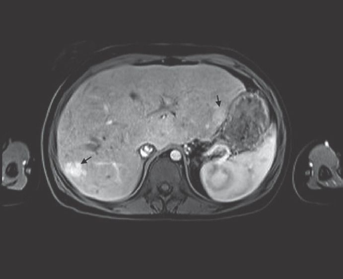
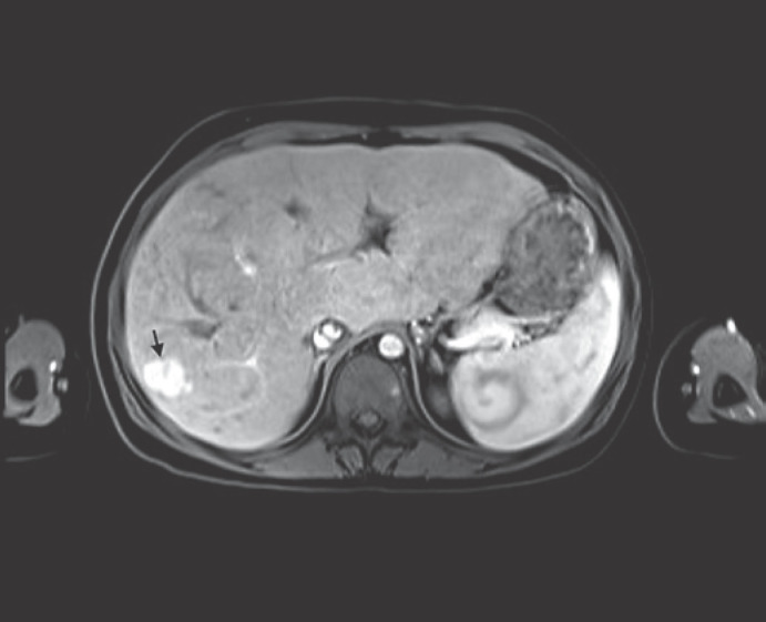
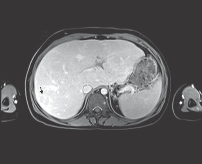
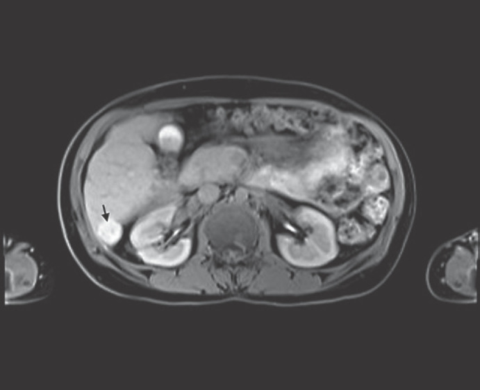
MRI reported >12 nodules, maximum diameter of 27 mm, isointense in T1-weighted sequences, hypointense in T2, with no restricted diffusion (shown in Fig. 1). Some were halo-surrounded, while others displayed a central scar. Most displayed hyper-enhancement in the hepatic arterial phase (shown in Fig. 2), becoming isointense in the portal phase and hypointense in the delayed one, a worrisome feature known as washout (shown in Fig. 3). Using hepatobiliary contrast, all nodules showed hyperintensity (shown in Fig. 4). Bloodwork revealed normal alpha-fetoprotein (AFP). Therefore, the final diagnosis of multiple focal nodular hyperplasia (FNH)-like in a FALD background was made, and the patient kept under surveillance.
Fig. 1.
Multiple de novo liver nodules are seen (arrows).
Fig. 2.
Hepatic arterial phase shows enhancement of nodule in segment V (arrow).
Fig. 3.
Late phase shows washout in the same nodule (arrow).
Fig. 4.
Hepatobiliary phase shows hyperintensity of nodule in segment VI (arrow).
The Fontan procedure is a palliative surgery for patients with an anatomic or functional single-ventricular congenital heart disease, consisting of a total extracardiac cavopulmonary connection created by anastomosing the superior vena cava to the right pulmonary artery (PA) and insertion of an extracardiac conduit between the inferior vena cava and the PA [1, 2]. The consequent chronic liver congestion and ischemia result in FALD, leading to cirrhosis in 1–5% of patients per year and hepatocellular carcinoma (HCC) in 1.3% [1, 2, 3]. Indeed, annual and early liver surveillance is mandatory [1].
Multiple FNH is a rare entity which has been associated with some vascular diseases and treatments [4, 5]. This clinical case highlights an association between multiple FNH and FALD. Given the inherent risk of HCC in FALD and similar MRI findings between FNH and HCC in this background, their differential diagnosis becomes challenging [1, 2]. In this context, AFP and MRI hepatobiliary contrast are key [2, 3]. Nonetheless, biopsy should be considered in dubious and atypical nodules [1].
Statement of Ethics
The study did not require ethics approval. Informed consent was obtained from the patient for publication of this case report and any accompanying images.
Conflict of Interest Statement
The authors have no conflicts of interest to declare.
Funding Sources
There are no funding sources to declare.
Author Contributions
All authors participated in the design, construction, and revision of the paper.
Data Availability Statement
All data generated or analyzed during this study are included in this article.
Funding Statement
There are no funding sources to declare.
References
- 1.Perucca G, de Lange C, Franchi-Abella S, Napolitano M, Riccabona M, Ključevšek D, et al. Surveillance of fontan-associated liver disease: current standards and a proposal from the European society of paediatric radiology abdominal task force. Pediatr Radiol. 2021;51((13)):2598–25606. doi: 10.1007/s00247-021-05173-x. [DOI] [PMC free article] [PubMed] [Google Scholar]
- 2.Wells ML, Hough DM, Fidler JL, Kamath PS, Poterucha JT, Venkatesh SK. Benign nodules in post-Fontan livers can show imaging features considered diagnostic for hepatocellular carcinoma. Abdom Radiol. 2017 Nov;42((11)):2623–2631. doi: 10.1007/s00261-017-1181-9. [DOI] [PubMed] [Google Scholar]
- 3.Téllez L, Rodríguez de Santiago E, Minguez B, Payance A, Clemente A, Baiges A, et al. Prevalence, features and predictive factors of liver nodules in Fontan surgery patients: the VALDIG Fonliver prospective cohort. J Hepatol. 2020 Apr;72((4)):702–710. doi: 10.1016/j.jhep.2019.10.027. [DOI] [PubMed] [Google Scholar]
- 4.Busireddy KK, Ramalho M, AlObaidy M, Matos AP, Burke LM, Dale BM, et al. Multiple focal nodular hyperplasia: MRI features. Clin Imaging. 2018 May-Jun;49:89–96. doi: 10.1016/j.clinimag.2017.11.010. [DOI] [PubMed] [Google Scholar]
- 5.Kayhan A, Venu N, Lakadamyalı H, Jensen D, Oto A. Multiple progressive focal nodular hyperplasia lesions of liver in a patient with hemosiderosis. World J Radiol. 2010 Oct 28;2((10)):405–409. doi: 10.4329/wjr.v2.i10.405. [DOI] [PMC free article] [PubMed] [Google Scholar]
Associated Data
This section collects any data citations, data availability statements, or supplementary materials included in this article.
Data Availability Statement
All data generated or analyzed during this study are included in this article.


