Abstract
Simple Summary
Codon 61 RAS mutations are rare in metastatic colorectal cancer. Despite being associated with primary and acquired resistance to anti-EGFR agents, little is known about their phenotype and prognostic impact. We retrospectively investigated the clinicopathological features and prognoses of 50 patients with tumors harboring codon 61 RAS mutations compared to 648 codon 61 RAS wild-type tumors. We identified a significant correlation between codon 61 RAS mutations and metastatic involvement of the peritoneum and ovary and a negative prognostic impact. This is the first evidence of an impact of RAS mutational status on the metastatization pattern. These results are of great interest given the high frequency of codon 61 RAS mutations as mechanisms of secondary resistance to anti-EGFRs and the advent of RAS inhibitors. This is the widest codon 61 RAS-mutated cohort reported so far; nevertheless, these findings must be validated in larger studies.
Abstract
RAS mutations involving codon 61 are rare in metastatic colorectal cancer (mCRC), accounting for only 1–4%, but they have recently been identified with high frequency in the circulating tumor DNA (ctDNA) of patients with secondary resistance to anti-EGFRs. This retrospective monocentric study aimed to investigate the clinical phenotype and prognostic performance of codon 61 RAS-mutated mCRC. Fifty patients with codon 61 RAS-mutated mCRC treated at our institution between January 2013 and December 2021 were enrolled. Additional datasets of codon 61 RAS wild-type mCRCs (648 patients) were used as comparators. The endpoint for prognostic assessment was overall survival (OS). Metastatic involvement of the peritoneum or ovary was significantly more frequent in codon 61 RAS-mutated mCRC compared to codon 61 RAS wild-type (54 vs. 28.5%), non-codon 61 RAS-mutated (35.6%), BRAF V600E-mutated (25%), and RAS/BRAF wild-type (20.5%) cohorts. At a median follow up of 96.2 months, the median OS for codon 61 RAS-mutated patients was significantly shorter compared to RAS/BRAF wild-type (26.9 vs. 36.0 months, HR 0.56) patients, while no significant difference was observed compared to non-codon 61 RAS-mutated and BRAF V600E-mutated patients. We showed a negative prognostic impact and a statistically significant correlation between codon 61 RAS mutations and metastatic involvement of the peritoneum and ovary.
Keywords: colorectal adenocarcinoma, RAS, metastatic colorectal cancer, RAS signaling, Codon 61, MAPK pathway, RAS effectors, KRAS inhibitors, resistance
1. Introduction
Nowadays, while the molecular classification of colorectal cancer (CRC) is becoming more and more complex [1], rat sarcoma virus (RAS) mutational status remains a key determinant in every turning point in patients’ therapeutic algorithm [2]. Together with NRAS and HRAS, KRAS is a gene belonging to the RAS family, which encodes guanosine-5′-triphosphate (GTP)-binding proteins, important effectors of ligand-bound epidermal growth factor receptor (EGFR) signaling through the mitogen-activated protein kinase (MAPK) axis [3]. KRAS mutations affect approximately 30–40% of metastatic CRC (mCRC), with mutations involving codons 12 and 13 being the most represented, occurring in about 85–90% of cases [3,4]. Several studies have demonstrated their role as predictive biomarkers of resistance to anti-EGFR agents [5,6,7]. Hence, all patients diagnosed with mCRC require RAS profiling before the administration of anti-EGFRs agents (cetuximab or panitumumab) [8,9,10,11,12,13,14].
Codon 61 mutations are less prevalent, affecting 1–4% of patients with mCRC. Similarly to other RAS mutations, these alterations are responsible for a constitutive activation of the RAS/RAF/MAPKs pathway, resulting in oncogenic activity and cell proliferation [15]. Furthermore, in KRAS codon 12 and 13, wild-type mCRCs codon 61 mutations have been linked to resistance to anti-EGFR therapies [16,17]. Recently, codon 61 variants have been identified with high frequency in the circulating tumor DNA (ctDNA) of patients with mCRC with secondary resistance to anti-EGFR agents [18,19,20,21], with a prevalence of 50% in the CHRONOS trial [18]. Other rare KRAS mutations involve exon 4, codon 117, and codon 146. Similarly to more frequent RAS mutations, mutations involving codon 117 and 146 have been associated with resistance to anti-EGFRs therapies [22,23]. Moreover, a large analysis showed a higher incidence of codon 117 and 146 in older patients [24].
Despite its growing clinical relevance, little is known about the clinicopathological and molecular features and prognosis of mCRCs harboring RAS codon 61 mutations and their differences with more common codon 12 and 13 mutations, as well as and their impact on prognosis. In 2014, a cohort study by Imamura et al. [25] reported the clinicopathological and molecular features of 19 KRAS codon 61-mutated mCRC to be similar to KRAS codon 12- and 13-mutated mCRCs. Another study found a weak tendency for peritoneum localization in a population of 14 patients with codon 61 RAS-mutated CRC [26]. In our study, we aimed to further investigate the clinical characteristics and prognosis of patients with mCRC harboring RAS codon 61 mutations treated at our institution compared to those harboring other non-codon 61 RAS-mutated and wild-type tumors.
2. Materials and Methods
This is an observational, retrospective, monocentric study. The study was approved by the local Ethics Committee of Fondazione Policlinico Universitario Agostino Gemelli IRCCS, Rome, Italy (protocol number 0054049/2019 18 December 2019). The objective of the study was to investigate and describe clinical phenotype and prognostic performance of mCRCs harboring RAS codon 61 mutations.
We examined the medical records of patients diagnosed with mCRC who were treated at our center from January 2013 through December 2021. Eligible subjects were those patients whose tumors carried mutations involving codon 61 of RAS gene and were evaluable for survival after at least one line of therapy. We collected data regarding bbaseline demographic and clinical characteristics, first-line treatment, and survival from medical records, while histological reports were used to gather pathological and molecular data. The following baseline demographic and clinical characteristics were collected: sex, age, Eastern Cooperative Oncology Group performance status (ECOG PS) at diagnosis, primary tumor location, onset of metastatic disease, number of metastatic sites, site of metastases, presence of peritoneal and/or adnexal metastases, mucinous histology, grade of differentiation, RAS/BRAF mutational status, microsatellite instability/mismatch repair (MSI/MMR) status, treatments received (surgery, neoadjuvant or adjuvant chemotherapy, first-line chemotherapy), investigator-assessed best response according to Response Evaluation Criteria in Solid Tumors (RECIST) 1.1 criteria, and survival. RAS and BRAF mutational status was assessed by means of next-generation sequencing (NGS) or pyrosequencing on formalin-fixed, paraffin-embedded (FFPE) archival tumor tissue samples from primary tumor or metastases. Expression of MMR proteins (MLH1, MSH2, MSH6, and PMS2) was performed via immunohistochemistry. MSI status was assessed via NGS.
Additional datasets of patients affected by mCRC without codon 61 RAS mutations (codon 61 RAS wild-type) treated at out center during the same time frame were used as comparators. Among this group of patients, we identified three different molecular subgroups, which included, respectively, patients with an RAS-mutated disease not involving codon 61 (non-codon 61 RAS-mutated mCRCs), patients with mCRC harboring a BRAF V600E mutation (BRAF V600E-mutated mCRCs), and patients with an RAS and BRAF wild-type disease (RAS/BRAF wild-type mCRCs).
For categorial data, counts and percentages were reported using a descriptive method; for continuous variables, median and range were provided. Fisher’s exact test or the chi-square test, when applicable, were used to compare group differences for categorical variables. Overall survival (OS), defined as the time occurring between the diagnosis of metastatic disease to the date of death from any cause, was the endpoint for prognostic analysis. All patients were followed up until death or the time of database lock (January 2023). Patients not experiencing events were censored at the date of last follow up. Survivals were estimated with the Kaplan–Meier method and compared using the log-rank test. Statistical significance was set at p = 0.05. Statistical analyses were performed using MedCalc version 14.8.1.
3. Results
Between January 2013 and January 2023, a total of 50 patients with a diagnosis of mCRC harboring an RAS codon 61 mutation were included in our analysis. Of those, 28 mutations (56%) affected KRAS and 22 (44%) NRAS. Patients and disease characteristics are summarized in Table 1.
Table 1.
Codon 61 RAS-mutated patients characteristics.
| Characteristics | N = 50 (%) | KRAS (n = 28) | NRAS (n = 22) | |
|---|---|---|---|---|
|
Age (at metastatic diagnosis),
median (range) |
65 yrs (34–86 yrs) | 65 yrs (41–86 yrs) | 63 yrs (34–84 yrs) | |
| ECOG PS | 0 | 25 (50%) | 15 (53%) | 10 (45%) |
| 1 | 19 (38%) | 8 (28%) | 11 (50%) | |
| 2 | 6 (12%) | 5 (19%) | 1 (5%) | |
| Sex | Male | 19 (38%) | 10 (36%) | 9 (41%) |
| Female | 31 (62%) | 18 (64%) | 13 (59%) | |
| Previous surgery | Y | 40 (80%) | 22 (79%) | 18 (82%) |
| N | 10 (20%) | 6 (21%) | 4 (18%) | |
| Metastatic at diagnosis | Y | 33 (66%) | 19 (68%) | 14 (64%) |
| N | 17 (34%) | 9 (32%) | 8 (36%) | |
| Primary tumor location | Right | 14 (28%) | 8 (29%) | 6 (27%) |
| Left | 36 (72%) | 20 (71%) | 16 (73%) | |
| Sites of metastatic disease at diagnosis | Liver | 24 (48%) | 12 (43%) | 12 (54%) |
| Lung | 11 (22%) | 7 (25%) | 4 (18%) | |
| Nodes | 15 (30%) | 8 (28%) | 7 (32%) | |
| Peritoneum/Ovary | 16 (32%) | 7 (25%) | 9 (41%) | |
| Other | 5 (10%) | 3 (10%) | 2 (9%) | |
| Peritoneal and/or ovarian metastasis | Y | 27 (54%) | 13 (46%) | 14 (64%) |
| N | 23 (46%) | 15 (54%) | 8 (36%) | |
| First line chemotherapy regimen | FOLFOXIRI +/− bevacizumab | 3 (6%) | 0 | 3 (14%) |
| FOLFOX +/− bevacizumab | 29 (58%) | 20 (71%) | 9 (41%) | |
| FOLFIRI +/− bevacizumab | 9 (18%) | 3 (11%) | 6 (27%) | |
| Other | 9 (18%) | 5 (18%) | 4 (18%) | |
| Total number of treatment lines | 1 | 20 (40%) | 15 (53%) | 5 (23%) |
| 2 | 10 (20%) | 5 (18%) | 5 (23%) | |
| 3 | 13 (26%) | 6 (21%) | 7 (32%) | |
| 4 | 5 (10%) | 1 (4%) | 4 (18%) | |
| 5 | 2 (4%) | 1 (4%) | 1 (4%) | |
| RAS mutation | KRAS | 28 (56%) | ||
| Q61X | 15 (30%) | |||
| Q61H | 6 (12%) | |||
| Q61L | 3 (6%) | |||
| Q61R | 2 (4%) | |||
| G61X | 2 (4%) | |||
| NRAS | 22 (44%) | |||
| Q61R | 8 (16%) | |||
| Q61K | 8 (16%) | |||
| Q61L | 5 (10%) | |||
| G61H | 1 (2%) | |||
ECOG PS: Eastern Cooperative Oncology Group performance status; N: no; Y: yes; yrs: years.
Median age at diagnosis was 65 years (range 34–86 years). Nineteen patients were males (38%) and thirty-one were females (62%). Patients were mainly in good clinical conditions at the time of diagnosis (88% with an ECOG PS 0 or 1). Thirty-six patients (72%) had a left-sided primary tumor, and thirty-three patients (66%) had a synchronous metastatic disease. The most frequent site of metastases was liver (24 patients, 48%), followed by peritoneum or ovary (16 patients, 32%), lymph nodes (15 patients, 30%), and lungs (11 patients, 22%). Moreover, 27 patients (54%) developed metastases involving the peritoneum or ovary during their clinical history. The majority of patients received resection of primary tumor (40 patients, 80%). Twenty nine patients (58%) underwent a first-line therapy which included bevacizumab. As chemotherapy regimen, twenty nine patients (58%) received mFOLFOX6 (with or without bevacizumab), while FOLFIRI (with or without bevacizumab) was administered in nine patients (18%). Only three patients were treated with FOLFOXIRI plus bevacizumab (6%), whereas nine patients (18%) received other regimens (such as a fluoropyrimidine, alone or in combination with bevacizumab). Twenty patients (40%) received only one line of therapy, while ten patients (20%) received two lines, thirteen patients (26%) received three lines, five patients (10%) received four lines and, only two patients (4%) received five lines of therapy.
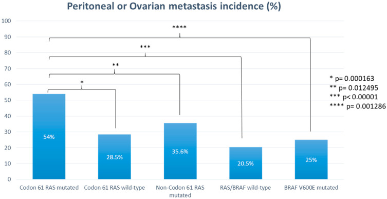
The comparator dataset included 648 consecutive patients with codon 61 RAS wild-type mCRC treated at our institution during the same time frame. This group included 326 patients (50.3%) with an RAS-mutated disease not involving codon 61 (non-codon 61 RAS-mutated mCRCs), 254 patients (39.2%) with an RAS and BRAF wild-type disease (RAS/BRAF wild-type mCRCs), and 68 patients (10.5%) with a BRAF V600E-mutated disease (BRAF V600E-mutated mCRCs). The probability of experiencing peritoneal or ovarian metastases was statistically significantly higher in patients with codon 61 RAS-mutated mCRC than in patients with codon 61 RAS wild-type mCRC (54% vs. 28.5%, p = 0.000163) (Figure 1). More specifically, the rate of peritoneal or ovarian metastases was higher in the codon 61 RAS-mutated cohort also when compared to the non-codon 61 RAS-mutated cohort (54% vs. 35.6%, p = 0.012495), BRAF V600E-mutated cohort (54% vs. 25%, p = 0.001286), and RAS/BRAF all wild-type cohort (54% vs. 20.5%, p < 0.00001) (Figure 1).
Figure 1.
Probability of experiencing peritoneal or ovarian metastases according to RAS and BRAF mutational status.
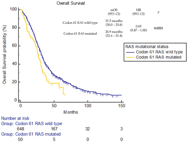
At a median follow up of 96.2 months (95% confidence interval (CI), 92.4–109.0 months), 40 death events were reported in the codon 61 RAS-mutated cohort and 556 in the comparator dataset. Median OS (mOS) was 26.9 months (95%CI 21.6–31.4 months) for the codon 61 RAS-mutated cohort and 31.5 months (95%CI 30.0–33.8 months) for the codon 61 RAS wild-type dataset (hazard ratio (HR) 0.69, 95%CI 0.47–1.00; p = 0.0221) (Figure 2).
Figure 2.
Overall survival according to codon 61 RAS mutational status.
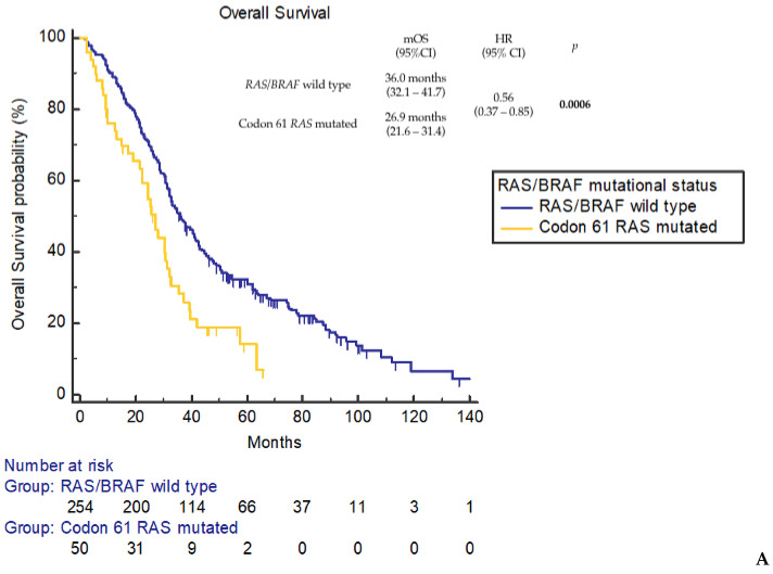
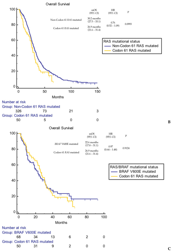
Moreover, dissecting the comparator dataset in accordance with RAS and BRAF mutational status, mOS was confirmed to be significantly shorter for the codon 61 RAS-mutated cohort compared to the RAS and BRAF wild-type cohort (mOS 36.0 months, 95%CI 32.1–41.7 months; HR 0.56, 95%CI 0.37–0.85; p = 0.0006) (Figure 3A). On the contrary, no statistically significant difference was observed compared to the non-codon 61 RAS-mutated cohort (mOS 30.2 months, 95%CI 27.5–33.1 months; HR 0.76, 95%CI 0.52–1.09; p = 0.0993) (Figure 3B) and the BRAF V600E-mutated cohort (mOS 22.6 months, 95%CI 17.8–31.1 months; HR 0.97, 95%CI 0.64–1.48; p = 0.9124) (Figure 3C).
Figure 3.
Overall survival according to codon 61 RAS mutational status. (A) Codon 61 RAS-mutated vs. RAS/BRAF wild-type patients. (B) Codon 61 RAS-mutated vs. non-codon 61 RAS-mutated patients. (C) Codon 61 RAS-mutated vs. BRAF V600E-mutated patients.
4. Discussion
In our study, we demonstrated that codon 61 RAS-mutated mCRCs display a tropism for metastatic spread to the peritoneum and ovary and have a negative prognostic impact.
We found out that patients with mCRC harboring codon 61 RAS mutation are more likely to experience peritoneal or ovarian metastases during their clinical history. Indeed, the incidence of peritoneal or ovarian involvement was significantly higher in the codon 61 RAS-mutated cohort than in the comparator dataset including mCRC without codon 61 RAS mutations (54 vs. 28.5%, p = 0.000163). The higher tropism for the peritoneum and ovary of codon 61 RAS-mutated mCRCs retained statistical significance when compared to all molecular subgroups of the control dataset (p = 0.012495, p = 0.001286, and p < 0.00001, compared to non-codon 61 RAS-mutated, BRAF V600E-mutated, and RAS/BRAF all wild-type cohort, respectively). This feature might be related to the worse prognostic impact. To our knowledge, this is the first evidence of an impact of RAS mutational status on metastatization pattern in colorectal tumors. Although involving a small population, this evidence might lead to a more accurate surveillance for peritoneal spread, such as diagnostic laparoscopy before primary tumor resection or routine peritoneal washing sampling. Moreover, this evidence might have pivotal implication in the era of neoadjuvant treatment of colon cancer that we are currently approaching [27]. Confirmation of a peritoneal or ovarian tropism could support therapeutic approaches such as prophylactic hyperthermic intraperitoneal chemotherapy (HIPEC) in combination for stage II–III primary tumor resection or in combination with cytoreductive surgery (CRS) for a stage IV disease in this category of patients. Thus, whether this evidence were validated, codon 61 RAS status should be taken into account in a routine clinical approach and might be used as a stratification factor when planning surgical trials (either prophylactic or therapeutic). The COLOPEC trial failed to show the efficacy of adjuvant HIPEC with oxaliplatin, delivered at the time of primary tumor resection or within 5–8 weeks, for T4 or perforated stage II–III colon cancer [28]. Compared to the control arm, there was no difference in the peritoneal-free survival rate at 18-months [28]. Accordingly, the PROPHYLOCHIP trial did not show benefit in terms of disease free-survival for second surgical look combined with HIPEC compared to surveillance in patients at a high risk of developing peritoneal metastases [29]. Concerning stage IV disease, the PRODIGE 7 trial failed to show an additional benefit, in terms of OS and disease-free survival, of combining oxaliplatin-based HIPEC with CRS [30]. Based on this evidence, HIPEC is not currently recommended, neither in adjuvant settings nor in combination with CRS for stage IV disease [2,31]. We postulate that codon 61 RAS mutations might be used as stratification factors or even inclusion criteria to optimize the selection of patients that can benefit from adjuvant or therapeutic HIPEC in future studies.
Furthermore, recently published analyses of colorectal peritoneal metastases microenvironment demonstrated a predominance of the consensus molecular subtype (CMS) 4, which is associated with the infiltration of regulatory T cells and macrophages that inhibit immune response [32]. This could unveil a role for immunotherapy regimens in this setting in order to overcome these inhibitory mechanisms and to control peritoneal disease. Patterns of tumor-infiltrating lymphocyte expression in peritoneal nodes seem also to be associated with a better surgical outcome and improved OS, particularly in the case of low-volume disease, providing a possible patient selection for peritoneal cytoreductive surgery and HIPEC, as well as novel pathways for effective immunotherapy [33].
Our data showed a negative prognostic impact of codon 61 RAS mutations compared to RAS/BRAF wild-type disease, while no difference in terms of OS was observed compared to other non-codon 61 RAS-mutated tumors and BRAF V600E-mutated tumors. After a median follow up of 96.2 months (95%CI 92.4–109.0), median OS was significantly shorter in tumors harboring RAS codon 61 mutations compared to those harboring wild-type codon 61 (26.9 vs. 31.5 months, p = 0.0221). The negative prognostic impact of codon 61 RAS mutations was retained compared to RAS/BRAF wild-type tumors (26.9 vs. 36.0 months, p = 0.0006). This negative prognostic role in colon cancer differs from what is observed in other diseases such as pancreatic adenocarcinoma, where RAS codon 61 mutations showed a significantly improved survival [34].
We showed that mCRCs harboring RAS codon 61 mutations have distinct clinical and biological behaviors. This is of great interest given the high frequency of codon 61 RAS mutations as mechanism of secondary resistance to anti-EGFR agents and the advent of RAS inhibitors [35]. The acquired RAS codon 61 mutations could play a role in developing resistance to EGFR inhibitors, being enriched in the setting of secondary resistance in mCRCs treated with anti-EGFR agents [36]. Notably, the incidence of acquired RAS codon 61 mutations differs according to the treatment line and to the administration in combination with doublet cytotoxic chemotherapy. Indeed, the analysis of paired plasma samples from patients with RAS/BRAF wild-type mCRC treated with anti-EGFR agents showed a low incidence of acquired KRAS codon 61 mutations in patients treated in the first line in combination with chemotherapy. On the contrary, patients treated with single-agent anti-EGFR in the third line were more likely to develop acquired mutations. Of those, 63% were KRAS codon 61 mutations [37]. In the CRICKET trial [38], RAS mutations were identified in 48% of liquid biopsy samples collected at the baseline of the anti-EGFR rechallenge; of those, 17% involved codon 61. Furthermore, codon 61 variants have been recently identified with high frequency in the ctDNA of patients with mCRC with secondary resistance to anti-EGFR agents [18,19,20,21], with a prevalence of 50% in the CHRONOS trial [18].
Despite many years of effort, only lately have anti-RAS therapies reached clinical application. This is probably linked to the great complexity of RAS, not only in CRC but also in other tumors. The RAS gene isoforms display notable variations in the frequency of mutations at each of the three hotspots (G12, G13, and Q61), which have distinct structural and biochemical defects [39]. Recently, novel KRAS G12C inhibitors, alone or in combination with EGFR inhibitors, showed promising results [40,41,42,43,44]. Finally, the phase III CodeBreaK 300 trial showed that dual KRAS G12C and EGFR blockade with sotorasib and panitumumab in refractory RAS G12C-mutated mCRCs is associated with longer progression-free survival and a higher response rate than the standard treatment [45].
Our study has several limitations. First of all, the small sample size and the mono-institutional design do not allow us to extend our conclusions to the general population. Nevertheless, we should point out that this is the widest codon 61 RAS-mutated cohort reported so far. Moreover, selection biases are inevitable, given the retrospective nature of our analysis. Wider, multicentric, and prospective trials are warranted to confirm our results and investigate the possible prophylactic and therapeutic implications.
5. Conclusions
In our study, we identified a statistically significant correlation between codon 61 RAS mutations and metastatic involvement of the peritoneum and ovary. This is the first evidence of an impact of RAS mutational status on the metastatization pattern in colorectal tumors. This evidence could lead to new prophylactic applications in preventing peritoneal spreading in this specific group of patients.
Differently to what we have thought for years, “not all RAS mutants are created equal”, as Hobbs et al. stated [36], and our aim in the future is to better characterize each of them, leading to new therapeutic strategies.
Acknowledgments
The authors thank all the patients and their relatives.
Author Contributions
Conceptualization: M.A.C., M.B. (Michele Basso), F.S., A.A. and G.T. (Giampaolo Tortora); Methodology: M.A.C., M.B. (Michele Basso), F.S. and A.A.; Software: M.A.C., M.B. (Michele Basso), F.S. and A.A.; Validation: M.A.C., M.B. (Michele Basso), F.S. and A.A.; Formal Analysis: M.A.C., M.B. (Michele Basso), F.S. and A.A.; Investigation: M.A.C., M.B. (Michele Basso), F.S., A.A., A.M., A.U. and L.M.L.; Resources: M.A.C., M.B. (Michele Basso), F.S., A.A., A.M., A.U. and L.M.L.; Data Curation: all authors; Writing—Original Draft Preparation: M.A.C., F.S. and A.A.; Writing—Review and Editing: all authors; Visualization: M.A.C., F.S. and A.A.; Supervision: M.A.C., M.B. (Michele Basso), F.S., A.A., C.P., L.S. and G.T. (Giampaolo Tortora); Project Administration: M.A.C., M.B. (Michele Basso), F.S., A.A., C.P., L.S. and G.T. (Giampaolo Tortora); Funding Acquisition: L.S. and G.T. (Giampaolo Tortora). All authors have read and agreed to the published version of the manuscript.
Institutional Review Board Statement
The study was conducted in accordance with the Declaration of Helsinki and approved by the local Ethics Committee of Fondazione Policlinico Universitario Agostino Gemelli IRCCS, Rome, Italy (protocol code: 0054049/2019; date of approval 18 December 2019).
Informed Consent Statement
Patient consent was waived due to the retrospective nature of the analysis. This is in accordance with Italian legislation (“Linee guida per i trattamenti di dati personali nell’ambito delle sperimentazioni cliniche di medicinali” 24 July 2008 GUN 190 14 August 2008) and the Ethics Committee of Fondazione Policlinico Universitario “Agostino Gemelli” IRCCS.
Data Availability Statement
Data are contained within the article.
Conflicts of Interest
The authors declare no conflicts of interest.
Funding Statement
This research was supported by the Associazione Italiana per la Ricerca sul Cancro (AIRC) under My first AIRC (grant number: 27367) to L.S.
Footnotes
Disclaimer/Publisher’s Note: The statements, opinions and data contained in all publications are solely those of the individual author(s) and contributor(s) and not of MDPI and/or the editor(s). MDPI and/or the editor(s) disclaim responsibility for any injury to people or property resulting from any ideas, methods, instructions or products referred to in the content.
References
- 1.Guinney J., Dienstmann R., Wang X., de Reyniès A., Schlicker A., Soneson C., Marisa L., Roepman P., Nyamundanda G., Angelino P., et al. The consensus molecular subtypes of colorectal cancer. Nat. Med. 2015;21:1350–1356. doi: 10.1038/nm.3967. [DOI] [PMC free article] [PubMed] [Google Scholar]
- 2.Cervantes A., Adam R., Roselló S., Arnold D., Normanno N., Taïeb J., Seligmann J., De Baere T., Osterlund P., Yoshino T., et al. Metastatic colorectal cancer: ESMO Clinical Practice Guideline for diagnosis, treatment and follow-up. Ann. Oncol. 2023;34:10–32. doi: 10.1016/j.annonc.2022.10.003. [DOI] [PubMed] [Google Scholar]
- 3.De Roock W., De Vriendt V., Normanno N., Ciardiello F., Tejpar S. KRAS, BRAF, PIK3CA, and PTEN mutations: Implications for targeted therapies in metastatic colorectal cancer. Lancet Oncol. 2011;12:594–603. doi: 10.1016/S1470-2045(10)70209-6. [DOI] [PubMed] [Google Scholar]
- 4.Rosty C., Young J.P., Walsh M.D., Clendenning M., Walters R.J., Pearson S., Pavluk E., Nagler B., Pakenas D., Jass J.R., et al. Colorectal carcinomas with KRAS mutation are associated with distinctive morphological and molecular features. Mod. Pathol. 2013;26:825–834. doi: 10.1038/modpathol.2012.240. [DOI] [PubMed] [Google Scholar]
- 5.Karapetis C.S., Khambata-Ford S., Jonker D.J., O’Callaghan C.J., Tu D., Tebbutt N.C., Simes R.J., Chalchal H., Shapiro J.D., Robitaille S., et al. K-rasMutations and Benefit from Cetuximab in Advanced Colorectal Cancer. N. Engl. J. Med. 2008;359:1757–1765. doi: 10.1056/NEJMoa0804385. [DOI] [PubMed] [Google Scholar]
- 6.Amado R.G., Wolf M., Peeters M., Van Cutsem E., Siena S., Freeman D.J., Juan T., Sikorski R., Suggs S., Radinsky R., et al. Wild-type KRAS is required for panitumumab efficacy in patients with metastatic colorectal cancer. J. Clin. Oncol. 2008;26:1626–1634. doi: 10.1200/JCO.2007.14.7116. [DOI] [PubMed] [Google Scholar]
- 7.De Roock W., Claes B., Bernasconi D., De Schutter J., Biesmans B., Fountzilas G., Kalogeras K.T., Kotoula V., Papamichael D., Laurent-Puig P., et al. Effects of KRAS, BRAF, NRAS, and PIK3CA mutations on the efficacy of cetuximab plus chemotherapy in chemotherapy-refractory metastatic colorectal cancer: A retrospective consortium analysis. Lancet Oncol. 2010;11:753–762. doi: 10.1016/S1470-2045(10)70130-3. [DOI] [PubMed] [Google Scholar]
- 8.Bardelli A., Siena S. Molecular mechanisms of resistance to cetuximab and panitumumab in colorectal cancer. J. Clin. Oncol. 2010;28:1254–1261. doi: 10.1200/JCO.2009.24.6116. [DOI] [PubMed] [Google Scholar]
- 9.Tran N.H., Cavalcante L.L., Lubner S.J., Mulkerin D.L., LoConte N.K., Clipson L., Matkowskyj K.A., Deming D.A. Precision medicine in colorectal cancer: The molecular profile alters treatment strategies. Ther. Adv. Med. Oncol. 2015;7:252–262. doi: 10.1177/1758834015591952. [DOI] [PMC free article] [PubMed] [Google Scholar]
- 10.Van Cutsem E., Lenz H.-J., Köhne C.-H., Heinemann V., Tejpar S., Melezínek I., Beier F., Stroh C., Rougier P., van Krieken J.H., et al. Fluorouracil, leucovorin, and irinotecan plus cetuximab treatment and RAS mutations in colorectal cancer. J. Clin. Oncol. 2015;33:692–700. doi: 10.1200/JCO.2014.59.4812. [DOI] [PubMed] [Google Scholar]
- 11.Allegra C.J., Jessup J.M., Somerfield M.R., Hamilton S.R., Hammond E.H., Hayes D.F., McAllister P.K., Morton R.F., Schilsky R.L. American society of clinical oncology provisional clinical opinion: Testing for KRAS gene mutations in patients with metastatic colorectal carcinoma to predict response to anti–epidermal growth factor receptor monoclonal antibody therapy. J. Clin. Oncol. 2009;27:2091–2096. doi: 10.1200/JCO.2009.21.9170. [DOI] [PubMed] [Google Scholar]
- 12.Tejpar S., Celik I., Schlichting M., Sartorius U., Bokemeyer C., Van Cutsem E. Association of KRAS G13D tumor mutations with outcome in patients with metastatic colorectal cancer treated with first-line chemotherapy with or without cetuximab. J. Clin. Oncol. 2012;30:3570–3577. doi: 10.1200/JCO.2012.42.2592. [DOI] [PubMed] [Google Scholar]
- 13.Bokemeyer C., Köhne C.-H., Ciardiello F., Lenz H.-J., Heinemann V., Klinkhardt U., Beier F., Duecker K., van Krieken J., Tejpar S. FOLFOX4 plus cetuximab treatment and RAS mutations in colorectal cancer. Eur. J. Cancer. 2015;51:1243–1252. doi: 10.1016/j.ejca.2015.04.007. [DOI] [PMC free article] [PubMed] [Google Scholar]
- 14.Douillard J.Y., Siena S., Cassidy J., Tabernero J., Burkes R., Barugel M., Humblet Y., Bodoky G., Cunningham D., Jassem J., et al. Final results from PRIME: Randomized phase III study of panitumumab with FOLFOX4 for first-line treatment of metastatic colorectal cancer. Ann. Oncol. 2014;25:1346–1355. doi: 10.1093/annonc/mdu141. [DOI] [PubMed] [Google Scholar]
- 15.Buhrman G., Wink G., Mattos C. Transformation Efficiency of RasQ61 Mutants Linked to Structural Features of the Switch Regions in the Presence of Raf. Structure. 2007;15:1618–1629. doi: 10.1016/j.str.2007.10.011. [DOI] [PMC free article] [PubMed] [Google Scholar]
- 16.Loupakis F., Ruzzo A., Cremolini C., Vincenzi B., Salvatore L., Santini D., Masi G., Stasi I., Canestrari E., Rulli E., et al. KRAS codon 61, 146 and BRAF mutations predict resistance to cetuximab plus irinotecan in KRAS codon 12 and 13 wild-type metastatic colorectal cancer. Br. J. Cancer. 2009;101:715–721. doi: 10.1038/sj.bjc.6605177. [DOI] [PMC free article] [PubMed] [Google Scholar]
- 17.Douillard J.-Y., Oliner K.S., Siena S., Tabernero J., Burkes R., Barugel M., Humblet Y., Bodoky G., Cunningham D., Jassem J., et al. Panitumumab–FOLFOX4 Treatment and RAS Mutations in Colorectal Cancer. N. Engl. J. Med. 2013;369:1023–1034. doi: 10.1056/NEJMoa1305275. [DOI] [PubMed] [Google Scholar]
- 18.Sartore-Bianchi A., Pietrantonio F., Lonardi S., Mussolin B., Rua F., Crisafulli G., Bartolini A., Fenocchio E., Amatu A., Manca P., et al. Circulating tumor DNA to guide rechallenge with panitumumab in metastatic colorectal cancer: The phase 2 CHRONOS trial. Nat. Med. 2022;28:1612–1618. doi: 10.1038/s41591-022-01886-0. [DOI] [PMC free article] [PubMed] [Google Scholar]
- 19.Misale S., Yaeger R., Hobor S., Scala E., Janakiraman M., Liska D., Valtorta E., Schiavo R., Buscarino M., Siravegna G., et al. Emergence of KRAS mutations and acquired resistance to anti-EGFR therapy in colorectal cancer. Nature. 2012;486:532–536. doi: 10.1038/nature11156. [DOI] [PMC free article] [PubMed] [Google Scholar]
- 20.Siena S., Sartore-Bianchi A., Garcia-Carbonero R., Karthaus M., Smith D., Tabernero J., Van Cutsem E., Guan X., Boedigheimer M., Ang A., et al. Dynamic molecular analysis and clinical correlates of tumor evolution within a phase II trial of panitumumab-based therapy in metastatic colorectal cancer. Ann. Oncol. 2018;29:119–126. doi: 10.1093/annonc/mdx504. [DOI] [PMC free article] [PubMed] [Google Scholar]
- 21.Ali M., Kaltenbrun E., Anderson G.R., Stephens S.J., Arena S., Bardelli A., Counter C.M., Wood K.C. Codon bias imposes a targetable limitation on KRAS-driven therapeutic resistance. Nat. Commun. 2017;8:15617. doi: 10.1038/ncomms15617. [DOI] [PMC free article] [PubMed] [Google Scholar]
- 22.Lavacchi D., Fancelli S., Roviello G., Castiglione F., Caliman E., Rossi G., Venturini J., Pellegrini E., Brugia M., Vannini A., et al. Mutations matter: An observational study of the prognostic and predictive value of KRAS mutations in metastatic colorectal cancer. Front. Oncol. 2022;12:1055019. doi: 10.3389/fonc.2022.1055019. [DOI] [PMC free article] [PubMed] [Google Scholar]
- 23.Zeng J., Fan W., Li J., Wu G., Wu H. KRAS/NRAS Mutations Associated with Distant Metastasis and BRAF/PIK3CA Mutations Associated with Poor Tumor Differentiation in Colorectal Cancer. Int. J. Gen. Med. 2023;16:4109–4120. doi: 10.2147/IJGM.S428580. [DOI] [PMC free article] [PubMed] [Google Scholar]
- 24.Serebriiskii I.G., Connelly C., Frampton G., Newberg J., Cooke M., Miller V., Ali S., Ross J.S., Handorf E., Arora S., et al. Comprehensive characterization of RAS mutations in colon and rectal cancers in old and young patients. Nat. Commun. 2019;10:1–12. doi: 10.1038/s41467-019-11530-0. [DOI] [PMC free article] [PubMed] [Google Scholar]
- 25.Imamura Y., Lochhead P., Yamauchi M., Kuchiba A., Qian Z.R., Liao X., Nishihara R., Jung S., Wu K., Nosho K., et al. Analyses of clinicopathological, molecular, and prognostic associations of KRAS codon 61 and codon 146 mutations in colorectal cancer: Cohort study and literature review. Mol. Cancer. 2014;13:135. doi: 10.1186/1476-4598-13-135. [DOI] [PMC free article] [PubMed] [Google Scholar]
- 26.Morris V.K., Lucas F.A.S., Overman M.J., Eng C., Morelli M.P., Jiang Z.-Q., Luthra R., Meric-Bernstam F., Maru D., Scheet P., et al. Clinicopathologic characteristics and gene expression analyses of non-KRAS 12/13, RAS-mutated metastatic colorectal cancer. Ann. Oncol. 2014;25:2008–2014. doi: 10.1093/annonc/mdu252. [DOI] [PMC free article] [PubMed] [Google Scholar]
- 27.Morton D., Seymour M., Magill L., Handley K., Glasbey J., Glimelius B., Palmer A., Seligmann J., Laurberg S., Murakami K., et al. Preoperative Chemotherapy for Operable Colon Cancer: Mature Results of an International Randomized Controlled Trial. J. Clin. Oncol. 2023;41:1541–1552. doi: 10.1200/JCO.22.00046. [DOI] [PMC free article] [PubMed] [Google Scholar]
- 28.Klaver C.E.L., Wisselink D.D., Punt C.J.A., Snaebjornsson P., Crezee J., Aalbers A.G.J., Brandt A., Bremers A.J.A., Burger J.W.A., Fabry H.F.J., et al. Adjuvant hyperthermic intraperitoneal chemotherapy in patients with locally advanced colon cancer (COLOPEC): A multicentre, open-label, randomised trial. Lancet Gastroenterol. Hepatol. 2019;4:761–770. doi: 10.1016/S2468-1253(19)30239-0. [DOI] [PubMed] [Google Scholar]
- 29.Goéré D., Glehen O., Quenet F., Guilloit J.-M., Bereder J.-M., Lorimier G., Thibaudeau E., Ghouti L., Pinto A., Tuech J.-J., et al. Second-look surgery plus hyperthermic intraperitoneal chemotherapy versus surveillance in patients at high risk of developing colorectal peritoneal metastases (PROPHYLOCHIP–PRODIGE 15): A randomised, phase 3 study. Lancet Oncol. 2020;21:1147–1154. doi: 10.1016/S1470-2045(20)30322-3. [DOI] [PubMed] [Google Scholar]
- 30.Quénet F., Elias D., Roca L., Goéré D., Ghouti L., Pocard M., Facy O., Arvieux C., Lorimier G., Pezet D., et al. Cytoreductive surgery plus hyperthermic intraperitoneal chemotherapy versus cytoreductive surgery alone for colorectal peritoneal metastases (PRODIGE 7): A multicentre, randomised, open-label, phase 3 trial. Lancet Oncol. 2021;22:256–266. doi: 10.1016/S1470-2045(20)30599-4. [DOI] [PubMed] [Google Scholar]
- 31.Argilés G., Tabernero J., Labianca R., Hochhauser D., Salazar R., Iveson T., Laurent-Puig P., Quirke P., Yoshino T., Taieb J., et al. Localised colon cancer: ESMO Clinical Practice Guidelines for diagnosis, treatment and follow-up. Ann. Oncol. 2020;31:1291–1305. doi: 10.1016/j.annonc.2020.06.022. [DOI] [PubMed] [Google Scholar]
- 32.Lenos K.J., Bach S., Moreno L.F., Hoorn S.T., Sluiter N.R., Bootsma S., Braga F.A.V., Nijman L.E., Bosch T.v.D., Miedema D.M., et al. Molecular characterization of colorectal cancer related peritoneal metastatic disease. Nat. Commun. 2022;13:4443. doi: 10.1038/s41467-022-32198-z. [DOI] [PMC free article] [PubMed] [Google Scholar]
- 33.Garland-Kledzik M., Uppal A., Naeini Y.B., Stern S., Erali R., Scholer A.J., Khader A.M., Santamaria-Barria J.A., Cummins-Perry K., Zhou Y., et al. Prognostic Impact and Utility of Immunoprofiling in the Selection of Patients with Colorectal Peritoneal Carcinomatosis for Cytoreductive Surgery (CRS) and Heated Intraperitoneal Chemotherapy (HIPEC) J. Gastrointest. Surg. 2021;25:233–240. doi: 10.1007/s11605-020-04886-y. [DOI] [PMC free article] [PubMed] [Google Scholar]
- 34.Witkiewicz A.K., McMillan E.A., Balaji U., Baek G., Lin W.-C., Mansour J., Mollaee M., Wagner K.-U., Koduru P., Yopp A., et al. Whole-exome sequencing of pancreatic cancer defines genetic diversity and therapeutic targets. Nat. Commun. 2015;6:6744. doi: 10.1038/ncomms7744. [DOI] [PMC free article] [PubMed] [Google Scholar]
- 35.Nusrat M., Yaeger R. KRAS inhibition in metastatic colorectal cancer: An update. Curr. Opin. Pharmacol. 2023;68:102343. doi: 10.1016/j.coph.2022.102343. [DOI] [PMC free article] [PubMed] [Google Scholar]
- 36.Vangala D., Ladigan S., Liffers S.T., Noseir S., Maghnouj A., Götze T.-M., Verdoodt B., Klein-Scory S., Godfrey L., Zowada M.K., et al. Secondary resistance to anti-EGFR therapy by transcriptional reprogramming in patient-derived colorectal cancer models. Genome Med. 2021;13:116. doi: 10.1186/s13073-021-00926-7. [DOI] [PMC free article] [PubMed] [Google Scholar]
- 37.Parseghian C.M., Sun R., Woods M., Napolitano S., Lee H.M., Alshenaifi J., Willis J., Nunez S., Raghav K.P., Morris V.K., et al. Resistance Mechanisms to Anti–Epidermal Growth Factor Receptor Therapy in RAS/RAF Wild-Type Colorectal Cancer Vary by Regimen and Line of Therapy. J. Clin. Oncol. 2023;41:460–471. doi: 10.1200/JCO.22.01423. [DOI] [PMC free article] [PubMed] [Google Scholar]
- 38.Cremolini C., Rossini D., Dell’aquila E., Lonardi S., Conca E., Del Re M., Busico A., Pietrantonio F., Danesi R., Aprile G., et al. Rechallenge for Patients with RAS and BRAF Wild-Type Metastatic Colorectal Cancer with Acquired Resistance to First-line Cetuximab and Irinotecan: A Phase 2 Single-Arm Clinical Trial. JAMA Oncol. 2019;5:343–350. doi: 10.1001/jamaoncol.2018.5080. [DOI] [PMC free article] [PubMed] [Google Scholar]
- 39.Hobbs G.A., Der C.J., Rossman K.L. RAS isoforms and mutations in cancer at a glance. J. Cell Sci. 2016;129:1287–1292. doi: 10.1242/jcs.182873. [DOI] [PMC free article] [PubMed] [Google Scholar]
- 40.Sacher A., LoRusso P., Patel M.R., Miller W.H., Garralda E., Forster M.D., Santoro A., Falcon A., Kim T.W., Paz-Ares L., et al. Single-Agent Divarasib (GDC-6036) in Solid Tumors with a KRAS G12C Mutation. N. Engl. J. Med. 2023;389:710–721. doi: 10.1056/NEJMoa2303810. [DOI] [PubMed] [Google Scholar]
- 41.Yaeger R., Weiss J., Pelster M.S., Spira A.I., Barve M., Ou S.-H.I., Leal T.A., Bekaii-Saab T.S., Paweletz C.P., Heavey G.A., et al. Adagrasib with or without Cetuximab in Colorectal Cancer with Mutated KRAS G12C. N. Engl. J. Med. 2023;388:44–54. doi: 10.1056/NEJMoa2212419. [DOI] [PMC free article] [PubMed] [Google Scholar]
- 42.Fakih M.G., Kopetz S., Kuboki Y., Kim T.W., Munster P.N., Krauss J.C., Falchook G.S., Han S.-W., Heinemann V., Muro K., et al. Sotorasib for previously treated colorectal cancers with KRASG12C mutation (CodeBreaK100): A prespecified analysis of a single-arm, phase 2 trial. Lancet Oncol. 2022;23:115–124. doi: 10.1016/S1470-2045(21)00605-7. [DOI] [PubMed] [Google Scholar]
- 43.Kuboki Y., Yaeger R., Fakih M., Strickler J., Masuishi T., Kim E., Bestvina C., Langer C., Krauss J., Puri S., et al. 315O Sotorasib in combination with panitumumab in refractory KRAS G12C-mutated colorectal cancer: Safety and efficacy for phase Ib full expansion cohort. Ann. Oncol. 2022;33:S680–S681. doi: 10.1016/j.annonc.2022.07.453. [DOI] [Google Scholar]
- 44.Hong D.S., Fakih M.G., Strickler J.H., Desai J., Durm G.A., Shapiro G.I., Falchook G.S., Price T.J., Sacher A., Denlinger C.S., et al. KRASG12C Inhibition with Sotorasib in Advanced Solid Tumors. N. Engl. J. Med. 2020;383:1207–1217. doi: 10.1056/NEJMoa1917239. [DOI] [PMC free article] [PubMed] [Google Scholar]
- 45.Fakih M.G., Salvatore L., Esaki T., Modest D.P., Lopez-Bravo D.P., Taieb J., Karamouzis M.V., Ruiz-Garcia E., Kim T.-W., Kuboki Y., et al. Sotorasib plus Panitumumab in Refractory Colorectal Cancer with Mutated KRAS G12C. N. Engl. J. Med. 2023;389:2125–2139. doi: 10.1056/NEJMoa2308795. [DOI] [PubMed] [Google Scholar]
Associated Data
This section collects any data citations, data availability statements, or supplementary materials included in this article.
Data Availability Statement
Data are contained within the article.


