Abstract
Phytopathogenic fungi normally secrete large amounts of CWDEs to enhance infection of plants. In this study, we identified and characterized a secreted glycosyl hydrolase 5 family member in Sclerotinia sclerotiorum (SsGH5, Sclerotinia sclerotiorum Glycosyl Hydrolase 5). SsGH5 was significantly upregulated during the early stages of infection. Knocking out SsGH5 did not affect the growth and acid production of S. sclerotiorum but resulted in decreased glucan utilization and significantly reduced virulence. In addition, Arabidopsis thaliana expressing SsGH5 became more susceptible to necrotrophic pathogens and basal immune responses were inhibited in these plants. Remarkably, the lost virulence of the ΔSsGH5 mutants was restored after inoculating onto SsGH5 transgenic Arabidopsis. In summary, these results highlight that S. sclerotiorum suppresses the immune responses of Arabidopsis through secreting SsGH5, and thus exerts full virulence for successful infection.
Keywords: Sclerotinia sclerotiorum, glycosyl hydrolase, virulence, immune response
1. Introduction
Sclerotinia sclerotiorum (Lib.) de Bary is a destructive ascomycete fungus with a wide host range [1,2]. This necrotrophic fungus can infect more than 700 species of plants and cause significant yield losses in many crops [3,4]. Equally important, the complexity of the disease cycle and pathogenic mechanism makes this fungus difficult to control [5,6]. Study of the pathogenesis of S. sclerotiorum will help to develop environmentally friendly strategies for Sclerotinia disease control.
Sclerotinia sclerotiorum has long evolved a powerful arsenal to combat plants, and numerous studies have shown that the successful infection of this fungus is closely linked to the production of oxalic acid (OA), cell-wall-degrading enzymes (CWDEs) and effectors [7,8,9]. OA plays important roles in many aspects of the plant immune response, such as providing a low pH environment for invasion, calcium sequestration, impairing the oxidative burst of the plant, induction of programmed cell death (PCD), and inhibition of autophagy [10,11,12,13,14,15,16]. Moreover, small secretory proteins are also involved in the pathogenesis of S. sclerotiorum. A large number of secreted proteins are predicted for S. sclerotiorum and the functions of some of them have been empirically studied. For example, S. sclerotiorum-secreted proteins SsCP1, SsITL1, SsSSVP1, and SsPINE1 interact with different plant important proteins, thereby inhibiting plant immunity and promoting infection [17,18,19,20]. Likewise, importantly, this fungus is induced by the host to secrete large amounts of CWDEs to break down plant cell walls and digest plant cells, ultimately facilitating successful infection [21,22,23,24].
A large number of studies have shown that CWDEs play key roles in the infection of many phytopathogens. For example, pectate lyases enzymes are important virulence factors in the pathogenic fungi Colletotrichum coccodes and Verticillium dahliae [25,26]. In addition, some CWDEs, showing xylanase and hemicellulase activities are essential for the virulence of Valsa mali and S. sclerotiorum [27,28]. In some cases, although CWDEs are very important to the virulence, they can also be recognized by host plants. For example, PsXEG1 is a protein of the GH12 family and an important virulence factor in Phytophthora sojae. It exerts xyloglucanase activity and recognized as PAMP by the RXEG1-BAK1 protein complex of plants [29,30]. Not coincidentally, a GH17 family protein CfGH17 in tomato pathogen Cladosporium fulvum has β-1,3-glucanase activity, and its expression in tomato can cause plant cell death [31]. It has also been reported that xylanases of V. dahliae containing a GH11 domain are important for the virulence of V. dahliae, and can cause cell death in Nicotiana benthamiana leaves [26]. Interestingly, recent studies have shown that CWDEs can help pathogens avoid host plant recognition. For instance, MoChia1 belonging to the GH18 family can not only regulate the growth of M. oryzae, but also binds to its own chitin to escape plant immunity during infection [32]. A GH17 extrafamily β-1,3-glucanase also can help M. oryzae escape plant recognition by hydrolyzing β-glucan during early infection [33]. These studies show that the mechanism of CWDEs in the process of pathogen infection is diversified. Although S. sclerotiorum secretes large amounts of CWDEs during infection, detailed information on the virulence-related functions of CWDEs has been rarely reported, and their role and mechanism in infection need to be further clarified.
In this study, we identified a secretory CWDE of S. sclerotiorum, which contains a GH5 family domain and is significantly upregulated during the early stage of infection. We named it SsGH5 (Sclerotinia sclerotiorum Glycosyl Hydrolase 5). By phenotyping SsGH5 knockout mutants and SsGH5 transgenic Arabidopsis, we found that SsGH5 is essential for the full virulence of S. sclerotiorum, and this protein can suppress the immune responses of Arabidopsis. This study first confirms the biological function of a core CWDE of S. sclerotiorum, which contributes to further understanding the virulence mechanism of S. sclerotiorum and thus to developing methods to address plant diseases caused by this destructive pathogen.
2. Results
2.1. Secretary Protein SsGH5 Is Conserved in Ascomycetes
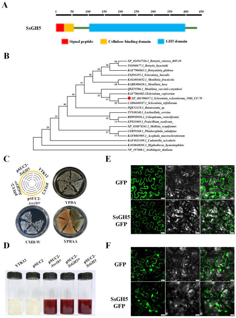
Recent studies have shown that the function of GH family proteins in different plant pathogens is very important. Our previous study showed that SsGH5 expression was significantly upregulated during S. sclerotiorum infection [9]. SsGH5 encodes a 440 aa protein, amino acids 1–19 at the N-terminus of SsGH5 are a predicted signal peptide (SP), amino acids 23–50 are a predicted cellulose-binding domain, and residues 104–402 contain a typical glycosyl hydrolase 5 family domain (Figure 1A). Phylogenetic analysis showed that SsGH5 and its direct homologs from different Ascomycetes fungi are evolutionarily conserved and that its arginine and lysine are highly conserved among the direct homologs (Figure 1B and Figure S3). To determine that the SsGH5 signaling peptide has normal secretory activity, we performed experimental validation by the yeast secretion trap system [34]. YTK12 with a pSUC2-Avr1bsp vector was used as a positive control, and YTK12 and YTK12 with a pSUC2 vector were used as a negative control (Figure S1a). YTK12 strains transformed with pSUC2-SsGH5sp or pSUC2-SsGH5 vectors grew normally on YPRAA medium and produced insoluble red triphenylformazan when treated with 2, 3, 5 chlorotriphenyltetrazole (TTC) (Figure 1C,D). To clarify the subcellular localization of SsGH5, we expressed a fusion protein of SsGH5-GFP in N. benthamiana leaves, and green fluorescence appeared in the periphery of the cells as observed by laser confocal microscopy (Figure 1E). We further treated the N. benthamiana leaves with 1 M NaCl to observe plasmolysis, and the fluorescence of SsGH5-GFP expression aggregated in the apoplast of N. benthamiana cells (Figure 1F). These results strongly suggested that SsGH5 is a secreted protein conserved in Ascomycete fungus.
Figure 1.
Secretary protein SsGH5 is conserved in Ascomycetes. (A) SsGH5 (red circle) is a secreted glycosyl hydrolase family 5 protein containing 440 amino acids, including a signal peptide (SP), a cellulose-binding domain and a glycosyl hydrolase family 5 structural domain. (B) Phylogenetic relationships of SsGH5 with other fungal homologs were determined by maximum likelihood. Branch length is proportional to the average probability of the change in features on that branch. Phylogenies were constructed using Mega X with the neighbor-joining method. (C) Secretory function of the SsGH5 signal peptide was verified by the yeast secretion trap screen assay. The signal peptide and intact protein of SsGH5 were fused in frame with the yeast invertase sequence in a pSUC2 vector and expressed in the YTK12 strain. The functional signal peptide of Avr1b was used as a positive control, while the YTK12 and pSUC2 empty plasmids were used as negative controls. (D) Invertase activity in TTC solution. TTC encountered sucrose breakdown products to produce triphenylformazan, which showed a red reaction to confirm that the functional signal peptide enables a sucrose-converting enzyme to be secreted. (E) SsGH5 green fluorescent fusion protein, expressed in N. benthamiana leaves by the Agrobacterium system to observe the subcellular localization of SsGH5, and GFP protein expressed as a control. (F) Subcellular localization of SsGH5 green fluorescent fusion protein in N. benthamiana leaves under plasmolysis, and GFP protein expressed as a control. The pictures were taken 48 h post-agroinfiltration with confocal laser scanning microscopy. And N. benthamiana cells were treated with 1 M NaCl for 3 min to observe plasmolysis. Bars, 20 μm.
2.2. SsGH5 Is a Glycosyl Hydrolase Specifically Induced by the Host
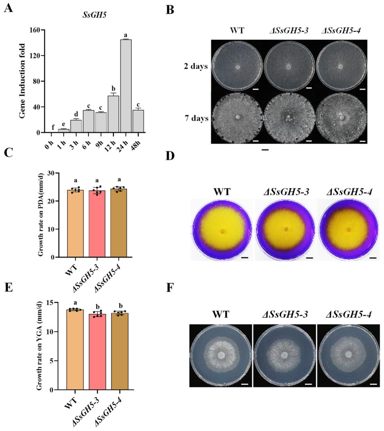
To further clarify the expression pattern of SsGH5, real-time quantitative PCR (RT-qPCR) was performed. Results indicated that when S. sclerotiorum was inoculated onto the leaves of the host, the transcript level of SsGH5 increased rapidly and reached the peak (145-fold) at 24 h post-inoculation (hpi) (Figure 2A). This result suggested that the transcription of SsGH5 is specifically induced by the host and this gene may play a significant role during S. sclerotiorum infection. To further investigate the function of SsGH5, we obtained two SsGH5 deletion mutants (ΔSsGH5-3 and ΔSsGH5-4) by targeting the replacement gene with a hygromycin resistance cassette and validated them at the DNA and RNA levels (Figure S1b–d). The growth rate, colony morphology, and oxalic acid production capacity of the two ΔSsGH5 mutants were similar to those of the wild-type strain 1980 (Figure 2B–D), suggesting that SsGH5 is not required for normal growth and does not affect oxalic acid production. SsGH5 belongs to glycosyl hydrolase family 5, a family of proteins commonly reported to have cellulase activity [35]. Therefore, we hypothesized that SsGH5 may have the ability to hydrolyze cellulose. β-glucan is a type of cellulose and one of the main components of plant cell walls [36]. When the WT and the SsGH5 knockout mutant strains were cultured on the medium with yeast extract, β-glucan and agar (YGA), the growth rate was lower than that of the WT (Figure 2E), but the colony morphology of these strains was not significantly different (Figure 2F). These results indicated that SsGH5 is a glycosyl hydrolase that is specifically upregulated at the stage of infection.
Figure 2.
SsGH5 is a glycosyl hydrolase specifically induced by the host at the S. sclerotiorum infection stage. (A) Relative levels of SsGH5 transcript accumulation were determined by RT-qPCR in inoculated Arabidopsis plants. Levels of β-tubulin transcripts of S. sclerotiorum were used to normalize different samples. Values are similar in three independent experiments. (B) Measurement of the wild-type (WT), ΔSsGH5-3 and ΔSsGH5-4 strain growth rates utilizing the cross-over method at 20 °C on potato dextrose agar (PDA). Upper graph bar, 1 cm. (C) Colony morphology of the WT, ΔSsGH5-3 and ΔSsGH5-4 strains grown at 20 °C for 15 days on PDA. (D) Acid-producing ability of the WT, ΔSsGH5-3 and ΔSsGH5-4 strains was determined on PDA containing 0.005% (w/v) bromophenol blue dye as an indicator. The yellow color appearing in the medium indicated acid production by mycelia. Upper graph bar, 1 cm. (E) Measurement of the WT, ΔSsGH5-3 and ΔSsGH5-4 strain growth rates utilizing the cross-over method at 20 °C on YGA. (F) Colony morphology of the WT, ΔSsGH5-3 and ΔSsGH5-4 strains grown at 20 °C for 2 days on YGA. Upper graph bar, 1 cm. These values are the mean ± S.E. Different letters on the same graph indicate statistical significance at p < 0.01 using one-way ANOVA.
2.3. SsGH5 Is Essential for the Full Virulence of S. sclerotiorum
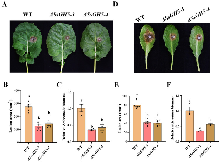
To clarify the function played by SsGH5 during the infection by S. sclerotiorum, we inoculated the WT, ΔSsGH5-3 and ΔSsGH5-4 strains onto 45-day-old Brassica napus (Westar) leaves, and photographed and counted lesion sizes after 36 h. The results showed that the ΔSsGH5 knockout strains had a significantly lower lesion area compared to the WT (Figure 3A). The mean lesion area of the WT was 276 mm2, while that of the ΔSsGH5-3 and ΔSsGH5-4 strains was only 124 mm2 and 143 mm2, respectively (Figure 3B). And the biomass statistics showed that the ΔSsGH5 knockout strains also had significantly lower biomass than the WT (Figure 3C). Similarly, virulence assay of these strains on 4-week-old A. thaliana (Col-0) leaves showed that the virulence of SsGH5 knockout strains was significantly reduced compared to that of the WT (Figure 3D–F). In conclusion, these results confirmed the essential role of SsGH5 in promoting full virulence during S. sclerotiorum infection.
Figure 3.
SsGH5 is essential for the full virulence of S. sclerotiorum. (A) Virulence assay of the S. sclerotiorum WT, ΔSsGH5 mutants ΔSsGH5-3 and ΔSsGH5-4 on B. napus. The symptoms were photographed at 36 hpi. (B) Measurement of lesion area by the cross-over method. (C) Equal area samples from infected sites were used to extract DNA, and the relative biomass was analyzed by RT-qPCR. (D) Virulence assay of the S. sclerotiorum WT, ΔSsGH5 mutants ΔSsGH5-3 and ΔSsGH5-4 on Arabidopsis Col-0. (E) Measurement of lesion area by the cross-over method. (F) Fixed mass samples of susceptible parts were selected and DNA was then extracted for subsequent detection of the amount of pathogen and plant endogenous genes using RT-qPCR, and we then performed relative biomass analyses. These values are the mean ± S.E. Different letters on the same graph indicate statistical significance at p < 0.01 using one-way ANOVA.
2.4. SsGH5 Inhibits Plant Immunity and Makes Plants More Susceptible to Necrotrophic Fungi
To further investigate the role of SsGH5 in the infection of plants by S. sclerotiorum, stable transgenic plants (35S:SsGH5-3 × Flag) constitutively expressing SsGH5 were generated in wild-type Arabidopsis (Col-0). The expression of SsGH5 in transgenic Arabidopsis was confirmed through Western blot (Figure S2a). Compared with WT Col-0, the size of SsGH5 transgenic lines (oxSsGH5-3, oxSsGH5-6, oxSsGH5-14) is slightly smaller (Figure S2b,c).
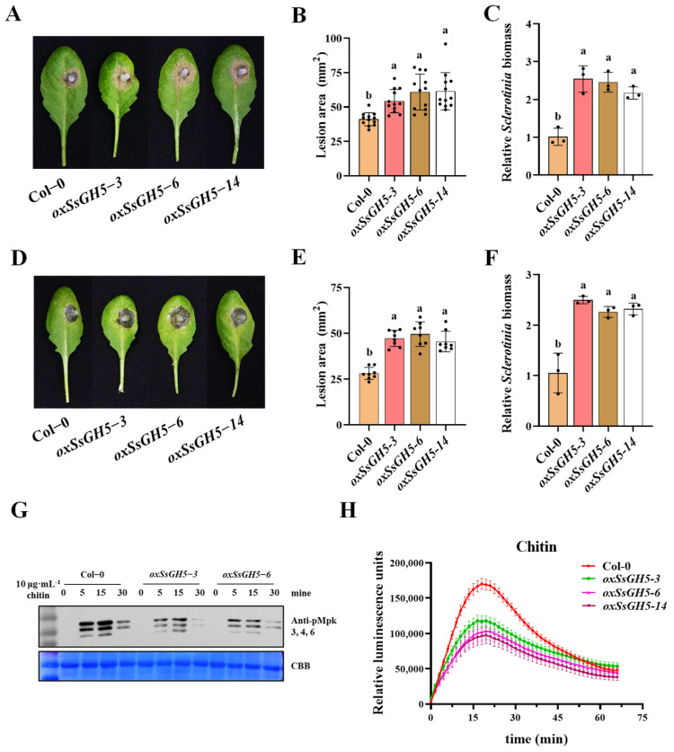
We first inoculated SsGH5 transgenic plants with S. sclerotiorum, and the result showed that transgenic Arabidopsis expressing SsGH5 was more susceptible to t S. sclerotiorum (Figure 4A), the lesion area of the transgenic Arabidopsis increased by 20% to 30% compared with that of Col-0 (Figure 4B), and the relative biomass statistics of lesion area showed consistent trends (Figure 4C). Likewise, the resistance of SsGH5 transgenic plants to B. cinerea, another necrotrophic fungal pathogen, was also significantly reduced (Figure 4D–F). In addition, SsGH5 transgenic plants showed a remarkably significant reduction in both chitin-triggered ROS burst and MAPK activation (Figure 4G,H). Taken together, SsGH5 transgenic A. thaliana exhibited significantly diminished immunity and heightened susceptibility to necrotrophic fungi.
Figure 4.
SsGH5 sensitizes plants to necrotrophic fungi and inhibits plant immunity. (A) Virulence assay of the S. sclerotiorum WT 1980 strain on Col-0 and SsGH5−3 × Flag-expressing (35S:SsGH5) lines at 30 hpi. (B) Measurement of lesion area by the cross-over method. (C) Equal area samples from infected sites were used to extract DNA, and the relative biomass was analyzed by RT-qPCR. (D) Virulence assay of the B. cinerea WT B05.10 strain on Col-0 and SsGH5−3 × Flag-expressing (35S:SsGH5) lines at 36 hpi. (E) Measurement of lesion area by the cross-over method. (F) Equal area samples from infected sites were used to extract DNA, and the relative biomass was analyzed by RT-qPCR. These values are the mean ± S.E. Different letters on the same graph indicate statistical significance at p < 0.01 using one-way ANOVA. (G) Expression of SsGH5 inhibited chitin-induced phosphorylation of MAPKs. MAPK activation was detected by immunoblotting with α-PERK antibody (top). Protein loading is shown by Coomassie Brilliant Blue (bottom). (H) Detection of chitin-induced ROS burst in Col-0 and SsGH5 transgenic lines. Leaf discs from 4-week-old plants were treated with 10 μg·mL−1 chitin and values represent the means ± SE (n = 12 biological replicates).
2.5. SsGH5 Is an Important Pathogenic Factor of S. sclerotiorum Infection
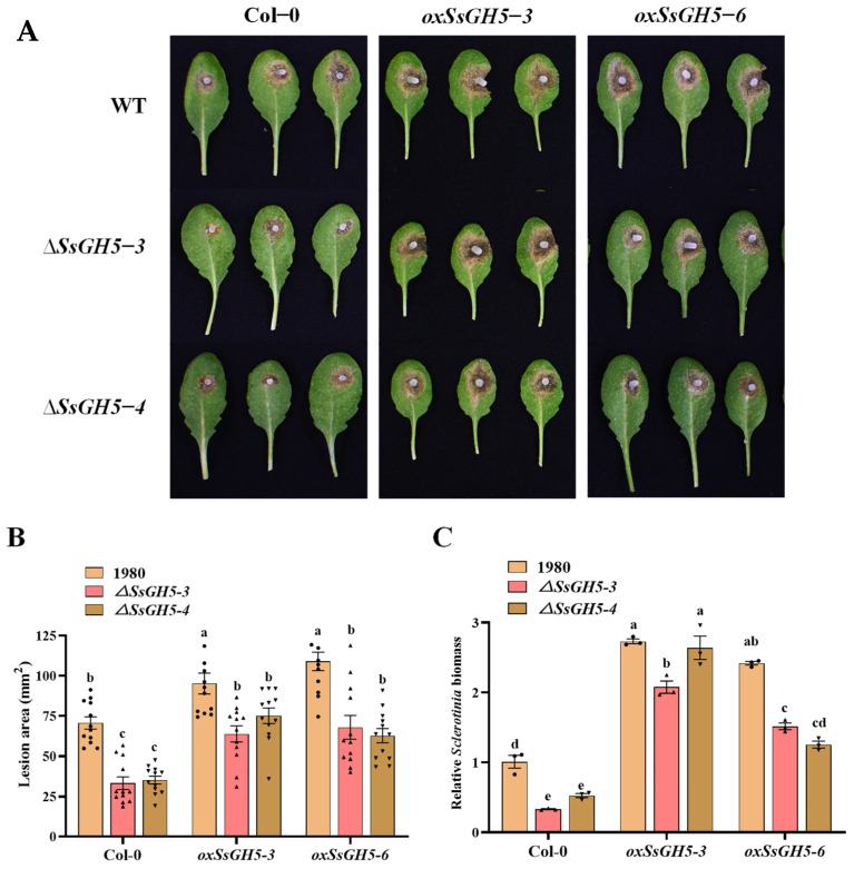
To further confirm that SsGH5 plays an important role during S. sclerotiorum infection and is involved in the interaction with plants, we inoculated the WT, ΔSsGH5-3 and ΔSsGH5-4 onto the leaves of Col-0 and SsGH5 transgenic lines (oxSsGH5-3 and oxSsGH5-6) for virulence assays. Statistical results showed that ΔSsGH5-3 and ΔSsGH5-4 resulted in a significantly lower lesion area on Col-0 compared to the WT strain, but the ΔSsGH5 knockout mutants appeared to show a significant recovery of virulence on SsGH5 transgenic lines, and the lesion area caused by them recovered to the level of the WT strain infesting Col-0 (Figure 5A,B). And the biomass statistics also show that the Sclerotinia biomass of ΔSsGH5 knockout mutants significantly recovered after inoculating onto SsGH5 transgenic lines (Figure 5C). The above results demonstrate that SsGH5 is important for the virulence of S. sclerotiorum and is likely involved in interaction between S. sclerotiorum and host plants.
Figure 5.
SsGH5 is an important pathogenic factor of S. sclerotiorum infection. (A) Virulence assay of the S. sclerotiorum WT and ΔSsGH5 mutants on Col-0 and 35S:SsGH5 Arabidopsis lines at 36 hpi. (B) Measurement of lesion area by the cross-over method. (C) Equal area samples from infected sites were used to extract DNA, and the relative biomass was analyzed by RT-qPCR. These values are the mean ± S.E. Different letters on the same graph indicate statistical significance at p < 0.01 using one-way ANOVA.
3. Discussion
Glycosyl hydrolase 5 family proteins have been studied for decades and are involved in many biomolecular hydrolysis processes, it has been found that the substrates of these enzymes are xylan, xyloglucan, dextran, cellulose and hemicellulose [35,37,38]. In the present study, we sought to investigate the function of SsGH5 in S. sclerotiorum. We found that SsGH5 is a secreted protein that is highly conserved in Ascomycetes (Figure 1). SsGH5 deletion mutants showed a reduced growth rate on medium with β-glucan as the sole carbon source, suggesting that SsGH5 functions as a glucanase. (Figure 2E). CWDEs are generally induced to be expressed by the host during the infection stage of plant pathogens, and the transcriptomic data also suggested that SsGH5 might be significantly induced as a CWDE of S. sclerotiorum during the infection stage [8,9]. RT-qPCR results further comfirmed that SsGH5 gene expression was indeed induced during S. sclerotiorum infection of Arabidopsis (Figure 2A). This further suggests that SsGH5 is a CWDE associated with pathogenesis in S. sclerotiorum.
Numerous studies have shown that CWDEs are secreted by S. sclerotiorum to degrade plant cell walls for nutrients [21,39]. S. sclerotiorum produces several forms of pectinases, such as polygalacturonases (PGs) to degrade pectin, which are important for the pathogenicity of this fungus [40,41]. In addition, GH proteins predominate in CWDEs and are secreted by necrotrophic phytopathogenic fungi that are essential in degrading the cell wall and facilitating the pathogen’s full virulence [27,42,43]. In this study, we reported that SsGH5 was closely associated with the virulence of S. sclerotiorum. SsGH5 is not required for normal growth and does not affect oxalic acid production (Figure 2B–D), but SsGH5 knockout mutants showed significantly reduced virulence on both B. napus and Arabidopsis leaves (Figure 3A,D), indicating that this gene significantly affects the virulence of S. sclerotiorum. The importance of GH proteins for the virulence of plant pathogens is likely to be dependent on their hydrolase activity. Correspondingly, the glucanase activity of SsGH5 deletion mutants was decreased. It is noteworthy that the reduction in hydrolase activity observed in SsGH5 deletion mutants does not align consistently with the decrease in virulence. These results may be due to the existence of redundant genes in S. sclerotiorum, and SsGH5 may also have other functions. In fact, previous studies have shown that GH proteins were also involved in plant–pathogen interaction. For instance, Ebg1, as a glucanase in M. oryzae, can hydrolyze its own glucan, thereby achieving immune escape [33]. Interestingly, plants seem to have developed receptors capable of recognizing the GH family of proteins. Ebg1 also can be recognized by plants to trigger immune responses and cause cell death [33]. This is not an isolated example, XEG1 is important for virulence and can be recognized by plant RXEG1 as a PAMP and trigger intense cell death [29,30,44]. PpOPEL and VdXyn4 containing the GH domains could cause plant cell death, and plants pretreated with these proteins exhibit robust induced resistance [26,45]. These studies showed that the roles of GH in the pathogenesis are diverse and may be more complicated than we thought. Notably, we obtained transgenic Arabidopsis that stably expressed SsGH5 and did not observe cell death in SsGH5 transgenic plants (Figure S2). However, both the chitin-triggered ROS burst and the activation of MAPKs were significantly suppressed in the SsGH5 transgenic lines (Figure 4G,H), which supported the PTI of Arabidopsis was inhibited by SsGH5. As far as we know, this is the first report that GH protein of plant pathogen has the function of inhibiting the immunity of host plants. Most importantly, we found that SsGH5 transgenic Arabidopsis was significantly susceptibility to necrotrophic fungal pathogens including S. sclerotiorum and B. cinerea (Figure 4A–F). Furthermore, expression of SsGH5 in Arabidopsis plants restored the loss of virulence of the ΔSsGH5 mutants (Figure 5). The results further indicated that the SsGH5 could play important roles in plants during the pathogenic process of S. sclerotiorum.
Taken together, our results indicate that SsGH5 is not only essential for S. sclerotiorum virulence, but also has a significant effect on plant immunity, which are different from previous research in which GH family proteins caused plant cell death and activated plant immune responses. Moreover, the subcellular localization of SsGH5 suggests that it may interact with plant immunity-related proteins or receptors in the extracellular space to inhibit plant immunity and promote the colonization of S. sclerotiorum. The specific target of SsGH5 and the biological function and mechanism of its interaction need to be further studied.
4. Methods
4.1. Fungal Strains, Plant Materials and Growth Condition Conditions
The S. sclerotiorum wild-type strain 1980 (ATCC 18683) was obtained from diseased bean plants in Scottsbluff, NE [46]. And the WT strain was cultured on potato dextrose agar (PDA) (Lot 3199948, Becton Dickinson and Company, Franklin Lakes, NJ, USA) plates in a dark greenhouse at a constant temperature of 20 °C. The gene knockout mutants were cultured on PDA plates amended with hygromycin B. 100 μg·mL−1 (Sigma, St. Louis, MO, USA). The above PDA plates were placed in a 4 °C refrigerator (MPR-512HI, Alphavita, Dalian, China) for long-term storage.
All Arabidopsis plants used in this study were in the Columbia-0 (Col-0) genetic background. Arabidopsis lines were grown in growth chamber soil at 22 °C, 75 mE−2·s−1 (T5 LED tube light, 4000 K). The light/dark photoperiod was 12 h and the relative humidity was 40~60%.
4.2. Bioinformatics Analysis
The SsGH5 protein sequence (Ss1G_00746) was downloaded from the NCBI GenBank database. SIGNALP 4.0 and SIGNALP 4.1 were used for signal peptide prediction [47,48], and DeepTMHMM (https://dtu.biolib.com/DeepTMHMM, accessed on 15 June 2022) was used for prediction of trans-membrane helices. Phylogenetic analysis was performed to reconstruct the phylogenetic tree with MEGA X using the maximum likelihood method.
4.3. Plasmid Construction and Generation of Transgenic Plants
The coding sequence of SsGH5 was amplified from S. sclerotiorum cDNA with primers containing homologous fragments of the pCNF3 vector. The fragment was then cloned into CaMV 35S promoter-driven binary expression vectors pCNF3 with 3 × flag and pTF101 with GFP tags fused at the C terminus for the SsGH5-GFP localization observation in N. benthamiana and Agrobacterium-mediated floral dipping of Arabidopsis. SsGH5 and SsGH5sp subcloned into the pSUC2 vector for expression in the YTK12 yeast strain to verify secretion using the same methods via homologous recombination (Figure S1a) (Vazyme, Cat. C113-02).
Agrobacterium tumefaciens GV3101 carrying pCNF3-SsGH5−3 × flag was cultured overnight in LB liquid medium containing 50 mg·mL−1 rifampicin and 50 mg·mL−1 kanamycin. After centrifugation at 4000 rpm for 5 min, the bacteria were suspended in Agrobacterium infiltration buffer containing 5% sucrose and 0.04% (v/v) silwetL-77 at an optical density (OD) 600 = 0.8. Arabidopsis buds were thoroughly immersed in the bacterial suspension, and plants were moisturized for 8 h after immersion. Plants were then maintained at 22 °C and 45% relative humidity with a 16 h light/8 h dark photoperiod, conditions for seed harvesting.
Transgenic Arabidopsis was screened with 1/2 MS (Lot P17261.01, Duchefa Biochemie, Haarlem, The Netherland) containing 50 mg·mL−1 kanamycin and further confirmed by immunoblotting using anti-flag antibody (F1804, Sigma, St. Louis, MO, USA).
4.4. Gene Knockout and Complementation of S. sclerotiorum
Obtaining gene knockout mutants of the SsGH5 gene from S. sclerotiorum was accomplished by the split-marker technique [49]. The knockout strategy of this technique is illustrated in Supplementary Figure (Figure S1b). Two fragments of approximately 1000 bp, SsGH5-5′ and SsGH5-3′, were amplified from genomic DNA by PCR reactions with primers P1/P2 (both containing Sal I site) and P3/P4 (both containing Xba I site) using KOD DNA Polymerase (TOYOBO KMM-101, Japan) (Table S1). These products were cloned into the Sal I and Xba I sites in the pUCH18 vector containing the hygromycin-resistant cassette, respectively [18]. The pUCH18-SsGH5-5′ and pUCH18-SsGH5-3′ were obtained after successful cloning. The fusion sequences SsGH5-5′ and SsGH5-3′ with two truncated hygromycin-resistant genes, SsGH5-5′-HY and YG-SsGH5-3′, were mass amplified with primers P1/HY and YG/P4, respectively (Table S1). After the amount of purified SsGH5-5′-HY and YG-SsGH5-3′ DNA fragments both reached 10 μg or more, they were mixed in equimolar amounts and used for transformation.
For preparation and transformation of S. sclerotiorum protoplasts, the methodology of the previous study was adopted [18]. Mycelial balls grown in PDB for 36 h were lysed with lysing enzymes from Trichoderma harzianum (Cat. L1412, Sigma, St. Louis, MO, USA) to obtain fresh protoplasts. Fragments of SsGH5-5′-HY and YG-SsGH5-3′ were transferred into the protoplasts of WT strains covered with RM (0.7 M Sucrose, 0.5 g·L−1 Yeast Extract, 10 g·L−1 Agar) medium containing 200 μg·mL−1 hygromycin B. Putative transformants with hygromycin B resistance were obtained after 5 to 7 days. After verification of correct substitution of site according to the knockout strategy schematic, they were induced to produce sexual state ascospores, which were screened for hygromycin resistance in order to obtain pure syngeneic knockout mutant strains. The pure knockout mutant strains were validated by PCR and RT-PCR (Figure S1c,d).
4.5. Determination of the Biological Characteristics of S. sclerotiorum Transformants
Transformant strains of S. sclerotiorum (ΔSsGH5 mutant and WT strains) were characterized for growth rate, colony morphology, acid production capacity and virulence. The WT and knockout strains were inoculated in the center of PDA plates for 3 days at 20 °C to measure the growth rate and incubated continuously until 14 days to record their colony morphology by photograph. To assay the ability of oxalic acid production, the wild-type strain and deletion mutant strains were inoculated into the center of PDA plates containing 0.005% (w/v) bromophenol blue dye and grown at 20 °C for 36 h, and their acid production ability was qualitatively characterized by vitual observation. The WT strain and the knockout strains were inoculated into the center of YGA plates and incubated at 20 °C for 3 days, and the growth rate was measured and the colony morphology was recorded by photographs (YGA medium: 0.5% (w/v) yeast extract, 1% (w/v) β-glucan (Aladdin SKU G304913) and 1% (w/v) agar).
4.6. The Fungal Inoculation Assay
For the fungal inoculation assay, agar discs (2 mm diameter) were punched from the actively growing edge of the fungal 2 × SY plates (SY medium: 0.5% (w/v) sucrose and yeast extract, 1% (w/v) agar) and inoculated on leaves of 4–5-week-old Arabidopsis plants and incubated at 22 °C. Eight to twelve biological replicates per treatment were taken from eight Arabidopsis plants. At the same time, the true leaves of B. napus grown for 30~40 days were selected for inoculation with fungi for virulence determination. Fungal virulence was assessed in two ways: macroscopically by measuring the long and short axes of the lesion with calipers, and molecularly by measuring the ratio of S. sclerotiorum DNA to host plant DNA in infected leaves. Disease lesion area was calculated based on the elliptical area formula. Relative pathogen biomass was based on the ratio of pathogen DNA to host plant DNA, fixed-mass samples from all infected sites were selected to extract DNA and amplified by quantitative PCR of genomic DNA for the S. sclerotiorum β-Tubulin gene or B. cinerea actin gene, and the A. thaliana UBQ5 gene or B. napus actin gene (Table S1 for primers) by quantitative PCR of genomic DNA. Three replicates (each replicate contained 2–4 diseased leaves) were set up for each treatment, and each experiment was performed three times.
4.7. ROS Production Analysis and the MAPK Activation Assay
The third or fourth pair of true leaves of 4-week-old soil-grown Arabidopsis plants were cut into leaf discs (5 mm in diameter) and further cut into leaf strips. The leaves were left to float in 96-well plates with 100 µL ddH2O and shaken gently overnight to eliminate the wounding effect. For the assay, ddH2O was replaced by 100 µL reaction solution containing 50 µM L-012 (CAS:143556-24-5, Wako, Japan), 10 µg·mL−1 horseradish peroxidase (Sigma Cat. P6782) and chitin. Measurements were performed immediately after the addition of the reaction solution on a Multimode Reader Platform (SPARK 10M, Tecan Austria GmbH, Männedorf, Switzerland) and the values generated by ROS represent the relative light units of different plants.
Arabidopsis seedlings grown on 1/2 MS plates for 10 days were transferred to 1 mL sterilized ddH2O, recovered overnight, and then treated with the indicated concentrations of chitin for 0, 5, 15 and 30 min. The total protein of the samples was extracted with protein extraction buffer (20-mM Tris-HCl, pH 7.5, 100-mM NaCl, 1-mM EDTA, 10% glycerol and 1% Triton X-100) and the samples were kept at 95 °C for 10 min. The supernatant was collected after centrifugation at 12,000 rpm for 2 min, and the protein samples containing 1 × SDS buffer were loaded onto 10% (v/v) SDS-PAGE gels and immunoblotted with anti-PERK1/2 antibody to detect pMPK3, pMPK4 and pMPK6 (Cat. 9101S, CST, Boston, MA, USA).
4.8. The Secretion Trap Screen Assay
In this study, the predicted signal peptide fragment of the SsGH5 gene was fused to the N-terminus of the secretion-defective convertase gene (suc2) in the vector pSUC2 and then transformed into the yeast strain YTK12. Candidate yeast transformants were screened and cultured on CMD-W medium (Coolaber, Beijing, China) and YPRAA (10 g·L−1 yeast extract, 20 g·L−1 peptone, 20 g·L−1 raffinose, 2 mg·L−1 antimycin A, and 2% agar) medium (Macklin, Shanghai, China), and strains with secretion activity were able to grow on YPRAA. TTC (Sigma-Aldrich CAS NO 298-96-4) was used to assay the secretion sucrase activity of the candidate yeast transformants. The candidate yeast transformants were incubated in 10% sucrose solution at 30 °C for 35 min, then the supernatant was centrifuged and the final concentration of 0.1% TTC reagent was added and left at room temperature for 5 min to observe the color change in the test tube. The positive reaction changed from colorless to dark red, and the Avr1bsp transformant, YTK12-pUSC2 and YTK12 strains were used as positive and negative controls, respectively.
4.9. RNA Isolation, cDNA Synthesis and RT-qPCR Analysis
Fungal samples were ground to a powder in liquid nitrogen, total RNA was extracted using trizol, and DNA was removed using RNase-free recombinant DNase I (2270A, Takara, Beijing, China). To examine the expression patterns of the SsGH5 and WT strains, mycelium was cultured on PDA for 36 h and inoculated onto Arabidopsis leaves. Mycelia were harvested at 0, 1, 3, 6, 9,12, 24 and 48 h to extract nucleic acids. The concentration of total RNA was quantified using a spectrophotometer (Thermo, Waltham, MA, USA), and first-strand cDNA was synthesized using Easy Script One-Step gDNA Removal and cDNA Synthesis SuperMix (AE311-02, Transgen, Beijing, China). RT-PCR was performed in a CFX96 RT-PCR Detection System and TransStart Green qPCR SuperMix (Transgen AQ101-01). RNA samples for each RT-PCR were normalized with the β-tubulin gene Sstub1 of S. sclerotiorum. For the SsGH5 gene, RT-PCR assays were repeated at least twice, with three biological replicates each time.
5. Conclusions
In summary, our study determined that SsGH5, a GH5-family-secreted protein, as significantly upregulated during infection of S. sclerotiorum. SsGH5 is indispensable for the full virulence of S. sclerotiorum. In addition, SsGH5 can suppress Arabidopsis immunity and cause Arabidopsis to be more susceptible to S. sclerotiorum and B. cinerea. These results showed that SsGH5 is likely to be involved in the interaction between S. sclerotiorum and the host plant. Further screening the target protein of SsGH5 and exploring the biological function and mechanism may reveal a new model for pathogen–plant interactions.
Supplementary Materials
The following supporting information can be downloaded at: https://www.mdpi.com/article/10.3390/ijms25052693/s1.
Author Contributions
J.C. and X.L. designed the experiments and analyzed the data; H.Z. performed gene complement, identification and partial vector construction; J.C. and X.L. wrote the manuscript. J.X., Y.F., B.L., X.Y., T.C., Y.L. and D.J. provided constructive inputs in manuscript preparation. All authors have read and agreed to the published version of the manuscript.
Institutional Review Board Statement
Not applicable.
Informed Consent Statement
Not applicable.
Data Availability Statement
The sequence of SsGH5 was deposited in the GenBank database, with accession numbers XP_001598657.1 (hypothetical protein SS1G_00746 [Sclerotinia sclerotiorum 1980 UF-70]—Protein—NCBI (nih.gov)).
Conflicts of Interest
The authors declare that the research was conducted in the absence of any commercial or financial relationships that could be construed as a potential conflict of interest.
Funding Statement
This research was supported by the National Nature Science Foundation of China (32172368, 32130087), the earmarked fund of the China Agriculture Research System (CARS-12), and the Huazhong Agricultural University Scientific and Technological Self-innovation Foundation (2021ZKPY014). The funders were not involved in study design, data collection and analysis, the decision to publish, or manuscript preparation.
Footnotes
Disclaimer/Publisher’s Note: The statements, opinions and data contained in all publications are solely those of the individual author(s) and contributor(s) and not of MDPI and/or the editor(s). MDPI and/or the editor(s) disclaim responsibility for any injury to people or property resulting from any ideas, methods, instructions or products referred to in the content.
References
- 1.Boland G.J., Hall R. Index of plant hosts of Sclerotinia sclerotiorum. Can. J. Plant Pathol. 1994;16:93–108. doi: 10.1080/07060669409500766. [DOI] [Google Scholar]
- 2.Bolton M.D., Thomma B.P.H.J., Nelson B.D. Sclerotinia sclerotiorum (Lib.) de Bary: Biology and molecular traits of a cosmopol itan pathogen. Mol. Plant Pathol. 2006;7:1–16. doi: 10.1111/j.1364-3703.2005.00316.x. [DOI] [PubMed] [Google Scholar]
- 3.Chitrampalam P., Figuli P.J., Matheron M.E., Subbarao K.V., Pryor B.M. Biocontrol of Lettuce Drop Caused by Sclerotinia sclerotiorum and S. minor in Desert Agroecosystems. Plant Dis. 2008;92:1625–1634. doi: 10.1094/PDIS-92-12-1625. [DOI] [PubMed] [Google Scholar]
- 4.Derbyshire M., Denton-Giles M., Hegedus D., Seifbarghy S., Rollins J., van Kan J., Seidl M.F., Faino L., Mbengue M., Navaud O., et al. The Complete Genome Sequence of the Phytopathogenic Fungus Sclerotinia sclerotiorum Reveals Insights into the Genome Architecture of Broad Host Range Pathogens. Genome Biol. Evol. 2017;9:593–618. doi: 10.1093/gbe/evx030. [DOI] [PMC free article] [PubMed] [Google Scholar]
- 5.Zhou F., Zhang X.-L., Li J.-L., Zhu F.-X. Dimethachlon Resistance in Sclerotinia sclerotiorum in China. Plant Dis. 2014;98:1221–1226. doi: 10.1094/PDIS-10-13-1072-RE. [DOI] [PubMed] [Google Scholar]
- 6.Hou Y.-P., Mao X.-W., Qu X.-P., Wang J.-X., Chen C.-J., Zhou M.-G. Molecular and biological characterization of Sclerotinia sclerotiorum resistant to the anilinopyrimidine fungicide cyprodinil. Pestic. Biochem. Physiol. 2018;146:80–89. doi: 10.1016/j.pestbp.2018.03.001. [DOI] [PubMed] [Google Scholar]
- 7.Marciano P., Lenna P.D., Magro P. Oxalic acid, cell wall-degrading enzymes and pH in pathogenesis and their significance in the virulence of two Sclerotinia sclerotiorum isolates on sunflower. Physiol. Plant Pathol. 1983;22:339–345. doi: 10.1016/S0048-4059(83)81021-2. [DOI] [Google Scholar]
- 8.Guyon K., Balagué C., Roby D., Raffaele S. Secretome analysis reveals effector candidates associated with broad host range necrotrophy in the fungal plant pathogen Sclerotinia sclerotiorum. BMC Genom. 2014;15:336. doi: 10.1186/1471-2164-15-336. [DOI] [PMC free article] [PubMed] [Google Scholar]
- 9.Lyu X., Shen C., Fu Y., Xie J., Jiang D., Li G., Cheng J. Comparative genomic and transcriptional analyses of the carbohydrate-active enzymes and secretomes of phytopathogenic fungi reveal their significant roles during infection and development. Sci. Rep. 2015;5:15565. doi: 10.1038/srep15565. [DOI] [PMC free article] [PubMed] [Google Scholar]
- 10.Cessna S.G., Sears V.E., Dickman M.B., Low P.S. Oxalic Acid, a Pathogenicity Factor for Sclerotinia sclerotiorum, Suppresses the Oxidative Burst of the Host Plant. Plant Cell. 2000;12:2191–2199. doi: 10.1105/tpc.12.11.2191. [DOI] [PMC free article] [PubMed] [Google Scholar]
- 11.Guimarães R.L., Stotz H.U. Oxalate Production by Sclerotinia sclerotiorum Deregulates Guard Cells during Infection. Plant Physiol. 2004;136:3703–3711. doi: 10.1104/pp.104.049650. [DOI] [PMC free article] [PubMed] [Google Scholar]
- 12.Kim K.S., Min J.-Y., Dickman M.B. Oxalic Acid Is an Elicitor of Plant Programmed Cell Death during Sclerotinia sclerotiorum Disease Development. Mol. Plant-Microbe Interact. 2008;21:605–612. doi: 10.1094/MPMI-21-5-0605. [DOI] [PubMed] [Google Scholar]
- 13.Williams B., Kabbage M., Kim H.-J., Britt R., Dickman M.B. Tipping the Balance: Sclerotinia sclerotiorum Secreted Oxalic Acid Suppresses Host Defenses by Manipulating the Host Redox Environment. PLoS Pathog. 2011;7:e1002107. doi: 10.1371/journal.ppat.1002107. [DOI] [PMC free article] [PubMed] [Google Scholar]
- 14.Heller A., Witt-Geiges T. Oxalic Acid Has an Additional, Detoxifying Function in Sclerotinia sclerotiorum Pathogenesis. PLoS ONE. 2013;8:e72292. doi: 10.1371/journal.pone.0072292. [DOI] [PMC free article] [PubMed] [Google Scholar]
- 15.Kabbage M., Williams B., Dickman M.B. Cell Death Control: The Interplay of Apoptosis and Autophagy in the Pathogenicity of Sclerotinia sclerotiorum. PLoS Pathog. 2013;9:e1003287. doi: 10.1371/journal.ppat.1003287. [DOI] [PMC free article] [PubMed] [Google Scholar]
- 16.Xu L., Xiang M., White D., Chen W. pH dependency of sclerotial development and pathogenicity revealed by using genetically defined oxalate-minus mutants of Sclerotinia sclerotiorum. Environ. Microbiol. 2015;17:2896–2909. doi: 10.1111/1462-2920.12818. [DOI] [PubMed] [Google Scholar]
- 17.Lyu X., Shen C., Fu Y., Xie J., Jiang D., Li G., Cheng J. A Small Secreted Virulence-Related Protein Is Essential for the Necrotrophic Interactions of Sclerotinia sclerotiorum with Its Host Plants. PLoS Pathog. 2016;12:e1005435. doi: 10.1371/journal.ppat.1005435. [DOI] [PMC free article] [PubMed] [Google Scholar]
- 18.Yang G., Tang L., Gong Y., Xie J., Fu Y., Jiang D., Li G., Collinge D.B., Chen W., Cheng J. A cerato-platanin protein SsCP1 targets plant PR1 and contributes to virulence of Sclerotinia sclerotiorum. New Phytol. 2018;217:739–755. doi: 10.1111/nph.14842. [DOI] [PubMed] [Google Scholar]
- 19.Tang L., Yang G., Ma M., Liu X., Li B., Xie J., Fu Y., Chen T., Yu Y., Chen W., et al. An effector of a necrotrophic fungal pathogen targets the calcium-sensing receptor in chloroplasts to inhibit host resistance. Mol. Plant Pathol. 2020;21:686–701. doi: 10.1111/mpp.12922. [DOI] [PMC free article] [PubMed] [Google Scholar]
- 20.Wei W., Xu L., Peng H., Zhu W., Tanaka K., Cheng J., Sanguinet K.A., Vandemark G., Chen W. A fungal extracellular effector inactivates plant polygalacturonase-inhibiting protein. Nat. Commun. 2022;13:2213. doi: 10.1038/s41467-022-29788-2. [DOI] [PMC free article] [PubMed] [Google Scholar]
- 21.Riou C., Freyssinet G., Fevre M. Production of Cell Wall-Degrading Enzymes by the Phytopathogenic Fungus Sclerotinia sclerotiorum. Appl. Environ. Microbiol. 1991;57:1478. doi: 10.1128/aem.57.5.1478-1484.1991. [DOI] [PMC free article] [PubMed] [Google Scholar]
- 22.Issam S.M., Mohamed G., Dominique L.M., Thierry M., Farid L., Nejib M. A β-Glucosidase From Sclerotinia sclerotiorum Biochemical Characterization and Use in Oligosaccharide Synthesis. Appl. Biochem. Biotechnol. 2004;112:63. doi: 10.1385/ABAB:112:2:63. [DOI] [PubMed] [Google Scholar]
- 23.Ellouze O.E., Loukil S., Marzouki M.N. Cloning and molecular characterization of a new fungal xylanase gene from Sclerotinia sclerotiorum S2. BMB Rep. 2011;44:653–658. doi: 10.5483/BMBRep.2011.44.10.653. [DOI] [PubMed] [Google Scholar]
- 24.Gupta N.C., Yadav S., Arora S., Mishra D.C., Budhlakoti N., Gaikwad K., Rao M., Prasad L., Rai P.K., Sharma P. Draft genome sequencing and secretome profiling of Sclerotinia sclerotiorum revealed effector repertoire diversity and allied broad-host range necrotrophy. Sci. Rep. 2022;12:21855. doi: 10.1038/s41598-022-22028-z. [DOI] [PMC free article] [PubMed] [Google Scholar]
- 25.Ben-Daniel B.H., Bar-Zvi D., Tsror L. Pectate lyase affects pathogenicity in natural isolates of Colletotrichum coccodes and in pelA gene-disrupted and gene-overexpressing mutant lines. Mol. Plant Pathol. 2011;13:187–197. doi: 10.1111/j.1364-3703.2011.00740.x. [DOI] [PMC free article] [PubMed] [Google Scholar]
- 26.Wang D., Chen J.-Y., Song J., Li J.-J., Klosterman S.J., Li R., Kong Z.-Q., Subbarao K.V., Dai X.-F., Zhang D.-D. Cytotoxic function of xylanase VdXyn4 in the plant vascular wilt pathogen Verticillium dahliae. Plant Physiol. 2021;187:409–429. doi: 10.1093/plphys/kiab274. [DOI] [PMC free article] [PubMed] [Google Scholar]
- 27.Yu Y., Xiao J., Du J., Yang Y., Bi C., Qing L. Disruption of the Gene Encoding Endo-β-1, 4-Xylanase Affects the Growth and Virulence of Sclerotinia sclerotiorum. Front. Microbiol. 2016;7:1787. doi: 10.3389/fmicb.2016.01787. [DOI] [PMC free article] [PubMed] [Google Scholar]
- 28.Xu M., Gao X., Chen J., Yin Z., Feng H., Huang L. The feruloyl esterase genes are required for full pathogenicity of the apple tree canker pathogen Valsa mali. Mol. Plant Pathol. 2017;19:1353–1363. doi: 10.1111/mpp.12619. [DOI] [PMC free article] [PubMed] [Google Scholar]
- 29.Ma Z., Song T., Zhu L., Ye W., Wang Y., Shao Y., Dong S., Zhang Z., Dou D., Zheng X., et al. A Phytophthora sojae Glycoside Hydrolase 12 Protein Is a Major Virulence Factor during Soybean Infection and Is Recognized as a PAMP. Plant Cell. 2015;27:2057–2072. doi: 10.1105/tpc.15.00390. [DOI] [PMC free article] [PubMed] [Google Scholar]
- 30.Sun Y., Wang Y., Zhang X., Chen Z., Xia Y., Wang L., Sun Y., Zhang M., Xiao Y., Han Z., et al. Plant receptor-like protein activation by a microbial glycoside hydrolase. Nature. 2022;610:335–342. doi: 10.1038/s41586-022-05214-x. [DOI] [PubMed] [Google Scholar]
- 31.Ökmen B., Bachmann D., de Wit P.J. A conserved GH17 glycosyl hydrolase from plant pathogenic Dothideomycetes releases a DAMP causing cell death in tomato. Mol. Plant Pathol. 2019;20:1710–1721. doi: 10.1111/mpp.12872. [DOI] [PMC free article] [PubMed] [Google Scholar]
- 32.Yang C., Yu Y., Huang J., Meng F., Pang J., Zhao Q., Islam A., Xu N., Tian Y., Liu J. Binding of the Magnaporthe oryzae Chitinase MoChia1 by a Rice Tetratricopeptide Repeat Protein Allows Free Chitin to Trigger Immune Responses. Plant Cell. 2019;31:172–188. doi: 10.1105/tpc.18.00382. [DOI] [PMC free article] [PubMed] [Google Scholar]
- 33.Liu H., Lu X., Li M., Lun Z., Yan X., Yin C., Yuan G., Wang X., Liu N., Liu D., et al. Plant immunity suppression by an exo-β-1,3-glucanase and an elongation factor 1α of the rice blast fungus. Nat. Commun. 2023;14:5491. doi: 10.1038/s41467-023-41175-z. [DOI] [PMC free article] [PubMed] [Google Scholar]
- 34.Lee S.J., Kim B.D., Rose J.K.C. Identification of eukaryotic secreted and cell surface proteins using the yeast secretion trap screen. Nat. Protoc. 2006;1:2439. doi: 10.1038/nprot.2006.373. [DOI] [PubMed] [Google Scholar]
- 35.Li Z., Pei X., Zhang Z., Wei Y., Song Y., Chen L., Liu S., Zhang S.-H. The unique GH5 cellulase member in the extreme halotolerant fungus Aspergillus glaucus CCHA is an endoglucanase with multiple tolerance to salt, alkali and heat: Prospects for straw degradation applications. Extremophiles. 2018;22:675–685. doi: 10.1007/s00792-018-1028-5. [DOI] [PubMed] [Google Scholar]
- 36.Endler A., Persson S. Cellulose Synthases and Synthesis in Arabidopsis. Mol. Plant. 2011;4:199–211. doi: 10.1093/mp/ssq079. [DOI] [PubMed] [Google Scholar]
- 37.Uversky V.N., Couturier M., Féliu J., Bozonnet S., Roussel A., Berrin J.-G. Molecular Engineering of Fungal GH5 and GH26 Beta-(1,4)-Mannanases toward Improvement of Enzyme Activity. PLoS ONE. 2013;8:e79800. doi: 10.1371/journal.pone.0079800. [DOI] [PMC free article] [PubMed] [Google Scholar]
- 38.Ogunyewo O.A., Okereke O.E., Kumar S., Yazdani S.S. Characterization of a GH5 endoxylanase from Penicillium funiculosum and its synergism with GH16 endo-1,3(4)-glucanase in saccharification of sugarcane bagasse. Sci. Rep. 2022;12:17219. doi: 10.1038/s41598-022-21529-1. [DOI] [PMC free article] [PubMed] [Google Scholar]
- 39.Paolo A., Francesco F. Pectin-degrading enzymes and plant-parasite interactions. Eur. J. Plant Pathol. 1995;101:365–375. [Google Scholar]
- 40.Francesco F., Luca S., Renato D.O. Relationships among Endo-Polygalacturonase, Oxalate, pH, and Plant Polygalacturonase-Inhibiting Protein (PGIP) in the Interaction between Sclerotinia sclerotiorum and Soybean. Mol. Plant Microbe Interact. 2004;17:1402–1409. doi: 10.1094/MPMI.2004.17.12.1402. [DOI] [PubMed] [Google Scholar]
- 41.Kasza Z., Vagvolgyi C., Fevre M., Cotton P. Molecular Characterization and in planta Detection of Sclerotinia sclerotiorum Endopolygalacturonase Genes. Curr. Microbiol. 2004;48:208–213. doi: 10.1007/s00284-003-4166-6. [DOI] [PubMed] [Google Scholar]
- 42.Yang Y., Yang X., Dong Y., Qiu D. The Botrytis cinerea Xylanase BcXyl1 Modulates Plant Immunity. Front. Microbiol. 2018;9:2535. doi: 10.3389/fmicb.2018.02535. [DOI] [PMC free article] [PubMed] [Google Scholar]
- 43.Yu C., Li T., Shi X., Saleem M., Li B., Liang W., Wang C. Deletion of Endo-β-1,4-Xylanase VmXyl1 Impacts the Virulence of Valsa mali in Apple Tree. Front. Plant Sci. 2018;9:663. doi: 10.3389/fpls.2018.00663. [DOI] [PMC free article] [PubMed] [Google Scholar]
- 44.Xia Y., Ma Z., Qiu M., Guo B., Zhang Q., Jiang H., Zhang B., Lin Y., Xuan M., Sun L., et al. N-glycosylation shields Phytophthora sojae apoplastic effector PsXEG1 from a specific host aspartic protease. Proc. Natl. Acad. Sci. USA. 2020;117:27685–27693. doi: 10.1073/pnas.2012149117. [DOI] [PMC free article] [PubMed] [Google Scholar]
- 45.Chang Y.H., Yan H.Z., Liou R.F. A novel elicitor protein from Phytophthora parasitica induces plant basal immunity and systemic acquired resistance. Mol. Plant Pathol. 2014;16:123–136. doi: 10.1111/mpp.12166. [DOI] [PMC free article] [PubMed] [Google Scholar]
- 46.Godoy G., Steadman J., Dickman M., Dam R. Use of mutants to demonstrate the role of oxalic acid in pathogenicity of Sclerotinia sclerotiorum on Phaseolus vulgaris. Mol. Plant Pathol. 1990;37:179–191. doi: 10.1016/0885-5765(90)90010-U. [DOI] [Google Scholar]
- 47.Bendtsen J.D., Nielsen H., Heijne G.G.V., Brunak S. Improved Prediction of Signal Peptides: SignalP 3.0. J. Mol. Biol. 2004;340:783–795. doi: 10.1016/j.jmb.2004.05.028. [DOI] [PubMed] [Google Scholar]
- 48.Petersen T.N., Brunak S., Heijne G.V., Nielsen H. SIGNALP 4.0: Discriminating signal peptides from transmembrane regions. Nat. Methods. 2011;8:785–786. doi: 10.1038/nmeth.1701. [DOI] [PubMed] [Google Scholar]
- 49.Catlett N.L., Lee B.N., Yoder O.C., Turgeon B.G. Split-Marker Recombination for Efficient Targeted Deletion of Fungal Genes. Fungal Genet. Rep. 2003;50:9–11. doi: 10.4148/1941-4765.1150. [DOI] [Google Scholar]
Associated Data
This section collects any data citations, data availability statements, or supplementary materials included in this article.
Supplementary Materials
Data Availability Statement
The sequence of SsGH5 was deposited in the GenBank database, with accession numbers XP_001598657.1 (hypothetical protein SS1G_00746 [Sclerotinia sclerotiorum 1980 UF-70]—Protein—NCBI (nih.gov)).


