Abstract
Objectives
In SLE, deregulation of haematopoiesis is characterised by inflammatory priming and myeloid skewing of haematopoietic stem and progenitor cells (HSPCs). We sought to investigate the role of extramedullary haematopoiesis (EMH) as a key player for tissue injury in systemic autoimmune disorders.
Methods
Transcriptomic analysis of bone marrow (BM)-derived HSPCs from patients with SLE and NZBW/F1 lupus-prone mice was performed in combination with DNA methylation profile. Trained immunity (TI) was induced through β-glucan administration to the NZBW/F1 lupus-prone model. Disease activity was assessed through lupus nephritis (LN) histological grading. Colony-forming unit assay and adoptive cell transfer were used to assess HSPCs functionalities.
Results
Transcriptomic analysis shows that splenic HSPCs carry a higher inflammatory potential compared with their BM counterparts. Further induction of TI, through β-glucan administration, exacerbates splenic EMH, accentuates myeloid skewing and worsens LN. Methylomic analysis of BM-derived HSPCs demonstrates myeloid skewing which is in part driven by epigenetic tinkering. Importantly, transcriptomic analysis of human SLE BM-derived HSPCs demonstrates similar findings to those observed in diseased mice.
Conclusions
These data support a key role of granulocytes derived from primed HSPCs both at medullary and extramedullary sites in the pathogenesis of LN. EMH and TI contribute to SLE by sustaining the systemic inflammatory response and increasing the risk for flare.
Keywords: lupus erythematosus, systemic; hematology; lupus nephritis
WHAT IS ALREADY KNOWN ON THIS TOPIC
Haematopoietic stem and progenitor cells (HSPCs) in SLE exhibit deregulation of haematopoiesis with skewing towards the myeloid lineage and priming of HSPCs that exhibit a pro-inflammatory ‘trained immunity’ signature, priming them towards an inflammatory phenotype that fuels inflammation.
WHAT THIS STUDY ADDS
Molecular signatures of bone marrow-derived HSPCs from patients with severe SLE and lupus-prone mice are common.
In lupus-prone mice, widespread extramedullary haematopoiesis in the spleen correlates with renal inflammation and the severity of lupus nephritis (LN).
Further induction of trained immunity via β-glucan accentuates splenic extramedullary haematopoiesis, increases neutrophil production and worsens LN.
HOW THIS STUDY MIGHT AFFECT RESEARCH, PRACTICE OR POLICY
These data support a key role of granulocytes in the pathogenesis of LN and may account—at least in part—for the salutary effects of immunosuppressive agents targeting neutrophils such as cyclophosphamide in LN.
Extramedullary tissues such as the spleen may be providing the inflammatory license to HSPC-derived myeloid cells to promote disease flares and tissue injury in SLE.
Introduction
Recent advances in our understanding of SLE pathogenesis have culminated in the approval of novel targeted therapies for the first time in decades.1–3 Despite significant progress, the molecular and phenotypic heterogeneity of SLE remains to be defined, and many patients present with therapy-refractory disease. We have previously reasoned that dysregulation of haematopoietic stem and progenitor cells (HSPCs; Lin−Sca-1+c-Kit+), might contribute to SLE pathogenesis given that cells mediating inflammatory target organ damage originate from the bone marrow (BM)-derived HSPCs.4 5 Based on that premise, recent data from our group suggested that HSPCs are key cells for the adaptation and fine-tuning of inflammation in the SLE setting.6 Specifically, we demonstrated that HSPCs exhibit myeloid skewing and associated strong transcriptomic myeloid signatures, reminiscent of trained immunity. These findings were consistent across species in both SLE patient samples and murine lupus model.
Systemic inflammation is the hallmark of systemic autoimmune diseases including SLE. In order to meet the increased demand for effector cells in the periphery, systemic inflammation has been shown to promote medullary and extramedullary myelopoiesis.7 Specifically, HSPCs in the BM and in the spleen produce myeloid cells, such as neutrophils and monocytes, which in turn mediate a diverse set of tasks. To this end, recent evidence demonstrates that extramedullary haematopoietic (EMH) sites, such as the spleen, are active contributors to the peripheral effector cell pool, and can also shape the inflammatory potential of descendant effector cells. This has been demonstrated in systemic inflammatory diseases, such as inflammatory bowel disease and axial spondyloarthropathy.8 9
Here, we show that SLE disease severity is associated with dysregulated BM haematopoiesis, which is characterised by hypoxia and myelopoiesis transcriptomic signatures and that these effects are underlined by epigenetic imprints through DNA methylation. Induction of trained immunity enhances both central and splenic myelopoiesis and accelerates disease progression in SLE. Splenic EMH activity correlates with the severity of lupus nephritis (LN) while HSPCs exiting from the BM and expanding in the spleen of lupus-prone mice display an inflammatory phenotype. These data demonstrate that dysregulated BM and extramedullary haematopoiesis are present in SLE and play a central role in the production of pro-inflammatory cells that mediate target organ damage.
Materials and methods
Animal studies
C57BL/6 mice were purchased from the Jackson Laboratory. NZB/OlaHsd and NZW/OlaHsd mice were purchased from Envigo. NZB/OlaHsd and NZW/OlaHsd mice were used to produce NZB/W F1 mice. This experimental model is considered as ‘diseased’ (lupus, F1-L; aged 28–36 weeks), when exhibiting ≥300 mg/dL of urine protein level.10 Τhus, when mice are 12 weeks of age, they are considered ‘prediseased’ (F1-P). Proteinuria was assessed and scored semi-quantitatively with reagent strips for urinalysis (Healgen Scientific). Scores were as follows: grade 0, 0 mg/dL; grade±, <30 mg/dL; grade 1+, ≥30 mg/dL; grade 2+, ≥100 mg/dL; grade 3+, ≥300 mg/dL and grade 4+, ≥2000 mg/dL. C57BL/6 mice were used as non-autoimmune wild-type mice at 12 (B6-Y) and 28–36 (B6-O) weeks of age. For the adoptive transfer experiment, the highly immunodeficient NBSGW (NOD.Cg-KitW-41JTyr+PrkdcscidIl2rgtm1Wjl/ThomJ, Jackson Laboratory) mouse strain was used, which carries a hypomorphic mutation in the c-kit gene, allowing haematopoietic expansion without preconditioning. The maintenance of the animals took place in the animal facility of the Biomedical Research Foundation Academy of Athens. Only female mice were used and euthanised by cervical dislocation.
Human subjects
BM aspirates were obtained from individuals with SLE. Patients met the 1999 American College of Rheumatology revised criteria for the classification of SLE11 as described previously.6 We have grouped the patients to moderate and severe disease as previously described.12 More specifically, patients were included in this study if they were rated as British Isles Lupus Assessment Group-A (BILAG-A) or BILAG-B. Active manifestations classified as ‘BILAG-A’ were assigned as severe (eg, lupus rash, vasculitis and/or skin) involving >18% of body surface area; severe pleural effusion with hypoxaemia; thrombocytopenia with Systemic Lupus Erythematosus Disease Activity Index 2000 (SLEDAI-2K) ≥8 and/or Physician Global Assessment ≥1.5, necessitating increase or intensification of treatment as follows: (i) initiating glucocorticoids either oral at a dose of ≥20 mg/day (prednisone equivalent) and/or intravenous pulse methylprednisolone; (ii) increasing (at least doubling) the dosage of glucocorticoids and (iii) initiating immunosuppressive (including calcineurin inhibitors) or biological (including intravenous immunoglobulin) agents. The requirement for therapy intensification was introduced to ascertain medium/high disease activity. The patient’s clinical and serological characteristics are summarised in online supplemental table 1.
lupus-2023-001110supp001.pdf (718.4KB, pdf)
Mononuclear cell isolation and processing
BM mononuclear cells (BMMCs) from human individuals were isolated using Histopaque 1077 (Sigma-Aldrich). BMMCs were washed and erythrocytes were lysed with RBC buffer (420301, BioLegend). CD34+ cells were isolated using EasySep Human CD34 Positive Selection Kit II (18056, StemCell Technologies).
Tissue isolation and single-cell suspension
For the kidney, mechanical homogenisation was followed by enzymatic digestion in Digestion Medium containing 7 mg/mL collagenase D (#11088866001, Roche) and 4.7 mg/mL DNase I (D5025-150KU, Sigma). The mononuclear layer was isolated by density gradients of 40% and 80% Percoll (P1644, Sigma) solutions and stained with conjugated antibodies. In the case of the spleen and the BM, single-cell suspensions were prepared, stained and CD11b−Gr-1−Ter119−B220−CD16/32−Sca-1+c-Kit+ (HSPCs) cells were sorted. Cell purity was ≥95%. For spleen area measurement, spleens were dissected and photographed using appropriate scaling, and their size was measured via the ImageJ software.13
Colony-forming unit assay
Colony-forming unit-granulocyte (CFU-G), CFU-macrophage (CFU-M), CFU-granulocyte macrophage (CFU-GM) and CFU-granulocyte, erythrocyte, macrophage, megakaryocyte (CFU-GEMM) were assessed by culturing sorted HSPCs from BM or spleen or 7-AAD+Lin−CD45+ cells from kidneys in MethoCult GF (StemCell Technologies; M3434) in duplicates. The enumeration and identification of colonies was performed after 7–9 days. Normalisation was performed to colony numbers/103 HSPCs.
Adoptive transfer
Splenic HSPCs were transferred from F1-P (CD45.2) and F1-L (CD45.2) to NBSGW (CD54.1) mice. In total, 7–8×10⁴ HSPCs were transferred retro-orbitally to congenic CD45.1+ NBSGW mice that were 6–9 weeks of age. For control, mice which received only phosphate-buffered saline (PBS) were used. At 16 hours postinjection, cell infiltration of CD45.2+ HSPCs, myeloid progenitors (MyP) and myeloid cells to the BM, spleen and kidneys were investigated using flow cytometry.
In vivo β-glucan model
Female NZBW/F1 mice aged 12 weeks were injected intraperitoneally with either 1 mg of β-glucan peptide (Trametes versicolor/#tlrl-bgp, InvivoGen) in 200 μL of PBS or 200 μL PBS as a control every 15 days, until the age of 28 weeks. Repeated injections, starting at 12 weeks, were performed as it has previously been shown that administration of β-glucan in NZW/B F1 mice accelerates and exacerbates the onset and development of lupus.14 Mice were sacrificed when most exhibited ≥100 mg/dL (clinical stage of disease) urine proteinuria as measured by urine dipstick. Prospective measurements of body weight, proteinuria, antidouble stranded DNA (anti-dsDNA), nephritis staging scoring and flow cytometry for cell populations under study were performed.
Anti-dsDNA quantification
Sera of PBS-treated and β-glucan-treated F1-L mice were isolated. Quantification of anti-dsDNA was performed using the LBIS Mouse anti-dsDNA ELISA kit (#631-02699), according to the manufacturer’s instructions. The samples were measured spectrophotometrically (optical density 450 nm) in an ELISA reader and output data were processed in GraphPad Prism V.8.
Nephritis scoring system
To assess nephritis severity, we used the National Institutes of Health activity/chronicity scoring system by evaluating murine kidney histological preparations stained with H&E and Masson’s trichrome stain. In this system, glomerular features, including endocapillary proliferation, wire-loop/subendothelial deposits, necrosis/karyorrhexis and cellular crescents, were graded as follows: 0 (absent), 1+ (<25% of glomeruli affected), 2+ (25%–50% of glomeruli affected) and 3+ (>50% of glomeruli affected). Fibrinoid necrosis and cellular crescents were weighted double due to their more ominous prognostic importance. Neutrophil exudation was categorised as mild (1+), moderate (2+) or severe (3+) based on the presence of more than two neutrophils per glomerulus. Interstitial inflammation was graded as follows: 0 (absent), 1 (mild), 2 (moderate) and 3 (severe). The maximum activity score totaled 24.
A similar scoring system for chronicity was computed by summing individual scores (0 to 3+) for glomerular sclerosis, fibrous crescents, tubular atrophy and interstitial fibrosis. Glomerular sclerosis was graded as 1+ (segmental or global glomerulosclerosis in <25% of glomeruli), 2+ (in 25%–50% of glomeruli) or 3+ (>50% of glomeruli). Similarly, fibrous crescents involving <25% of glomeruli were graded as 1+, 25%–50% as 2+ and >50% as 3+. Interstitial fibrosis and tubular atrophy were categorised as mild (1+), moderate (2+) or severe (3+). Assessment was considered appropriate when >10 glomeruli could be evaluated per stain. Overall, 11 mice were sacrificed, all of which met the criteria for evaluation and were incorporated into our analysis.
Ethics study approval
This study followed the recommendations in the Animal Research: Reporting of In Vivo Experiments (ARRIVE) guidelines.
Patient and public involvement
Patients and/or the public were not involved in the design, or conduct, or reporting, or dissemination plans of this research.
Statistical analysis
Results are presented as mean±SD. Data were analysed using the two-tailed, Student’s t-test or the two-tailed Mann-Whitney U test, as appropriate (after testing for normality with the F-test). For comparisons of multiple groups, one-way analysis of variance followed by Tukey multiple comparison test was used. For correlation analysis involving small samples (defined as <30), non-parametric Spearman’s coefficient was calculated. All statistical analyses were performed on GraphPad Prism software V.8.0.1. P values <0.05 were considered to be statistically significant.
Results
Disease severity in SLE is associated with hypoxia and myelopoiesis signature
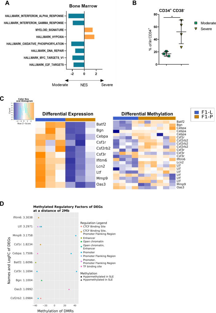
We have previously provided evidence for the dysregulation of haematopoiesis at the transcriptional level in SLE compared with control individuals, with skewing towards the myeloid lineage and a trained immunity-related signature of HSPCs.6 To further investigate the signature of BM-derived CD34+ cells from patients with SLE in correlation with disease severity, we analysed the transcriptomic data from patients with ‘severe versus moderate’ disease. We observed downregulation of the interferon (IFN)-related pathway and pathways associated with cell cycle and DNA repair, as well as oxidative phosphorylation in patients with severe disease, whereas the hypoxia pathway, linked to glycolysis,15 and the myeloid cell signatures were upregulated (figure 1A). The downregulation of oxidative phosphorylation (OXPHOS) and cell cycle-associated signatures, and the upregulation of hypoxia signature16 in cells from patients with severe disease imply a less differentiated phenotype. To confirm this, we performed flow cytometry analysis which showed that the percentage of the less differentiated CD38- HSPCs within the CD34+ population was increased in the peripheral blood of patients with high disease severity (figure 1B). These data propose that enhanced differentiation of CD38+ MyP in severe disease may lead to their decrease in the CD34+ progenitor pool, which is however associated with an upregulation of myeloid signature in the less differentiated CD38− cells. Taken together, these findings suggest that increased disease activity is positively associated with myeloid skewing in haematopoietic progenitors. In severe disease, the increased myelopoiesis may lead to an emergency HSPCs renewal resulting in a leakage of these ‘immature cells’ in the circulation of severe patients.
Figure 1.
Dysregulated BM haematopoiesis in SLE is marked by transcriptome reprogramming through DNA methylation. (A) Bar chart representing enriched GSEA pathways between BM-derived CD34+ cells from patients with SLE with ‘severe versus moderate disease’. IFNα response (NES=−2.65, FDR=0.0); IFNγ response (NES=−2.1, FDR=0.00); myeloid signature (NES=1.41, FDR=0.037); hypoxia (NES=1.7, FDR=0.002); oxidative phosphorylation (NES=−2.1, FDR=0.0003); DNA repair (NES=−1.5, FDR=0.02); Myc targets (NES=−1.93, FDR=0.0006); E2F targets (NES=−1.87, FDR=0.001). (B) Frequency of CD34+ CD38− cells gated in Lin−CD34+ from the peripheral blood of patients with SLE with severe and moderate disease (*p=0.0273). Lineage includes CD3/14/16/19/20/56. Data are represented as mean±SD. (C) Heatmap (left panel) of DEGs related to differentiation and heatmap (right panel) of DMRs that are found up to 2 Mb away between F1-L and F1-P. Orange/Blue gradient represents the row Z-score of upregulation/downregulation and hypermethylation/hypomethylation in F1-L compared with F1-P mice. (D) Dot plot where y-axis are the DEGs related to the differentiation with their logFC values that have the closest methylated RFs in a distance of 2 Mb. X-axis shows the methylation values of those RFs, and the shape and colour of each dot gives their methylation and annotation status, respectively. BM, bone marrow; DEG, differentially expressed genes; DMR, differentially methylated region; FC, fold change; FDR, false discovery rate; F1-L, lupus mice; F1-P, prediseased mice; GSEA, gene set enrichment analysis; IFN, interferon; NES, normalised enrichment score; RF, regulatory factor.
Changes in DNA methylation underlie dysregulated BM haematopoiesis in SLE
Previously, we have shown the deregulation of haematopoiesis at the transcriptional level in SLE, with skewing towards the myeloid lineage and a trained immunity-related signature of HSPCs.6 Since our data showed a pattern of trained immunity which to a large degree is underpinned by a change in the methylomics, we sought to further explore it in a murine model of SLE because of the sparsity of the human material. To further define the regulatory mechanisms, we compared the methylation profiles of BM-derived HSPCs between F1-P and F1-L mice, as epigenetic regulation is important for the regulation of HSPCs function and inflammation.10–12 Principal component analysis and clustering analysis using differentially methylated regions (DMRs) revealed that HSPCs derived from F1-L mice exhibit a distinct methylation profile than those from F1-P mice (online supplemental figure 1A) with 529 DMRs identified (|differential methylation level|>25% and multiple-testing corrected p<0.05). We observed 389 hypomethylated and 140 hypermethylated DMRs in HSPCs of F1-L compared with F1-P (online supplemental figure 1B). Target regions were enriched for promoters, introns, intergenic regions and CpG islands. Specifically, 57% of DMRs are CpG islands while 22.87% were promoters, 27.03% introns and 36.86% intergenic regions (online supplemental figure 1C—left panel). It was also observed that in F1-L methylation levels were lower compared with F1-P, as depicted for each annotated region, while the average methylation percentage is reported per group (online supplemental figure 1C—right panel).
Next, we sought to investigate the methylation status of specific genes related to HSPCs differentiation and myeloid skewing as they have been previously identified to be differentially expressed6 (figure 1A). In order to define the methylation profile of these genes, we studied the DMRs located in their 5’ and 3’ flanking regions (2 Mb distance).17 Specifically, the genes Blnk1, Rag1 and Rag2, which are lymphoid markers, were downregulated and their closest DMRs were hypermethylated in F1-L HSPCs compared with F1-P; in contrast, myeloid markers such as Csf1r and Cebpα were upregulated, and their closest DMRs were hypomethylated in F1-L HSPCs compared with F1-P (figure 1C). These genes consist of multiple differentially methylated regulatory elements such as transcription binding sites, promoter and CTCF (CCCTC-binding factor) (figure 1D). Taken together, these findings corroborate our previous transcriptomic data,6 which revealed skewing towards myeloid lineage in SLE and support the involvement of changes in DNA methylation and its regulation in the differentiation process.
Induction of inflammation enhances both central and splenic myelopoiesis and accelerates disease progression in SLE
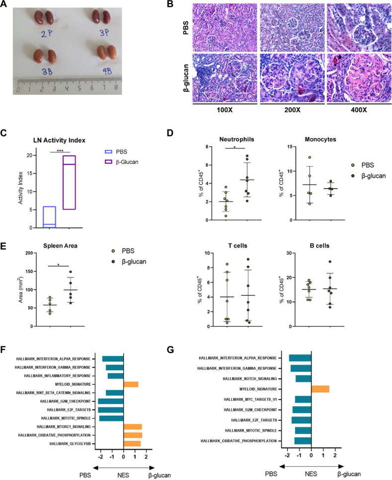
The upregulation of myeloid signature in CD34+ cells from patients with increased disease activity and the pronounced myeloid skewing of HSPCs observed in the BM of lupus-prone mice are reminiscent of the effects observed on induction of inflammatory trained immunity.6 18 To this end, we investigated the effect of β-glucan on F1-L mice. Progression of LN between the groups was assessed through kidney pathology findings using a standardised LN scoring system. β-Glucan administration resulted in increased severity of LN, as shown by the macroscopic appearance of the kidneys (figure 2A) and the histological findings (figure 2B,C), suggesting that induction of inflammation increased kidney disease severity, but demonstrated no significant difference in anti-dsDNA levels which were high in both groups (online supplemental figure 2A). Of note, we found no signs of nephritis in wild-type non-autoimmune-prone B6 mice treated for three consecutive months with the same dosing of β-glucan (data not shown).
Figure 2.
Induction of trained immunity enhances both central and splenic myelopoiesis and accelerates disease progression in SLE. (A) Macroscopic images of kidneys of F1-L mice treated with PBS (P) or β-glucan (B). (B) Microscopic images of kidney sections stained with H&E of PBS-treated or β-glucan-treated F1-L mice, showing in the latter case glomeruli with global endocapillary hyperplasia and global subendothelial deposits with wire loops and thrombi. Crescent in the right glomerulus (β-glucan, 200×). Glomeruli with global subendothelial deposits with wire loops and endocapillary thrombi (β-glucan, 400×). (C) LN activity index (see ‘Materials and methods’ section) in PBS-treated and β-glucan-treated F1-L mice (***p=0.003). (D) Frequencies of CD11b+ Ly6G+ neutrophils, CD11b+ Ly6C+ monocytes, CD3+ T and B220+ B cells in CD45+ cells in PBS-treated and β-glucan-treated F1-L mice (*p=0.0132). (E) Spleen area (mm2) of PBS-treated and β-glucan-treated F1-L mice (*p=0.0452). Data are represented as mean±SD. (F) Bar chart representing enriched pathways between BM-derived HSPCs of ‘β-glucan-treated versus PBS-treated’ F1-L mice. IFNα response (NES=−1.85, FDR=0.0); IFNγ response (NES=−1.54, FDR=0.017); inflammatory response (NES=−1.39, FDR=0.06); myeloid signature (NES=1.30, FDR=0.0); Wnt/- catenin signalling (NES=−1.53, FDR=0.015); G2M checkpoint (NES=−2.23, FDR=0.0); E2F targets (NES=−2.22, FDR=0.0); mitotic spindle (NES=−2.11, FDR=0.0); Mtorc1 signalling (NES=1.60, FDR=0.02); oxidative phosphorylation (NES=1.63, FDR=0.0); glycolysis (NES=1.49, FDR=0.041). (G) Bar chart representing enriched pathways between spleen-derived HSPCs of ‘β-glucan-treated versus PBS-treated’ F1-L mice. IFNα response (NES=−1.87, FDR=0.0); IFNγ response (NES=−1.74, FDR=0.0); Notch signalling (NES=−1.32, FDR=0.17); myeloid signature (NES=1.44, FDR=0.0); Myc targets (NES=−1.36, FDR=0.15); G2M checkpoint (NES=−1.56, FDR=0.02); E2F targets (NES=−1.65, FDR=0.0017); mitotic spindle (NES=−1.36, FDR=0.13); oxidative phosphorylation (NES=−1.42, FDR=0.124). BM, bone marrow; FDR, false discovery rate; F1-L, lupus mice; IFN, interferon; NES, normalised enrichment score; PBS, phosphate-buffered saline.
To investigate the relative contribution of various immune cells to the increased severity of LN observed in F1-L mice, we performed flow cytometry of the mononuclear cell fraction isolated from the kidneys. Notably, β-glucan administration resulted in an increased frequency of neutrophils (CD11b+Ly-6CintLy-6Ghi) in the kidneys, with no significant changes in monocytes (CD11b+Ly-6ChiLy-6G−), T (CD11b-CD3-e+) and B (CD11b−B220+) cells (figure 2D, online supplemental figure 2B), suggesting that enhanced neutrophilic inflammation might mediate kidney damage. Furthermore, the spleen of β-glucan-treated mice was significantly enlarged, a macroscopic finding consistent with enhanced inflammation and/or EMH (figure 2E). Despite that fact, there was no difference in the HSPC frequency in the BM and spleen (online supplemental figure 2C,F); Granulocyte-monocyte progenitors (GMPs) were decreased in both tissues of β-glucan-treated F1-L mice (online supplemental figure 2D,G), accompanied by an increase in neutrophil frequency in the BM (online supplemental figure 2E,H). Overall, our findings support that β-glucan treatment exaggerates LN and enhances myelopoiesis in the BM and spleen of F1-L mice, providing pro-inflammatory myeloid cells that potentially sustain and amplify inflammation in the periphery.
To study the molecular alterations that take place in HSPCs from F1-L mice during induction of inflammatory training, we performed RNA sequencing of BM-derived and spleen-derived HSPCs of F1-L mice, treated with either β-glucan or PBS. In the BM cells, we identified 699 differentially expressed genes (DEGs) (|fold change (FC)|≥1.5, p<0.05) in the β-glucan group compared with the control group (online supplemental figure 2I). Gene set enrichment analysis (GSEA) demonstrated that OXPHOS and glycolysis pathways were over-represented among the upregulated genes (figure 2F), suggesting a marked effect of trained immunity in the upregulation of metabolic genes.15 19 Similarly, we identified 813 DEGs in spleen-derived HSPCs (online supplemental figure 2J). In line with the aforementioned effects in severe disease, we observed downregulation of the IFN pathway in both tissues and upregulation of myeloid signature (figure 2F,G), which are concordant with the findings in humans. Together, these data suggest that both human and murine SLE HSPCs enhanced disease activity is positively associated with myeloid—and negatively associated with IFN—signatures in haematopoietic progenitors.
Splenic EMH activity strongly correlates with lupus nephritis severity
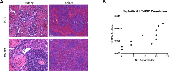
Since we observed splenic enlargement associated with increased disease activity in lupus-prone mice on induction of trained immunity, we sought to investigate EMH in SLE. LN is a major complication of SLE and is invariably present in the NZBW/F1 mouse model.20 Mice with severe nephritis presented histologically high activity scores and extensive splenic EMH, as evidenced by the significant red pulp expansion and overall disorganised splenic architecture with blurring of the red/white pulp demarcation (figure 3A). Spearman’s correlation coefficient analysis was performed in order to assess whether the extent of EMH correlates with the severity of LN. To this end, the frequency of spleen-derived long-term haematopoietic stem cell (LT-HSC), used as a surrogate of splenic EMH extent,21 was found to strongly correlate with LN activity index (R=0.9264, 95% CI 0.7252 to 0.9818; figure 3B). Taken together, these results show a strong correlation between the degree of splenic EMH with the severity of LN, which implies that splenic EMH may actively contribute to the peripheral immune cell pool by producing pro-inflammatory cells.
Figure 3.
Splenic extramedullary haematopoiesis activity strongly correlates with LN severity. (A) Representative kidney (200×) and corresponding spleen (40×) histological sections of mice with mild (NIH activity <5) and severe nephritis (NIH activity >15) (see ‘Materials and methods’ section) stained with H&E. Mice with mild nephritis had preserved splenic architecture (top right picture) while mice with severe nephritis had disorganised splenic architecture with poor demarcation of the red/white pulp and increased presence of megakaryocytes. (B) Correlation between frequency of spleen-derived LT-HSC (Lin-CD48−CD150+Sca-1+c-Kit+) and LN NIH activity index (Spearman’s correlation R=0.9264, 95% CI 0.7252 to 0.9818, p=0.0001; n=11). LN, lupus nephritis; LT-HSC, long-term haematopoietic stem cell; NIH, National Institutes of Health.
HSPCs expand in the spleen of F1-L mice displaying an inflammatory phenotype likely due to reduced BM retention via Cxcr4
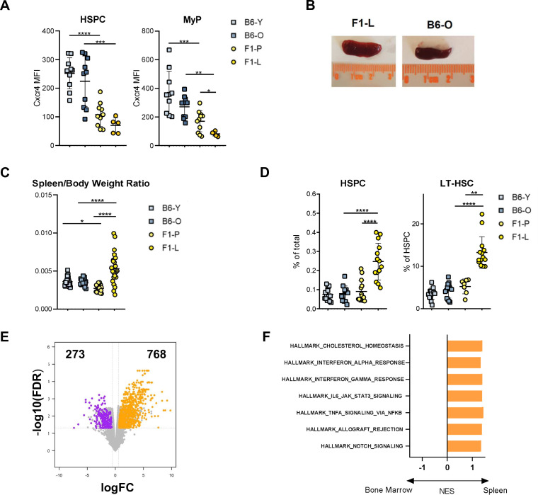
Studies in humans and mice have shown a high frequency of patrolling HSPCs in SLE with a potential to infiltrate into extramedullary sites, such as the spleen and kidneys, and contribute to peripheral tissue injury.6 22 To investigate the role of EMH in SLE, we initially studied the expression levels of CXCR4, a critical receptor mediating the retention of HSPCs in the BM using flow cytometry.23 24 Cxcr4 levels were downregulated in BM-derived HSPCs and MyP from F1-L mice compared with their aged-matched controls (figure 4A). C57BL/6 mice are justified as controls for experiments due to their stable genetic background,25 robust immune competence26 and translational relevance. This approach facilitates the identification of disease mechanisms and therapeutic interventions while minimising variability and enhancing the validity of experimental studies. Previous reports have indicated the presence of splenomegaly27 and splenic EMH in SLE.5 28 29 Thus, we hypothesised that the mobilised HSPCs could accumulate in the spleen of lupus-prone mice and establish hubs of haematopoiesis and differentiation. Splenomegaly was a prominent feature of F1-L mice (figure 4B), as evidenced by spleen/body weight ratio measurement (figure 4C). Furthermore, we demonstrated increased frequency of HSPCs in the spleen of F1-L mice using flow cytometry (figure 4D, online supplemental figure 3A). Among HSPC subpopulations, the percentage of CD48−CD150+ LT-HSCs was significantly increased in the spleen of F1-L mice (figure 4D). Taken together, these data indicate the presence of EMH in the spleen of F1-L mice.
Figure 4.
HSPCs expand in the spleen of F1-L mice displaying an inflammatory phenotype likely due to reduced BM retention via Cxcr4. (A) MFI of CXCR4 in BM-derived HSPC (Lin−Sca-1+c-Kit+, ****p<0.0001; ***p=0.0004) and MyP (Lin−Sca-1+c-Kit−, ***p=0.004; **p=0.0077; *p=0.0211) of F1-P, F1-L and their age-matched C57BL/6 mice (n=5–10). (B) Spleen size of F1-L and B6-O mice. (C) Spleen/Body weight ratio across the groups (****p<0.0001; *p=0.0333; n=20–27). (D) Frequencies of spleen-derived HSPC (Lin−Sca-1+c-Kit+, ****p<0.0001) and LT-HSC (Lin−CD48−CD150+Sca-1+c-Kit+, ****p<0.0001; **p=0.0035) from all conditions (n=7–14). (E) Volcano plot of DEGs between spleen-derived and BM-derived (Lin−Sca-1+c-Kit+) HSPCs of F1-L mice. Significantly upregulated and downregulated genes are coloured orange and purple, respectively. Grey points indicate genes with no significant difference in expression. (F) Bar chart representing enriched GSEA pathways between spleen-derived and BM-derived HSPCs of F1-L mice. Cholesterol homeostasis (NES=1.4, FDR=0.02), IFNα response (NES=1.32, FDR=0.1), IFNγ response (NES=1.4, FDR=0.03), IL6/JAK/STAT3 signalling (NES=1.38, FDR=0.03), TNF-α-NF-κβ signalling (NES=1.44, FDR=0.007), allograft rejection (NES=1.38, FDR=0.03), Notch signalling (NES=1.34, FDR=0.07). Data are represented as mean±SD. BM, bone marrow; DEG, differentially expressed genes; FC, fold change; FDR, false discovery rate; F1-L, lupus mice; F1-P, prediseased mice; GSEA, gene set enrichment analysis; HSPC, haematopoietic stem and progenitor cell; IFN, interferon; MFI, mean fluorescence intensity; NES, normalised enrichment score; NF-κβ, nuclear factor kappa B; TNF, tumour necrosis factor.
To shed light on the molecular alterations of HSPCs in SLE, we conducted a transcriptome analysis of BM-derived and spleen-derived HSPCs from F1-L mice. Differential expression analysis identified 1041 DEGs (|FC|≥1.5, false discovery rate (FDR) <0.05), of which 768 were upregulated and 273 were downregulated in the spleen-derived HSPCs compared with the BM-derived HSPCs of F1-L mice (figure 4E). To obtain insights into the biological properties of HSPCs between the two haematopoietic tissues, GSEA was performed. GSEA indicated positive enrichment in spleen-derived HSPCs for several biological pathways related to inflammation and the immune system, such as the IFNα (NES=1.32, FDR=0.1) and IFNγ responses (NES=1.4, FDR=0.03), IL6-JAK-STAT3 cascade (NES=1.38, FDR=0.03) and tumour necrosis factor-α signalling (NES=1.44, FDR=0.007), as well as the cholesterol homeostasis pathway (NES=1.4, FDR=0.02) (figure 4F). These findings demonstrate that HSPCs in the spleen of F1-L mice possess a prominent inflammatory signature.
Splenic extramedullary haematopoiesis is skewed towards granulopoiesis in SLE at the preclinical stage
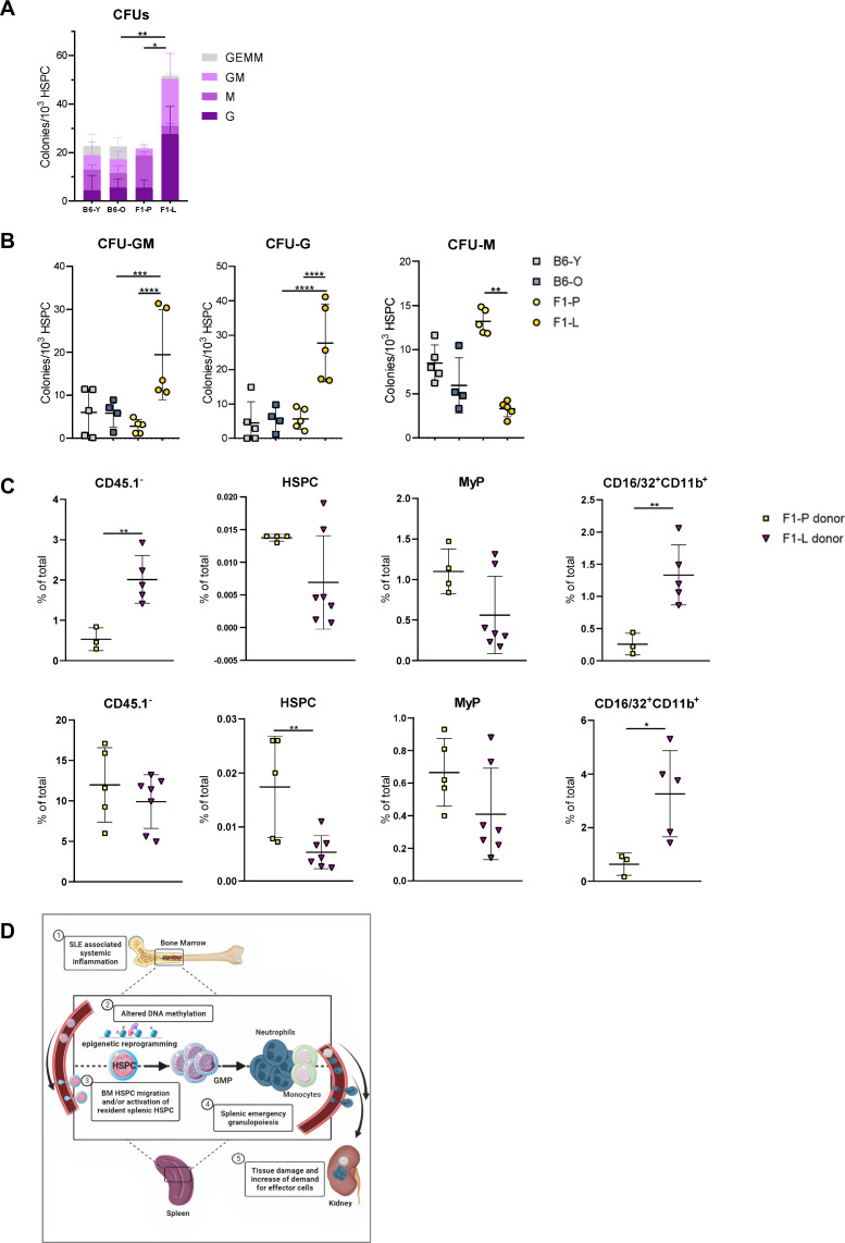
We then investigated whether the SLE-mediated chronic inflammatory state drives lineage bias of HSPCs, resulting in enhanced myelopoiesis. To this end, we performed CFU assay to assess the differentiation capacity of splenic HSPCs and their potential to contribute to myeloid cell accumulation in inflamed tissue. Notably, the number of total colonies per 103 HSPCs was increased in both the BM and spleen of F1-L (figure 5A, online supplemental figure 3B). This differentiation bias towards myelopoiesis was further supported by the increased number of CFU-GM and of CFU-G derived from the BM and the spleen of F1-L compared with F1-P and their age-matched counterparts (figure 5B, online supplemental figure 3B,C). As expected, BM-derived HSPCs generate a higher total number of colonies, compared with their splenic counterparts (online supplemental figure 3B). To further support these observations, we adoptively transferred HSPCs isolated from F1-P and F1-L (CD45.2) mice into NBSGW (CD45.1) recipient mice. HSPCs from both donors displayed the capacity to migrate in the BM and spleen and differentiate after 16 hours, as shown by flow cytometry (figure 5C). The significantly higher frequency of F1-L donor-derived CD45.1−CD16/32+CD11b+ myeloid cells in the spleen of the recipients suggests the differentiation of HSPCs into mature myeloid cells (figure 5C, online supplemental figure 4). Our findings support the high clonogenic capacity of spleen-derived HSPCs from F1-L mice, and their skewing towards granulopoiesis.
Figure 5.
Splenic extramedullary haematopoiesis is skewed towards granulopoiesis in SLE at the preclinical stage. (A) Total colonies per 103 spleen-derived HSPC (**p=0.0088; *p=0.0149). (B) CFU-G, GFU-GM and CFU-M numbers per 103 BM-derived HSPC (CFU-G: ****p<0.0001; CFU-GM: ****p<0.0001; **p=0.0002; CFU-M: **p=0.0052; n=5–6). (C) Frequencies of donor-derived (F1-P or F1-L) total cells (CD45.1-), Lin−Sca-1+c-Kit+ HSPCs, Lin−Sca-1+c-Kit− MyPs and CD16/32+CD11b+ cells in the BM (upper panels; **p=0.0096) and spleen (lower panels; **p=0.0090; *p=0.0357) of NBSGW recipients. Data are represented as mean±SD. (D) Graphical abstract showing that myeloid skewing of haematopoiesis through transcriptome and epigenetic regulation leads to migration and activation of HSPCs, resulting in emergency granulopoiesis and tissue damage in SLE. BM, bone marrow; CFU, colony-forming unit; CFU-G, CFU-granulocyte; CFU-GM, CFU-granulocyte macrophage; CFU-M, CFU-macrophage; F1-L, lupus mice; F1-P, prediseased mice; HSPC, haematopoietic stem and progenitor cell.
Discussion
HSPCs can sense inflammatory stimuli and respond with enhanced proliferation and generation of inflammatory differentiated cells.30 Furthermore, increasing evidence suggests that aberrations occurring in progenitor cells, can alter the fate of the resulting haematopoietic cells, priming them towards an inflammatory phenotype that fuels inflammation.8 9 18 31–33 We have previously shown that in SLE BM haematopoiesis is skewed towards myelopoiesis.6 Here, we show that the myeloid priming of HSPCs in patients with SLE is further enhanced in patients with severe disease phenotype. We demonstrate that disease severity in human SLE is associated with a strong myelopoiesis signature in haematopoietic cells, which is in part regulated by DNA methylation. Further induction of sterile inflammation in SLE-susceptible mice accentuated myeloid skewing and accelerated disease progression, sharing common features with high disease activity in patients with SLE. We have also observed that the disease activity and progression correlates with the extent of EMH, indicating that splenic EMH is an important and rather overlooked feature of the disease. An interesting finding was that enhanced disease activity both in human and in lupus-prone mice was negatively associated with the HSC IFN signature. This could be attributed to enhanced mobilisation and migration to the spleen of active HSPCs that carry this signature, especially considering that splenic HSPCs are characterised by IFNα/IFNγ response signature.
Our findings are in line with the enhanced mobilisation of HSPCs observed during infection, inflammation, stress and injury which disrupts the CXCR4/CXCL12 axis in the BM niche, a vital interaction for BM retention, resulting in HSPCs mobilisation.23 24 HSPCs subsequently infiltrate peripheral tissues, fostering the local production of haematopoietic cells, a phenomenon termed EMH.18 34 35 It is increasingly recognised that cells derived from EMH foci can demonstrate distinct attributes from their BM counterparts, disproportionately contributing to inflammatory processes.8 9 31 32 Here, we demonstrate the downregulation of Cxcr4 in F1-L mice HSPCs and MyPs, underlining their propensity for mobilisation. Taking these findings together, we propose that mobilisation of BM HSPCs with subsequent EMH initiation are emerging key components in SLE pathogenesis that drive inflammation through the supply of primed myeloid pro-inflammatory cells.
Innate immune training through β-glucan administration has been shown to reprogramme HSPCs and transcend into terminally differentiated myeloid cells in wild-type mice.18 Given that haematopoiesis in SLE shares various features with trained immunity, we asked whether induction of trained immunity could affect the disease progression in SLE. Our study is consistent with a previous report,14 showing the effect of prolonged β-glucan treatment on LN exaggeration in F1-L mice and the absence of such effect on non-autoimmune C57BL/6 mice. However, given the impact of trained immunity on haematopoietic cells, we investigated the effect of β-glucan administration on medullary and extramedullary haematopoiesis, the mononuclear cell fraction of the kidney and the molecular characteristics of BM-derived and spleen-derived HSPCs in the SLE setting. Specifically, we observed enhanced myelopoiesis in the BM and spleen of β-glucan-treated mice and increased presence of neutrophils in their kidneys. Our results further demonstrated that the activation of innate immunity leads to the accentuation of haematopoietic aberrancies observed and ultimately leads to the exaggeration of LN. On the molecular level, β-glucan administration was associated with alterations in metabolic, cell cycle, IFNα and ΙFNγ response pathways in HSPCs derived from the BM and spleen of F1-L mice. In the BM, the upregulation of glycolysis on β-glucan administration is in line with previous studies.18 In HSPCs though, the activation of OXPHOS is associated with loss of self-renewal potential, and induction of DNA damage.36 37 This loss of self-renewal potential is likely reflected in the negative enrichment of G2M checkpoint, E2F targets and mitotic spindle, suggesting the exhaustion of these HSPCs. This is also observed in the splenic HSPCs from β-glucan-treated mice.
In SLE, deregulation of haematopoiesis is characterised by myeloid skewing and priming of pro-inflammatory ‘immune-trained’ HSPCs resulting in enhanced granulopoiesis mediated by alterations in progenitor cells.18 38 Although a strong granulocytic signature in the peripheral blood and increased rates of NETosis have been reported in LN, very few intact neutrophils are present in kidney biopsies. Tissue factor-bearing and interleukin-17A (IL-17A)-bearing NETs are abundant in discoid skin lesions and in the glomerular and tubulointerstitial compartments of proliferative LN specimens. Active NETs represent scaffolds with high concentrations of bioactive IL-17A and tissue factor (TF) that remain in end-organ tissues even in the absence of intact neutrophils, activating resident cells and promoting thromboinflammation and fibrosis.39 We show here that patients with severe SLE such as nephritis have a strong myeloid signature indicative of activation of neutrophils, emergency myelopoiesis and EMH, which may sustain and amplify the inflammatory response and contribute to the risk of flare. These primed HSPCs promote granulopoiesis and support EMH in the spleen. In conclusion, based on our findings and prior studies, we propose a ‘feed-forward inflammatory loop’ model (figure 5D), according to which an initial triggering event sets in motion a vicious circle of inflammation and organ damage.30 In this model, HSPCs in the BM receive inflammatory cues and get activated, either through sensing pathogen-associated molecular patterns or indirectly via pro-inflammatory cytokines production.40 In this sequence of events, the spleen in turn acts as a hub that potentiates HSPCs and produces inflammation-primed myeloid cells that mediate target organ damage. The overall inflammatory environment perpetuates the loop by reprogramming trained immunity-related molecular pathways.
Acknowledgments
We thank Anastasia Apostolidou from the Flow Cytometry Facility, BRFAA for cell sorting; Dr Ioannis Vatsellas from the Greek Genome Center, BRFAA for the next-generation sequencing service; Pavlos Alexakos from the Laboratory Animal Facility for providing C57BL/6 mice.
Footnotes
Twitter: @abanos82
EZ, MG and SAD contributed equally.
AB, IM and DTB contributed equally.
Contributors: EZ, MG and SAD designed the study, performed experiments, analysed data, generated figures and wrote the manuscript. DY and PP performed methylation analysis. GG and JW performed the RRBS experiment. AF assisted with bioinformatic analysis of RNA-seq data. HG performed the histological evaluation of kidney sections. AB and DTB performed clinical evaluation of individuals with SLE, provided human samples, assisted in designing experiments and critically edited the manuscript. IM assisted in designing experiments, supervised the study and critically edited the manuscript. MG is responsible for the overall content as guarantor. All authors critically revised and gave final approval of the manuscript.
Funding: This work was supported by a research grant from the European Research Council under the European Union’s Horizon 2020 research and innovation programme (grant agreement number 742390). Computational time granted from the National Infrastructures for Research and Technology (GRNET) in the National HPC facility—ARIS—under project ID pr011006_taskp-BIOSLE and project ID pr007036_fat-SLEPHE. MG, AF and IM were supported by the Hellenic Foundation for Research and Innovation (HFRI) and the General Secretariat for Research and Technology (GSRT), under the HFRI 'Research Projects to Support Faculty Members & Researchers and Procure High-Value Research Equipment' grant (GA. 452) and General Secretariat for Research and Technology Management and Implementation Authority for Research, Technological Development and Innovation Actions (MIA-RTDI) (grant Τ2EDK-02288, MDS-TARGET).
Competing interests: None declared.
Patient and public involvement: Patients and/or the public were not involved in the design, or conduct, or reporting, or dissemination plans of this research.
Provenance and peer review: Not commissioned; externally peer reviewed.
Supplemental material: This content has been supplied by the author(s). It has not been vetted by BMJ Publishing Group Limited (BMJ) and may not have been peer-reviewed. Any opinions or recommendations discussed are solely those of the author(s) and are not endorsed by BMJ. BMJ disclaims all liability and responsibility arising from any reliance placed on the content. Where the content includes any translated material, BMJ does not warrant the accuracy and reliability of the translations (including but not limited to local regulations, clinical guidelines, terminology, drug names and drug dosages), and is not responsible for any error and/or omissions arising from translation and adaptation or otherwise.
Data availability statement
Data are available on reasonable request. Murine RNA sequencing (RNA-seq) and methylation data have been deposited to GEO under accession number (GSE218780). Human RNA-seq data have been deposited to the EGA database under study EGAS00001003679; dataset EGAD00001009744. Any additional information required to reanalyse the data reported in this paper is available from the corresponding authors on reasonable request.
Ethics statements
Patient consent for publication
Consent obtained directly from patient(s).
Ethics approval
This study was approved by Attikon University Hospital Ethical Committee (Athens, Greece, protocol 10|22-6-2017). All procedures in mice were in accordance with institutional guidelines and were reviewed and approved by the Greek Federal Veterinary Office (Athens, Greece, protocol 792312|041219). The study was performed in accordance with the ethical standards of the Declaration of Helsinki (1964) and its subsequent amendments. Informed consent was obtained from all patients prior to sample collection.
References
- 1. Furie R, Rovin BH, Houssiau F, et al. Two-year, randomized, controlled trial of belimumab in lupus nephritis. N Engl J Med 2020;383:1117–28. 10.1056/NEJMoa2001180 [DOI] [PubMed] [Google Scholar]
- 2. Morand EF, Furie R, Tanaka Y, et al. Trial of anifrolumab in active systemic lupus erythematosus. N Engl J Med 2020;382:211–21. 10.1056/NEJMoa1912196 [DOI] [PubMed] [Google Scholar]
- 3. Tsokos GC. Systemic lupus erythematosus. N Engl J Med 2011;365:2110–21. 10.1056/NEJMra1100359 [DOI] [PubMed] [Google Scholar]
- 4. King KY, Goodell MA. Inflammatory modulation of HSCs: viewing the HSC as a foundation for the immune response. Nat Rev Immunol 2011;11:685–92. 10.1038/nri3062 [DOI] [PMC free article] [PubMed] [Google Scholar]
- 5. Niu H, Fang G, Tang Y, et al. The function of hematopoietic stem cells is altered by both genetic and inflammatory factors in lupus mice. Blood 2013;121:1986–94. 10.1182/blood-2012-05-433755 [DOI] [PMC free article] [PubMed] [Google Scholar]
- 6. Grigoriou M, Banos A, Filia A, et al. Transcriptome reprogramming and myeloid skewing in haematopoietic stem and progenitor cells in systemic lupus erythematosus. Ann Rheum Dis 2020;79:242–53. 10.1136/annrheumdis-2019-215782 [DOI] [PMC free article] [PubMed] [Google Scholar]
- 7. Kim CH. Homeostatic and pathogenic extramedullary hematopoiesis. J Blood Med 2010;1:13–9. 10.2147/JBM.S7224 [DOI] [PMC free article] [PubMed] [Google Scholar]
- 8. Regan-Komito D, Swann JW, Demetriou P, et al. GM-CSF drives dysregulated hematopoietic stem cell activity and pathogenic extramedullary myelopoiesis in experimental spondyloarthritis. Nat Commun 2020;11:155. 10.1038/s41467-019-13853-4 [DOI] [PMC free article] [PubMed] [Google Scholar]
- 9. Griseri T, McKenzie BS, Schiering C, et al. Dysregulated hematopoietic stem and progenitor cell activity promotes interleukin-23-driven chronic intestinal inflammation. Immunity 2012;37:1116–29. 10.1016/j.immuni.2012.08.025 [DOI] [PMC free article] [PubMed] [Google Scholar]
- 10. Vlachou K, Mintzas K, Glymenaki M, et al. Elimination of granulocytic myeloid-derived suppressor cells in lupus-prone mice linked to reactive oxygen species-dependent extracellular trap formation. Arthritis Rheumatol 2016;68:449–61. 10.1002/art.39441 [DOI] [PubMed] [Google Scholar]
- 11. Hochberg MC. Updating the American College of Rheumatology revised criteria for the classification of systemic lupus erythematosus. Arthritis Rheum 1997;40:1725. 10.1002/art.1780400928 [DOI] [PubMed] [Google Scholar]
- 12. Gergianaki I, Fanouriakis A, Adamichou C, et al. Is systemic lupus erythematosus different in urban versus rural living environment? Data from the Cretan lupus epidemiology and surveillance registry. Lupus 2019;28:104–13. 10.1177/0961203318816820 [DOI] [PubMed] [Google Scholar]
- 13. Schneider CA, Rasband WS, Eliceiri KW. NIH image to Imagej: 25 years of image analysis. Nat Methods 2012;9:671–5. 10.1038/nmeth.2089 [DOI] [PMC free article] [PubMed] [Google Scholar]
- 14. Fagone P, Mangano K, Mammana S, et al. Acceleration of SLE-like syndrome development in NZBxNZW F1 mice by beta-glucan. Lupus 2014;23:407–11. 10.1177/0961203314522333 [DOI] [PubMed] [Google Scholar]
- 15. Cheng S-C, Quintin J, Cramer RA, et al. mTOR- and HIF-1Α-mediated aerobic glycolysis as metabolic basis for trained immunity. Science 2014;345:1250684. 10.1126/science.1250684 [DOI] [PMC free article] [PubMed] [Google Scholar]
- 16. Suda T, Takubo K, Semenza GL. Metabolic regulation of hematopoietic stem cells in the hypoxic niche. Cell Stem Cell 2011;9:298–310. 10.1016/j.stem.2011.09.010 [DOI] [PubMed] [Google Scholar]
- 17. van Heyningen V, Bickmore W. Regulation from a distance: long-range control of gene expression in development and disease. Philos Trans R Soc Lond B Biol Sci 2013;368:20120372. 10.1098/rstb.2012.0372 [DOI] [PMC free article] [PubMed] [Google Scholar]
- 18. Mitroulis I, Ruppova K, Wang B, et al. Modulation of myelopoiesis progenitors is an integral component of trained immunity. Cell 2018;172:147–61. 10.1016/j.cell.2017.11.034 [DOI] [PMC free article] [PubMed] [Google Scholar]
- 19. Arts RJW, Carvalho A, La Rocca C, et al. Immunometabolic pathways in BCG-induced trained immunity. Cell Rep 2016;17:2562–71. 10.1016/j.celrep.2016.11.011 [DOI] [PMC free article] [PubMed] [Google Scholar]
- 20. Wang Y, Hu Q, Madri JA, et al. Amelioration of lupus-like autoimmune disease in NZB/Wf1 mice after treatment with a blocking monoclonal antibody specific for complement component C5. Proc Natl Acad Sci U S A 1996;93:8563–8. 10.1073/pnas.93.16.8563 [DOI] [PMC free article] [PubMed] [Google Scholar]
- 21. Morita Y, Iseki A, Okamura S, et al. Functional characterization of hematopoietic stem cells in the spleen. Exp Hematol 2011;39:351–359. 10.1016/j.exphem.2010.12.008 [DOI] [PubMed] [Google Scholar]
- 22. Kokkinopoulos I, Banos A, Grigoriou M, et al. Patrolling human SLE haematopoietic progenitors demonstrate enhanced extramedullary colonisation; implications for peripheral tissue injury. Sci Rep 2021;11:15759. 10.1038/s41598-021-95224-y [DOI] [PMC free article] [PubMed] [Google Scholar]
- 23. Nie Y, Han Y-C, Zou Y-R. CXCR4 is required for the quiescence of primitive hematopoietic cells. J Exp Med 2008;205:777–83. 10.1084/jem.20072513 [DOI] [PMC free article] [PubMed] [Google Scholar]
- 24. Sugiyama T, Kohara H, Noda M, et al. Maintenance of the hematopoietic stem cell pool by CXCL12-CXCR4 chemokine signaling in bone marrow stromal cell niches. Immunity 2006;25:977–88. 10.1016/j.immuni.2006.10.016 [DOI] [PubMed] [Google Scholar]
- 25. Bryant CD. The blessings and curses of C57Bl/6 Substrains in mouse genetic studies. Ann N Y Acad Sci 2011;1245:31–3. 10.1111/j.1749-6632.2011.06325.x [DOI] [PMC free article] [PubMed] [Google Scholar]
- 26. Song HK, Hwang DY. Use of C57Bl/6N mice on the variety of immunological researches. Lab Anim Res 2017;33:119–23. 10.5625/lar.2017.33.2.119 [DOI] [PMC free article] [PubMed] [Google Scholar]
- 27. Harris AA, Kamishima T, Horita T, et al. Splenic volume in systemic lupus erythematosus. Lupus 2009;18:1119–20. 10.1177/0961203309104430 [DOI] [PubMed] [Google Scholar]
- 28. Schneider E, Moreau G, Arnould A, et al. Increased fetal and extramedullary hematopoiesis in Fas-deficient C57Bl/6-Lpr/Lpr mice. Blood 1999;94:2613–21. [PubMed] [Google Scholar]
- 29. Weindel CG, Richey LJ, Bolland S, et al. B cell autophagy mediates TLR7-dependent autoimmunity and inflammation. Autophagy 2015;11:1010–24. 10.1080/15548627.2015.1052206 [DOI] [PMC free article] [PubMed] [Google Scholar]
- 30. Chavakis T, Mitroulis I, Hajishengallis G. Hematopoietic progenitor cells as integrative hubs for adaptation to and fine-tuning of inflammation. Nat Immunol 2019;20:802–11. 10.1038/s41590-019-0402-5 [DOI] [PMC free article] [PubMed] [Google Scholar]
- 31. Wu C, Ning H, Liu M, et al. Spleen mediates a distinct hematopoietic progenitor response supporting tumor-promoting myelopoiesis. J Clin Invest 2018;128:3425–38. 10.1172/JCI97973 [DOI] [PMC free article] [PubMed] [Google Scholar]
- 32. Barrett TJ, Distel E, Murphy AJ, et al. Apolipoprotein AI) promotes atherosclerosis regression in diabetic mice by suppressing myelopoiesis and plaque inflammation. Circulation 2019;140:1170–84. 10.1161/CIRCULATIONAHA.119.039476 [DOI] [PMC free article] [PubMed] [Google Scholar]
- 33. Xu Y, Murphy AJ, Fleetwood AJ. Hematopoietic progenitors and the bone marrow niche shape the inflammatory response and contribute to chronic disease. IJMS 2022;23:2234. 10.3390/ijms23042234 [DOI] [PMC free article] [PubMed] [Google Scholar]
- 34. Granick JL, Simon SI, Borjesson DL. Hematopoietic stem and progenitor cells as effectors in innate immunity. Bone Marrow Res 2012;2012:165107. 10.1155/2012/165107 [DOI] [PMC free article] [PubMed] [Google Scholar]
- 35. Massberg S, Schaerli P, Knezevic-Maramica I, et al. Immunosurveillance by hematopoietic progenitor cells trafficking through blood, lymph, and peripheral tissues. Cell 2007;131:994–1008. 10.1016/j.cell.2007.09.047 [DOI] [PMC free article] [PubMed] [Google Scholar]
- 36. Takizawa H, Fritsch K, Kovtonyuk LV, et al. Pathogen-induced TLR4-TRIF innate immune signaling in hematopoietic stem cells promotes proliferation but reduces competitive fitness. Cell Stem Cell 2020;27:177. 10.1016/j.stem.2020.06.010 [DOI] [PubMed] [Google Scholar]
- 37. Takubo K, Nagamatsu G, Kobayashi CI, et al. Regulation of glycolysis by Pdk functions as a metabolic checkpoint for cell cycle quiescence in hematopoietic stem cells. Cell Stem Cell 2013;12:49–61. 10.1016/j.stem.2012.10.011 [DOI] [PMC free article] [PubMed] [Google Scholar]
- 38. de Laval B, Maurizio J, Kandalla PK, et al. C/EBPβ-dependent epigenetic memory induces trained immunity in hematopoietic stem cells. Cell Stem Cell 2023;30:112. 10.1016/j.stem.2022.12.005 [DOI] [PubMed] [Google Scholar]
- 39. Frangou E, Chrysanthopoulou A, Mitsios A, et al. Redd1/Autophagy pathway promotes thromboinflammation and fibrosis in human systemic lupus erythematosus (SLE) through nets decorated with tissue factor (TF) and Interleukin-17A (IL-17A). Ann Rheum Dis 2019;78:238–48. 10.1136/annrheumdis-2018-213181 [DOI] [PMC free article] [PubMed] [Google Scholar]
- 40. Demel UM, Lutz R, Sujer S, et al. A complex proinflammatory cascade mediates the activation of HSCs upon LPS exposure in vivo. Blood Adv 2022;6:3513–28. 10.1182/bloodadvances.2021006088 [DOI] [PMC free article] [PubMed] [Google Scholar]
Associated Data
This section collects any data citations, data availability statements, or supplementary materials included in this article.
Supplementary Materials
lupus-2023-001110supp001.pdf (718.4KB, pdf)
Data Availability Statement
Data are available on reasonable request. Murine RNA sequencing (RNA-seq) and methylation data have been deposited to GEO under accession number (GSE218780). Human RNA-seq data have been deposited to the EGA database under study EGAS00001003679; dataset EGAD00001009744. Any additional information required to reanalyse the data reported in this paper is available from the corresponding authors on reasonable request.


