Abstract
Objective
This study evaluated the root and canal morphology in permanent mandibular incisors teeth using cone-beam computer tomography imaging in a Spanish subpopulation, and compared these findings with ipsilateral (similarity) and contralateral (symmetry) incisors. In addition, the position of canal splitting was measured.
Methods
A total of 229 datasets comprising four mandibular teeth each (n=916 incisors) were analysed using Vertucci and Ahmed et al. classifications, and, the similarity and symmetry were calculated. The distance from the cemento-enamel junction (CEJ), and the most coronal canal divergence was measured (if present). The role of sex was also assessed. The Cochran Q Test, LOGIS PROC in SUDAAN, Chi-square, and Kappa were used for the different comparisons. A p-value of less than 0.05 was considered significant.
Results
All incisors were single-rooted and no significant differences regarding root canal morphology were found according to the sex of the subjects included in the database. The most common morphology was Vertucci type I/Ahmed et al. 1MI1(65.3% for central and 66.8% for lateral incisors respectively), followed by type III/1MI1–2–1 (31% for central and 30.6% for lateral incisors). 1.8% of the samples were considered as non-classifiable with Vertucci but were classified with codes using the Ahmed et al. system. Similarity values were 74.7% for the left side, and 74.2% for the right side, whereas symmetry values were 90% for central and 84.3% for lateral incisors. In the presence of divergences, the main (SD) distances from the CEJ were for type II/1MI1–2–1 3.8±0.8 (centrals) 4.0±0.7 mm (laterals); for type V/1MI1–2 this value ranged between 6.0±1.8 and 5.5±1.5 mm, whereas values for 1MI1–2–3–2–1 were 1.8 and 2.1 mm. No significant differences were found when the position of the most coronal divergence was compared between lateral and central incisors for the different morphologies.
Conclusion
A high prevalence of Vertucci I/Ahmed et al. 1MI1 configuration was present in mandibular incisors from Spanish individuals. Similarity and symmetry were common, particularly for central incisors. The position of the coronal splitting of the canals varied according to the root canal morphology.
Keywords: Ahmed, anatomy, cone-beam computed tomography, ethnicity, incisors, root canal, Vertucci
HIGHLIGHTS
One-third of mandibular incisors presented with morphologies different from Vertucci I/ Ahmed et al.' 1MI1.
The root canal morphology of seventeen (1.8% of the sample) mandibular incisors within the Spanish subpopulation studied was unclassifiable using Vertucci's system, but were classified using Ahmed et al. classification.
Ipsilateral and contralateral incisors may help to predict the morphology of the other mandibular incisors
The position of the most coronal root canal splitting in regards to the CEJ varies according to the morphology.
INTRODUCTION
Recent systematic reviews have reiterated the significant variability in root canal morphology globally when cone-beam computed tomography (CBCT) is used for assessment, including in mandibular incisors (1, 2). Studies based on clinical datasets demonstrated the differences in the number of canals and their configuration associated with the geographic location and ethnic backgrounds. The prevalence of two canals in mandibular incisors ranges between 2.7% and 45% (3–7).
An a priori understanding of root canal morphology, including the number of canals, is crucial for endodontic treatment, since missed canals are usually associated with persisting periapical lesions (8), and the need for additional treatments (9). Although available evidence suggests that the morphology of mandibular incisors is highly variable (3–7), endodontic case assessment tools consider these teeth to be of low difficulty, except in the presence of additional factors that would increase the difficulty level (10). Interestingly, one endodontic case assessment tool factors in the number of canals expected to be present in the tooth being considered for treatment, which may not be predictable based on two-dimensional imaging (11). It is worth noting that root canal morphology is of interest in subjects outside clinical dentistry such as anthropology (12).
There is limited evidence assessing root canal morphology and possible associations of mandibular incisors using CBCT when compared with other tooth types from Europe (1, 2). In addition, to the best of knowledge, there are no studies applying the system to classify root and root canal morphology proposed by Ahmed et al. (13) in this continent for this specific tooth type. Furthermore, mandibular incisors have not been previously assessed in the Spanish population. Though the Vertucci classification (14) has been used widely, the Ahmed et al. (13) classification has been purported to describe the root and canal morphology more accurately and practically (15), as allows to characterize all canal configurations and tooth anomalies (16). Similarly, the level of canal splitting has attracted attention due to its outstanding clinical relevance as it would support canal location when required (15, 17).
Therefore, the aim of the present study was three-fold; first, to assess the root canal morphology of mandibular incisors using Vertucci (14) and Ahmed et al. (13) classification systems (condition) based on available CBCT datasets in a Spanish subpopulation (context and subpopulation); second, to compare the morphology of central versus ipsilateral lateral incisors (similarity), and to compare central and lateral incisors versus their contralateral tooth (symmetry); and third, to investigate the distance between the cemento-enamel junction (CEJ) and the most coronal canal splitting (if present).
MATERIALS AND METHODS
The study was carried out and the current document was prepared following the "Preferred Reported items for Cross-Sectional Studies on Root and Canal anatomy using CBCT technology" (1) and Preferred Reporting items for root and canal anatomy in the human dentition (PROUD 2000) (2) guidelines. The present research was conducted in full accordance with ethical principles, including the World Medical Association Declaration of Helsinki (version 2013) and Ethical approval was granted by the University of Salamanca (20.10.2020-508).
Sample Size Calculation
Sample size calculation indicated that datasets from 103 patients were required to compare the morphology of central versus lateral incisors plus the mandibular quadrants with 80% power and 5% level of significance, and a 15% difference in prevalence in the outcomes in question. Nonetheless, the final minimum sample size was increased to 154 (103×1.5) to account for the sample set including pairs of teeth from the same subject.
Participants
For the selection process, a convenience sample of CBCT datasets from 285 subjects (165 female and 120 male) was included. The datasets were originally acquired in private practice in Salamanca (Spain) for reasons not related to the present study (e.g. implant surgery, orthodontic treatment, impacted teeth) from January 2020 to December 2021.
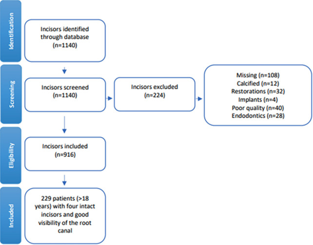
Inclusion criteria were as follows: subject age above 18 years; teeth with mature roots and with the outline of the root canals(s) visible on the datasets. Exclusion criteria were as follows: subjects with missing mandibular incisor(s); teeth with calcified canals, resorptive defects; developmental abnormalities, previous endodontic or restorative treatment and presence of implants in the anterior mandible; dataset not allowing complete visualization of all mandibular incisors. After applying all the inclusion and exclusion criteria, datasets from 229 patients were available, thus allowing an increased statistical power for the present study (Fig. 1).
Figure 1.
Flowchart of the cone-beam computed tomography dataset selection process
Image Acquisition and Evaluation
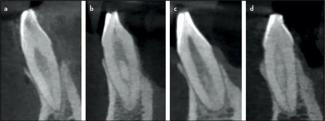
Datasets were acquired using a CS 8100 3D CBCT scanner (Carestream Dental, Rochester, NY, USA). The scanning setting was as follows: tube voltage 84 kV; tube current 4 mA; field of view of 8×9 cm; time of exposure 15 s; the voxel size was 150 µm. Datasets were reconstructed and visualized using the CS 3D imaging software. Axial, sagittal, and coronal planes were analysed. The number of roots, their morphology according to Vertucci classification (14), and supplemental configurations (Figs. 2, 3), were assessed according to the subject gender, plus similarity comparison "central versus lateral" and symmetry comparisons "left versus right" of the previous outcome measures were assessed. In parallel, root and canal morphologies were assessed based using the Ahmed et al. (13) system. In brief, this system encompasses the tooth number, the number of roots and their configuration, and, finally, the root canal configuration (13) (Fig. 2). Finally, the perpendicular distance between the CEJ and the first bifurcation was measured in roots with multiple canals, as previously described (17).
Figure 2.
Representative images of Vertucci's (14) and Ahmed et al. (13) morphology configurations, based on sagittal sections of CBCT datasets. A: type I/1MI1; B: type II/1MI1–2–1; C: type III/1MI1–2; D: Type VNC/1MI1–2–1–2–1
CBCT: Cone-beam computed tomography
Calibration and Assessment
Datasets were assessed by three experienced endodontists previously calibrated using 30 datasets, with interobserver agreement (kappa statistics) values ranging between 0.60 and 0.73 (depending on the assessors compared and the outcome measures), with a strength of agreement "moderate" or "substantial" (18). Two endodontists assessed the datasets contemporarily and any disagreement was solved with the help of the third endodontist until a decision was obtained by consensus.
Statistical Analysis
SPSS-Windows 21.0 (IBM Corp. Armonk, NY, USA) was used when the unit of analysis was the patient; SUDAAN 7.0 (RTI, RTP, NC, USA) was used when the unit was the tooth, to adjust for clustering. Confidence Interval Analysis (CIA) was used to calculate exact confidence intervals for percentages. Statistical tests and software are listed in the footnotes of the tables (results section). In all comparisons, a p-value of less than 0.05 was considered significant.
RESULTS
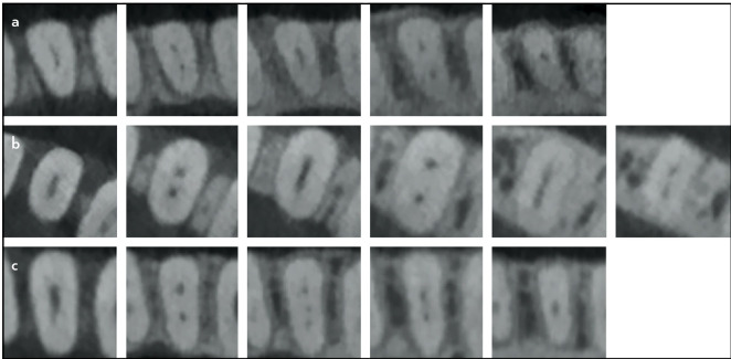
After the selection process, the sample size was 229 datasets (132 female and 97 male subjects), with a total of 916 incisors. All teeth were single-rooted and no significant differences were found based on sex (Table 1). The most common morphology was Vertucci type I/ Ahmed et al. 1MI1 (65.3% for central and 66.8% for lateral incisors respectively, followed by Vertucci type III/ Ahmed et al. 1MI1–2–1 (31% for central and 30.6% for lateral incisors), as well as configurations not listed originally by Vertucci were also evident (n=17; 1.8% of the total sample) (Table 1). Figures 2 and 3 illustrate various morphologies found in the datasets.
TABLE 1.
| Left side | Right side | Global comparison p |
|||||||
|---|---|---|---|---|---|---|---|---|---|
| Central | Lateral | Central | Lateral | ||||||
| n | % | n | % | n | % | n | % | ||
| Vertucci/Ahmed | |||||||||
| I/'MI1 | 150 | 65.5 | 1 55 | 67.7 | 1 49 | 65.1 | 1 51 | 65.9 | 0.733c |
| 111/1 Mi1-2-1 | 71 | 31 .0 | 67 | 29.3 | 71 | 31 .0 | 73 | 31.9 | |
| V/'MI1-2 | 3 | 1 .3 | 3 | 1.3 | 4 | 1 .7 | 2 | 0.9 | |
| VNC/1MI1-2-1-2-1 | 2 | 0.9 | 4 | 1.7 | 2 | 0.9 | 1 | 0.4 | |
| VNC/1MI1-2-1-2-1-2 | 2 | 0.9 | - | - | 2 | 0.9 | 2 | 0.9 | |
| VNC/1MI1-2-3-2-1 | 1 | 0.4 | - | - | 1 | 0.4 | - | - | |
: Chi-square corrected for clustering (4 teeth) after collapsing the last four categories (i.e., V, and so on). With CROSSTAB procedure in SUDAAN 7.0. A p-value of less than 0.05 was considered significant VNC: Vertucci non-classifiable
Figure 3.
Representative images of morphology configurations not classifiable based on Vertucci types. (a) 1MI1–2–1–2–1. (b) Type 1MI1–2–1–2–1–2. (c) 1MI1–2–3–2–1
When comparing the Vertucci type of central versus lateral incisors in the same quadrant (similarity), this coincided in 74.7% for the left side and 74.2% for the right side respectively, when all configurations were considered (Table 2). If single-canal roots are excluded from this analysis, the value is 65.8% for the left and 64.9% for the right sides.
TABLE 2.
Morphological similarity of mandibular incisor teeth included in the study using Vertucci (14) and Ahmed et al. (13) classification
| Root canal configurationsa Both incisors |
Mandibular left incisors | Mandibular right incisors | ||||||
|---|---|---|---|---|---|---|---|---|
| nb | % | (95%-CI)c | Kappa (95% CI)d |
n | % | (95%-CI) | Kappa (95% CI) |
|
| Configurations | 0.47 (0.36-0.59) | 0.45 (0.34-0.57) | ||||||
| Both type I ('MI1) | 127 | 55.5 | 0.50 (0.38-0.62) | 123 | 53.7 | 0.48 (0.36-0.60) | ||
| Both type III OMI1-2-1) | 43 | 18.8 | 0.46 (0.34-0.59) | 44 | 19.2 | 0.43 (0.31-0.56) | ||
| Both type V ('MI1-2) | 1 | 0.4 | NCe | 1 | 0.4 | NC | ||
| Both VNC ('MI1-2-1-2-1-2) | - | - | - | 2 | 0.9 | NC | ||
| I/III (1MI1/1MI1-2-1) | 47 | 20.5 | - | 51 | 22.3 | - | ||
| I/V (1MI1/1MI1-2) | 3 | 1.3 | - | 3 | 1.3 | - | ||
| I/VNC (1MI1/1MI1-2-1-2-1) | 1 | 0.4 | - | - | - | - | ||
| III/V (1MI1-2-1/1MI1-2) | 1 | 0.4 | - | 1 | 0.4 | - | ||
| III/VNC (1MI1-2-1/1MI1-2-1-2-1) | 3 | 1.3 | - | 3 | 1.3 | - | ||
| III/VNC (1MI1-2-1/1MI1-2-3-2-1) | 1 | 0.4 | - | 1 | 0.4 | - | ||
| VNC/VNC (1MI1-2-1-2-1/1MI1-2-1-2-1-2) | 2 | 0.9 | - | - | - | - | ||
| Same configuration | 171 | 74.7 | (69.0-80.3) | 170 | 74.2 | (68.6-79.9) | ||
| Different configurations | 58 | 25.3 | (19.7-31.0) | 59 | 25.8 | (20.1-31.4) | ||
: "Both" means a patient with both compared incisors having the same configuration; " / " is used to separate incisors with different configurations, b: Incisors pair, c: 95%-Confidence Interval, calculated with CIA v.1.0 statistical program, d: Kappa and 95% Confidence-Interval, calculated with SPSS-Windows 15.0, both global for each pair of compared incisors and some categories. Due to the limited sample size for some categories, categories for kappa calculation were collapsed, e: NC: Kappa non-calculable due to insufficient sample size. CI: Convidence interval
When comparing the same teeth types in different quadrants (symmetry), the morphology coincided for 90% of the central incisors and 84.3% of the lateral incisors (Table 3). If Vertucci type I/ Ahmed et al. 1MI1 roots are excluded from this analysis, the value is 87.9% for the central incisors and 78.9% for lateral incisors.
TABLE 3.
Morphological symmetry of mandibular incisor teeth included in the study using Vertucci (14) and Ahmed et al. (13) classification
| Root canal configurationsa Side 1/Side 2 |
Central Incisors | Lateral incisors | ||||||
|---|---|---|---|---|---|---|---|---|
| nb | % | (95%-CI)c | Kappa (95% CI)d |
n | % | (95%-CI) | Kappa (95% CI) |
|
| Configurations | 0.79 (0.71-0.87) | 0.68 (0.58-0.78) | ||||||
| Both type I (’MI1) | 140 | 61.1 | 0.82 (0.74-0.90) | 137 | 59.8 | 0.69 (0.58-0.79) | ||
| Both type III OMI1-2-1) | 60 | 26.2 | 0.78 (0.69-0.86) | 54 | 23.6 | 0.67 (0.57-0.78) | ||
| Both type V (’MI1-2) | 3 | 1.3 | NCe | 2 | 0.9 | NC | ||
| Both VNC (’MI1-2-1-2-1-2) | 2 | 0.9 | NC | - | - | NC | ||
| Both VNC (1MI1-2-3-2-1) | 18 | 7.9 | NC | - | - | NC | ||
| I/III (1MI1/1MI1-2-1) | 18 | 7.9 | - | 30 | 1 3.1 | - | ||
| I/V (1MI1/1MI1-2) | 1 | 0.4 | - | 1 | 0.4 | - | ||
| I/VNC (1MI1/1MI1-2-1-2-1) | - | - | - | 1 | 0.4 | - | ||
| III/VNC (1MI1-2-1/1MI1-2-1-2-1) | 4 | 1.7 | - | 2 | 0.9 | - | ||
| VNC/VNC (1MI1-2-1-2-1/1MI1-2-1-2-1-2) | - | - | - | 2 | 0.9 | - | ||
| Same configuration | 206 | 90.0 | (86.1-93.8) | 1 93 | 84.3 | (79.6-89.0) | ||
| Different configurations | 23 | 10.0 | (6.1-13.9) | 36 | 1 5.7 | (11.0-20.4) | ||
: "Both" means a patient with both incisors having the same root canal configuration: " / " is used to separate incisors with different configurations, b: Incisors pair, c: 95%-Confidence Interval, calculated with CIA v.1.0 statistical program, d: Kappa and 95% Confidence-Interval, calculated with SPSS-Windows 15.0, both global for each pair of compared incisors and for some categories. Due to the limited sample size for some categories, categories for kappa calculation were collapsed. e: NC: Kappa non calculable due to insufficient sample size
The distance between the CEJ and the most coronal bifurcation varied according to the morphology and tooth type (Table 4). No significant differences were found when the position of the most coronal divergence was compared between lateral and central incisors with type II/1MI1–2–1 morphology, having a mean (SD) as follows, 3.8±0.8 (centrals) 4.0±0.7 mm (laterals), or the other less frequent types (Table 4). For type V/1MI1–2 values ranged between 6.0±1.8 and 5.5±1.5 mm, whereas values for 1MI1–2–3–2–1 were 1.8 and 2.1 mm.
TABLE 4.
Distance in mm (mean and standard deviation) between the CEJ and most coronal canal bifurcation of mandibular incisor teeth included in the study using Ahmed et al. (13) classification
| Vertucci/Ahmed | Central | Lateral | |||
|---|---|---|---|---|---|
| n | Mean±SD | n | Mean±SD | pa | |
| III/1MI1–2–1 | 142 | 3.8±0.8 | 140 | 4.0±0.7 | 0.072 |
| Others (low frequency) | 17 | 4.0±1.9 | 12 | 3.9±1.9 | 0.752b |
| V/1MI1–2 | 7 | 5.8±1.2 | 5 | 5.7±1.4 | |
| VNC/1MI1–2–1–2–1 | 4 | 3.2±1.3 | 5 | 2.4±0.2 | |
| VNC/1MI1–2–1–2–1–2 | 4 | 2.7±1.2 | 2 | 2.9±1.0 | |
| VNC/1MI1–2–3–2–1 | 2 | 1.9±0.2 | – | – | |
: With procedure DESCRIPT (t-test) in SUDAAN 7.0, to account for clustering, i.e., multiple incisors per patient, b: Please, note that p-values are calculated only after collapsing all low-frequency categories. CEJ: Cemento-enamel junction
DISCUSSION
The results of the present study highlight the morphology of mandibular incisors in a Spanish subpopulation, with the majority of teeth assessed presenting a Vertucci type I/ Ahmed et al./ 1MI1 configuration, followed by types III/1MI1–2–1, V/1MI1–2 and others not described by Vertucci (configuration type 1–2–1–2–1, 1–2–1–2–1–2, 1–2–3–2–1), which can be described using Ahmed et al. classification system. The latter configurations are clinically important as they reiterate the crucial role irrigation during root canal treatment of mandibular incisors (19). A relatively high degree of similarity and symmetry was also found. These findings should improve the understanding of root canal morphology of maxillary incisors within the Spanish population and globally, when compared with other similar studies assessing other geographic regions and ethnic backgrounds. It is worth noting that the province of Salamanca has a relatively low gross immigration rate compared with the rest of the country (20).
The role of ethnicity in root canal morphology is corroborated by this study. A previous CBCT-based study using a database in Portugal, another European country that neighbours Spain, reported a Vertucci I/ Ahmed et al. 1MI1 prevalence of 72.6% and 70.1% for mandibular and central incisors (3), similar to the findings of the present study. Another study on an European subpopulation reported lower incidences of one canal, with 55% of central incisors and 57% of lateral incisors having a Vertucci I/ Ahmed et al. 1MI1 configuration in Italy (4). Two studies on Belgian and German populations reported a prevalence of 61.5% and 77.4% of single canal configuration in central incisors and 59.7% and 75.7% for lateral incisors, respectively (21, 22).
Most of the studies that examined the root and canal anatomy of mandibular incisors were undertaken on Asian populations. The results from the present study are in contrast with those arising from Eastern Asia (i.e. China and Taiwan), with a much higher proportion of mandibular incisors with single canals, ranging from 84.4% (both types of incisors) to 99.6% (central incisors) (3, 5, 23, 24), particularly in central incisors (3, 19). Nonetheless, other studies from the same region reported about 75% prevalence of single canals in mandibular lateral incisors (25, 26). Regarding studies from Western Asia, a study from Saudi Arabia reported the presence of one canal in 73.7% of central incisors and 69.2% of laterals (6). Contradictory findings were reported in two studies based in different regions in Turkey, with one study based in the Mediterranean area reporting a prevalence of 52.4% of single canals in mandibular incisors (27), whereas a second study from the Black Sea area reported higher values, 65.4% (28). Of two Iranian studies from different provinces, one reported that most central (84.6%) and lateral incisors (78.2%) had a single root canal (29), whereas a second study reported that 70.3% of incisors having a single canal (30). Another study from Israel found a prevalence of one canal in 59.5% of central incisors and 62.1% of laterals (7). Regarding the South Asia region, two studies from the same state in India report one canal in 66.5% and 65%, which are comparable (31, 32).
One study from the Americas originated from Chile, with a prevalence of one canal in 78.3% of central incisors and 79.1% for laterals (21). Supplementary configurations were also reported in other studies assessing mandibular incisors (7, 21, 22, 23, 27). It is obvious that countries have regions and cities with different and/or various ethnic groups that usually reflect on the tooth morphology types (2), including mandibular incisors (33).
There is currently a paucity of evidence regarding the other outcomes assessed in the present study. A limited number of two rooted mandibular incisors have been reported in previous studies (6, 21, 23), although their absence has been recorded in the present study and previous ones (29, 30). Differences in the number of canals in mandibular incisors associated with sex, not evident in the present study and another investigation (27), have been previously reported (6, 7, 23). Bilateral symmetry regarding root canal numbers has been calculated in a Saudi population as 91.2% for central incisors whereas for lateral was 85.8% (6), whereas values from a Chinese study were higher, at 95.2% and 93.8%, respectively (26). Another study from China reported symmetry for Vertucci III of 44.4% for central and 63.4% for lateral incisors, whilst for type V bilateral symmetry was 7.9% for central and 6.3% for lateral incisors (17). In addition, an Iranian study found no significant differences when comparing the presence/absence of a second canal in the left and right sides (29). Distinctly lower symmetry values (18–17.2%) were reported in a study from Germany.
Previous CBCT-based clinical studies compared the use of Vertucci and/or Ahmed et al. classifications in mandibular incisor teeth in Saudi (34, 35), South African (36), and Malaysian subpopulations (37). Thus, the present study is the first based in Europe. The study from Malaysia reported several non-classifiable root canal variations (37), as in the present study. Conversely, studies from Saudi Arabia (34) and South Africa were able to allocate all morphologies to Vertucci types (36).
The position of canal splitting has attracted particular attention recently with the findings of the current study being in agreement with a study from Beijing (China) (17). In the present study, the CEJ was used as the reference as this is a visible anatomic landmark (17). Therefore, the CEJ can be used as a reference for clinicians whilst aiming to locate further canals, which has a significant clinical translation, as missed canals are associated with treatment complications and the need of further treatment (8, 9). Only the most coronal splitting was considered in the present study, as a limited number of teeth having more than two canals was found. Overall, root canal split more commonly in the coronal and middle root thirds (17).
Differences in the findings can be attributed to methodological issues, including the CBCT machine used, imaging setting and the visualization process, apart from differences in the datasets of subjects included (22). The clinical translation of the above results is that the high similarity and symmetry of the configuration of the canals can guide the clinician, though, in the absence of perfect agreement, it should not be an anatomical diagnostic tool, and individual assessment for each tooth is crucial.
The present study fulfils the recently published guidelines listed in the "Preferred Reported items for Cross-Sectional Studies on Root and Canal anatomy using CBCT technology" and PROUD 2020 (1, 2). CBCT imaging has several advantages for this purpose, as previously reviewed, including its non-destructive nature, and relatively low cost (1, 2). In addition, when compared with micro-computed tomography, an ex-vivo contemporary alternative for the assessment of root canal morphology, it has been concluded that CBCT has high accuracy in the assessment of root canal configuration (38). One limitation of the present study is that a database from a single centre may have limited external validity even within the same city. A second limitation is that only subjects of age above 18 years were included and that the findings were not analyzed according to the age group of the subjects also considering that physiological dentine deposition related to ageing can alter internal root canal morphology, including the location of canal splitting (17). The inability to evaluate the effect of age was because of the anonymization of the datasets, which included the age of the participants, and the availability of CBCT datasets for younger subjects, which is relatively limited in the primary setting.
The findings of the present study support the recommendation from the American Association of Endodontists to consider CBCT imaging for the initial assessment of mandibular incisors scheduled for root canal treatment (39), as these have the potential for extra canals and a complex morphology should be suspected. An a priori understanding of root canal morphology and the likely position of canal splitting is crucial in decision-making and treatment planning. For example, access cavity design may be modified in the presence of a lingual canal (40), and non-surgical retreatment instead of surgical may be chosen if a missed canal is identified. These CBCT applications are aligned with those reported in recent surveys (41, 42). Further multi-centre studies to assess the variability in morphology of mandibular incisors using the Ahmed et al. classification are recommended globally to better understand the root and canal morphology of root types not classifiable based on the Vertucci classification.
CONCLUSION
One canal was present in two-thirds of mandibular incisor teeth in a Spanish subpopulation. In the presence of two canals, various configurations were found, and the position of the most coronal canal splitting was associated with the canal morphology. Symmetry and similarity were also prevalent in the database assessed.
Footnotes
Please cite this article as: Herrero-hernández S, Pablo ÓV, Bravo M, Conde A, Estevez R, Haddad Y, López-Valverde N, Rossi-Fedele G. Cone-beam Computed Tomography Analysis of the Root Canal Morphology of Mandibular Incisors Using Two Classification Systems in a Spanish Subpopulation: A Cross-Sectional Study. Eur Endod J 2024; 9: 106-13
Disclosures
Ethics Committee Approval
The study was approved by the University of Salamanca Ethics Committee (no: 508, date: 20/10/2020).
Authorship Contributions
Concept – N.L.V., S.H.H., M.B.; Design – S.H.H., O.V.P., A.C.; Supervision – N.L.V., R.E., A.C.; Funding – S.H.H., O.V.P., A.C.; Materials – R.E., M.B.; Data collection and/or processing – S.H.H., O.V.P.; Data analysis and/or interpretation – G.R.F., R.E., O.V.P., Y.H.; Literature search – N.L.V., S.H.H., M.B.; Writing – G.R.F., S.H.H.; Critical review – G.R.F., R.E., S.H.H., O.V.P., Y.H.
Conflict of Interest
All authors declared no conflict of interest.
Use of AI for Writing Assistance
Not declared.
Financial Disclosure
The authors declared that this study received no financial support.
Peer-review
Externally peer-reviewed.
References
- 1.Martins JNR, Kishen A, Marques D, Nogueira Leal Silva EJ, Caramês J, Mata A, et al. Preferred Reporting items for epidemiologic cross-sectional studies on root and root canal anatomy using cone-beam computed tomographic technology: a systematized assessment. J Endod. 2020;46(7):915–35. doi: 10.1016/j.joen.2020.03.020. [DOI] [PubMed] [Google Scholar]
- 2.Ahmed HMA, Rossi-Fedele G. Preferred Reporting items for root and canal anatomy in the human dentition (PROUD 2020)-a systematic review and a proposal for a standardized protocol. Eur Endod J. 2020;5(3):159–76. doi: 10.14744/eej.2020.88942. [DOI] [PMC free article] [PubMed] [Google Scholar]
- 3.Martins JNR, Gu Y, Marques D, Francisco H, Caramês J. Differences on the root and root canal morphologies between Asian and white ethnic groups analyzed by cone-beam computed tomography. J Endod. 2018;44(7):1096–104. doi: 10.1016/j.joen.2018.04.001. [DOI] [PubMed] [Google Scholar]
- 4.Valenti-Obino F, Di Nardo D, Quero L, Miccoli G, Gambarini G, Testarelli L, et al. Symmetry of root and root canal morphology of mandibular incisors: a cone-beam computed tomography study in vivo. J Clin Exp. 2019;11(6):e527–33. doi: 10.4317/jced.55629. [DOI] [PMC free article] [PubMed] [Google Scholar]
- 5.Wu YC, Cheng WC, Weng PW, Chung MP, Su CC, Chiang HS, et al. The presence of distolingual root in mandibular first molars ıs correlated with complicated root canal morphology of mandibular central incisors: a cone-beam computed tomographic study in a Taiwanese population. J Endod. 2018;44(5):711–6. doi: 10.1016/j.joen.2018.01.005. [DOI] [PubMed] [Google Scholar]
- 6.Mashyakhy M. Anatomical analysis of permanent mandibular incisors in a Saudi Arabian population: an in Vivo cone-beam computed tomography study. Niger J Clin Pract. 2019;22(1):1611–6. doi: 10.4103/njcp.njcp_291_19. [DOI] [PubMed] [Google Scholar]
- 7.Shemesh A, Kavalerchik E, Levin A, Ben Itzhak J, Levinson O, Lvovsky A, et al. Root canal morphology evaluation of central and lateral mandibular incisors using cone-beam computed tomography in an Israeli Population. J Endod. 2018;44(1):51–5. doi: 10.1016/j.joen.2017.08.012. [DOI] [PubMed] [Google Scholar]
- 8.Markvart M, Tibbelin N, Pigg M; EndoReCo; Fransson H. Frequency of additional treatments in relation to the number of root filled canals in molar teeth in the Swedish adult population. Int Endod. 2021;54(6):826–33. doi: 10.1111/iej.13478. Erratum Int Endod J 2021; 54(8):1419. [DOI] [PubMed] [Google Scholar]
- 9.Baruwa AO, Martins JNR, Meirinhos J, Pereira B, Gouveia J, Quaresma SA, et al. The influence of missed canals on the prevalence of periapical lesions in endodontically treated teeth: a cross-sectional study. J Endod. 2020;46(1):34–9. doi: 10.1016/j.joen.2019.10.007. [DOI] [PubMed] [Google Scholar]
- 10.British Endodontic Society BES Case Assessment Tool. Available at https://britishendodonticsociety.org.uk/professionals/bes_case_assessment_tool.aspx Accessed May 27, 2023.
- 11.American Association of Endodontists Case Assessments Tools. Available at https://www.aae.org/specialty/clinical-resources/treatment-planning/case-assessment-tools/ Accessed May 27, 2023.
- 12.Martins JNR, Marques D, Leal Silva EJN, Caramês J, Mata A, Versiani MA. Influence of demographic factors on the prevalence of a second root canal in mandibular anterior teeth-a systematic review and meta-analysis of cross-sectional studies using cone beam computed tomography. Arch Oral Biol. 2020;116:104749. doi: 10.1016/j.archoralbio.2020.104749. [DOI] [PubMed] [Google Scholar]
- 13.Ahmed HMA, Versiani MA, De-Deus G, Dummer PMH. A new system for classifying root and root canal morphology. Int Endod J. 2017;50(8):761–70. doi: 10.1111/iej.12685. [DOI] [PubMed] [Google Scholar]
- 14.Vertucci FJ. Root canal anatomy of the human permanent teeth. Oral Surg Oral Med Oral Pathol. 1984;58(5):58999. doi: 10.1016/0030-4220(84)90085-9. [DOI] [PubMed] [Google Scholar]
- 15.Saber SEDM, Ahmed MHM, Obeid M, Ahmed HMA. Root and canal morphology of maxillary premolar teeth in an Egyptian subpopulation using two classification systems: a cone beam computed tomography study. Int Endod J. 2019;52(3):267–78. doi: 10.1111/iej.13016. [DOI] [PubMed] [Google Scholar]
- 16.Ahmed HMA, Rossi-Fedele G, Dummer PMH. Critical analysis of a new system to classify root and canal morphology–a comprehensive review. Aust Endod J. 2023 doi: 10.1111/aej.12780. [Epub ahead of print] [DOI] [PubMed] [Google Scholar]
- 17.Zhu JX, Zhao Y, Dong YT, Wang ZH, Li G, Liu MQ, et al. Root canal morphology of mandibular incisors with double root canals in a Chinese population. Chin J Dent Res. 2020;23(3):199–204. doi: 10.3290/j.cjdr.a45224. [DOI] [PubMed] [Google Scholar]
- 18.Landis JR, Koch GG. The measurement of observer agreement for categorical data. Biometrics. 1977;33(1):159–74. doi: 10.2307/2529310. [DOI] [PubMed] [Google Scholar]
- 19.Rödig T, Koberg C, Baxter S, Konietschke F, Wiegand A, Rizk M. Micro-CT evaluation of sonically and ultrasonically activated irrigation on the removal of hard-tissue debris from isthmus-containing mesial root canal systems of mandibular molars. Int Endod J. 2019;52(8):1173–81. doi: 10.1111/iej.13100. [DOI] [PubMed] [Google Scholar]
- 20.Instituto Nacional de Estadística Foreign migration indicators. Available at https://www.ine.es/jaxiT3/Tabla.htm?t=5843&L=1 Accessed May 27, 2023.
- 21.Zhengyan Y, Keke L, Fei W, Yueheng L, Zhi Z. Cone-beam computed tomography study of the root and canal morphology of mandibular permanent anterior teeth in a Chongqing population. Ther Clin Risk Manag. 2015;23(12):19–25. doi: 10.2147/TCRM.S95657. [DOI] [PMC free article] [PubMed] [Google Scholar]
- 22.Liu J, Luo J, Dou L, Yang D. CBCT study of root and canal morphology of permanent mandibular incisors in a Chinese population. Acta Odontol Scand. 2014;72(1):26–30. doi: 10.3109/00016357.2013.775337. [DOI] [PubMed] [Google Scholar]
- 23.Wu YC, Cheng WC, Chung MP, Su CC, Weng PW, Cathy Tsai YW, et al. Complicated root canal morphology of mandibular lateral ıncisors ıs associated with the presence of distolingual root in mandibular first molars: a cone-beam computed tomographic study in a Taiwanese population. J Endod. 2018;44(1):73–9. doi: 10.1016/j.joen.2017.08.027. [DOI] [PubMed] [Google Scholar]
- 24.Lin Z, Hu Q, Wang T, Ge J, Liu S, Zhu M, et al. Use of CBCT to investigate the root canal morphology of mandibular incisors. Surg Radiol Anat. 2014;36(9):877–82. doi: 10.1007/s00276-014-1267-9. [DOI] [PubMed] [Google Scholar]
- 25.Torres A, Jacobs R, Lambrechts P, Brizuela C, Cabrera C, Concha G, et al. Characterization of mandibular molar root and canal morphology using cone beam computed tomography and its variability in Belgian and Chilean population samples. Imaging Sci Dent. 2015;45(2):95–101. doi: 10.5624/isd.2015.45.2.95. [DOI] [PMC free article] [PubMed] [Google Scholar]
- 26.Aslan H, Ertas H, Ertas EF, Kalabalik F, Saygili G, Capar ID. Evaluating root canal configuration of mandibular incisors with cone-beam computed tomography in a Turkish population. J Dent Sci. 2015;10(4):359–64. [Google Scholar]
- 27.Geduz G, Deniz Y, Zengin AZ, Eroglu E. Cone-beam computed tomography study of root canal morphology of permanent mandibular incisors in a Turkish sub-population. J Oral Maxillofac Radiol. 2015;3(1):7–10. [Google Scholar]
- 28.Saati S, Shokri A, Foroozandeh M, Poorolajal J, Mosleh N. Root morphology and number of canals in mandibular central and lateral incisors using cone beam computed tomography. Braz Dent J. 2018;29(3):239–44. doi: 10.1590/0103-6440201801925. [DOI] [PubMed] [Google Scholar]
- 29.Mirhosseini F, Tabrizizadeh M, Nateghi N, Shafiei Rad E, Derafshi A, Ahmadi B, et al. Evaluation of root canal anatomy in mandibular incisors using CBCT imaging technique in an Iranian population. J Dent (Shiraz) 2019;20(1):24–9. doi: 10.30476/DENTJODS.2019.44559. [DOI] [PMC free article] [PubMed] [Google Scholar]
- 30.Verma GR, Bhadage C, Bhoosreddy AR, Vedpathak PR, Mehrotra GP, Nerkar AC, et al. Cone beam computed tomography study of root canal morphology of permanent mandibular incisors in Indian subpopulation. Pol J Radiol. 2017;82(7):371–5. doi: 10.12659/PJR.901840. [DOI] [PMC free article] [PubMed] [Google Scholar]
- 31.Kamtane S, Ghodke M. Morphology of mandibular incisors: a study on CBCT. Pol J Radiol. 2016;13(81):15–6. doi: 10.12659/PJR.895694. [DOI] [PMC free article] [PubMed] [Google Scholar]
- 32.Baxter S, Jablonski M, Hülsmann M. Cone-beam-computed-tomography of the symmetry of root canal anatomy in mandibular incisors. J Oral Sci. 2020;62(2):180–3. doi: 10.2334/josnusd.19-0113. [DOI] [PubMed] [Google Scholar]
- 33.Herrero-Hernández S, López-Valverde N, Bravo M, Valencia de Pablo Ó, Peix-Sánchez M, Flores-Fraile J, et al. Root canal morphology of the permanent mandibular incisors by cone beam computed tomography: a systematic review. Appl Sci. 2020;10(14):4914. doi: 10.3390/app10144914. [DOI] [Google Scholar]
- 34.Iqbal A, Karobari MI, Alam MK, Khattak O, Alshammari SM, Adil AH, et al. Evaluation of root canal morphology in permanent maxillary an mandibular anterior teeth in Saudi subpopulation using two classification systems: a CBCT study. BMC Oral Health. 2022;22(1):171. doi: 10.1186/s12903-022-02187-1. [DOI] [PMC free article] [PubMed] [Google Scholar]
- 35.Alobaid MA, Alshahrani EM, Alshesri EM, Shaiban AM, Haralur SB, Chaturvedi S, et al. Radiographic assessment of root canal morphology of mandibular central incisors using new classification system. Medicine. 2022;101(37):e307501. doi: 10.1097/MD.0000000000030751. [DOI] [PMC free article] [PubMed] [Google Scholar]
- 36.Buchanan GD, Gamielden MY, Fabris-Rotelli I, van Schoor A, Uys A. Root and canal morphology of the permanent anterior dentition in a black South African population using cone-beam computed tomography and two classification systems. J Oral Sci. 2022;64(1):218–23. doi: 10.2334/josnusd.22-0027. [DOI] [PubMed] [Google Scholar]
- 37.Karobari MI, Noorani TY, Halim MS, Ahmed HMA. Root and canal morphology of the anterior permanent dentition in a Malaysian population using two classification systems: a CBCT clinical study. Aust Endod J. 2021;47(2):202–16. doi: 10.1111/aej.12454. [DOI] [PubMed] [Google Scholar]
- 38.Borges CC, Estrela C, Decurcio DA, PÉcora JD, Sousa-Neto MD, Rossi-Fedele G. Cone-beam and micro-computed tomography for the assessment of root canal morphology: a systematic review. Braz Oral Res. 2020;34:e056. doi: 10.1590/1807-3107bor-2020.vol34.0056. [DOI] [PubMed] [Google Scholar]
- 39.American Association of Endodontists The impact of cone beam computed tomography in endodontics: a new era in diagnosis and treatment planning. >Available at https://www.aae.org/specialty/wp-content/uploads/sites/2/2018/05/COL042Spring2018CBCTinDiagnosis.pdf Accessed Feb 13, 2024.
- 40.Mauger MJ, Waite RM, Alexander JB, Schindler WG. Ideal endodontic access in mandibular incisors. J Endod. 1999;25(3):206–7. doi: 10.1016/S0099-2399(99)80143-5. [DOI] [PubMed] [Google Scholar]
- 41.Mathew AI, Lee SC, Ha WN, Rossi-Fedele G, Doğramacı EJ. Cone-beam computed tomography-predictors and characteristics of usage in Australia and New Zealand, a multifactorial analysis. Aust Endod J. 2023;49(2):247–55. doi: 10.1111/aej.12663. [DOI] [PubMed] [Google Scholar]
- 42.Setzer FC, Hinckley N, Kohli MR, Karabucak B. A survey of cone-beam computed tomographic use among endodontic practitioners in the United States. J Endod. 2017;43(5):699–704. doi: 10.1016/j.joen.2016.12.021. [DOI] [PubMed] [Google Scholar]


