Abstract
Background
Flow starvation is a type of patient-ventilator asynchrony that occurs when gas delivery does not fully meet the patients’ ventilatory demand due to an insufficient airflow and/or a high inspiratory effort, and it is usually identified by visual inspection of airway pressure waveform. Clinical diagnosis is cumbersome and prone to underdiagnosis, being an opportunity for artificial intelligence. Our objective is to develop a supervised artificial intelligence algorithm for identifying airway pressure deformation during square-flow assisted ventilation and patient-triggered breaths.
Methods
Multicenter, observational study. Adult critically ill patients under mechanical ventilation > 24 h on square-flow assisted ventilation were included. As the reference, 5 intensive care experts classified airway pressure deformation severity. Convolutional neural network and recurrent neural network models were trained and evaluated using accuracy, precision, recall and F1 score. In a subgroup of patients with esophageal pressure measurement (ΔPes), we analyzed the association between the intensity of the inspiratory effort and the airway pressure deformation.
Results
6428 breaths from 28 patients were analyzed, 42% were classified as having normal-mild, 23% moderate, and 34% severe airway pressure deformation. The accuracy of recurrent neural network algorithm and convolutional neural network were 87.9% [87.6–88.3], and 86.8% [86.6–87.4], respectively. Double triggering appeared in 8.8% of breaths, always in the presence of severe airway pressure deformation. The subgroup analysis demonstrated that 74.4% of breaths classified as severe airway pressure deformation had a ΔPes > 10 cmH2O and 37.2% a ΔPes > 15 cmH2O.
Conclusions
Recurrent neural network model appears excellent to identify airway pressure deformation due to flow starvation. It could be used as a real-time, 24-h bedside monitoring tool to minimize unrecognized periods of inappropriate patient-ventilator interaction.
Supplementary Information
The online version contains supplementary material available at 10.1186/s13054-024-04845-y.
Keywords: Airway pressure deformation, Flow starvation, Patient–ventilator interaction, Asynchronies, Artificial intelligence algorithms
Background
In critically ill patients under invasive mechanical ventilation (IMV) on square-flow assisted ventilation, visual inspection of the ventilator waveforms allows the detection of patient-ventilator asynchronies. During inspiration, the depression or deformation of the airway pressure (Paw) waveform from the expected passive profile reflects flow starvation [1]. Flow starvation is a type of patient-ventilator asynchrony that occurs when gas delivery does not fully meet the patients’ ventilatory demand due to an insufficient airflow and/or a high inspiratory effort [2, 3]. Flow starvation leads to an additional load on patients and an elevated energy consumption by the respiratory muscles that can cause patient self-inflicted lung injury and concentric load-induced diaphragm injury [4, 5] due to increased transpulmonary pressures, lung strain and stress. Moreover, insufficient airflow produces dyspnea, particularly air hunger which is the most distressing type of dyspnea [6], and could induce harmful asynchronies like double triggering [7, 8]. Air hunger and dyspnea cause patient discomfort, increase anxiety, often leading to higher sedative doses, promoting delirium, and increased duration of IMV, intensive care unit (ICU) and hospital stay [9, 10].
The identification of abnormal patterns of Paw waveform at the bedside by visual inspection of the ventilator requires extensive knowledge of respiratory physiology, and is limited for short time periods of observation, leading to massive underdiagnosis [11]. Frequently, these anomalous patterns can be managed by adjusting the ventilator [12]. Automatic methods to continuously identify flow starvation through the identification of Paw waveform deformation could warn clinicians to modify the ventilator settings to limit discomfort and to minimize the development of potentially injurious asynchronies.
The aim of this study was to develop a supervised artificial intelligence (AI) algorithm for continuous identification and classification of Paw waveform deformation patterns in patient-triggered breaths, on square-flow assisted ventilation caused by a mismatch between the patient’s ventilatory demands and ventilator’s support. Additionally, we aimed to explore the association between the pattern of Paw deformation and the inspiratory effort evaluated by the esophageal pressure (Pes.)
Methods
Design
Ancillary analysis of two prospective cohort studies in adult critically ill patients receiving IMV. Patients admitted to the ICU (St. Michael's Hospital (Toronto, Canada) and Parc Taulí Hospital Universitari (Sabadell, Spain) receiving IMV > 24 h on square-flow assisted ventilation were included. Patients or their surrogate decision-makers provided informed consent to participate in the study collecting waveforms for processing and analysis.
Data collection
The data from St. Michael’s Hospital was part of the BEARDS study (NCT03447288) and included ventilator waveforms (airflow and Paw) and Pes from the first 7 days of IMV [13]. The data from Parc Taulí Hospital Universitari included ventilatory waveforms (airflow and Paw), from IMV patients, continuously recorded using the Better Care system (BCLink, Better Care, Sabadell, Spain. US patent No. 12/538,940) proceeding from several studies on patient-ventilator asynchronies (NCT02390024, NCT02714751, NCT03451461 and NCT05363332) from intubation to IMV liberation [17, 18]. Signals were pre-processed by MATLAB (The MathWorks, Inc., vR2018b, Natick, MA, USA). BEARDS signals were filtered with a Butterworth low-pass filter at 15 Hz to remove noise. All signals were decimated at a sampling rate of 40 Hz.
Two investigators (LS and VSP) with expertise in signal processing of ventilator waveforms visually inspected the tracings and selected breaths for the analysis. Eligible tracings were those: (1) with patient-triggered breaths, and (2) on square-flow volume assist-control ventilation. From those tracings, two subgroups of breaths were pre-selected. The subgroup 1 without inspiratory phase deformation, and the subgroup 2 with variable degree of deformation in the inspiratory phase on the Paw waveform as compared to normal breaths. Additionally, breaths were selected to have a balanced sampling at the beginning of IMV, in intermediate period and at the end of IMV. Finally, a sample of 6500 breaths of them were selected initially, and was estimated post-hoc based on the learning curve.
Exploration of the association between the pattern of Paw deformation and the inspiratory effort evaluated with the delta of Pes (ΔPes) was performed only in the subgroup of patients of the BEARDS study with esophageal pressure tracings.
Experts’ annotation of Paw deformation severity
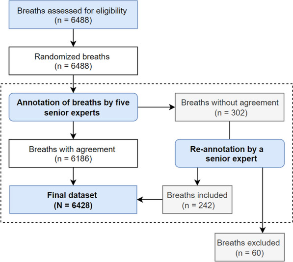
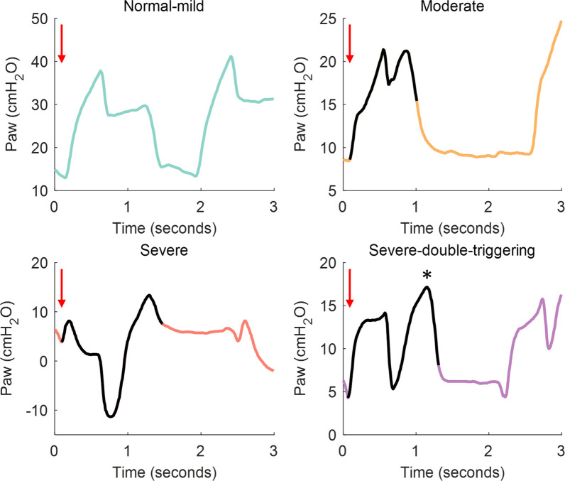
The selected ventilator tracings (Paw and flow) were visually inspected by five ICU senior physicians (LlB, RF, GMA, GM, CDH), with extensive clinical experience in IMV and management of asynchronies. They classified all breaths by identifying the amount of Paw deformation patterns as compared to a passive insufflation, which were stored in an interactive web application specifically developed for this purpose (Additional file 1: additional details in online data supplement Figure E1). Paw deformations were classified by the researchers in one of 3 pattern categories: normal (or with mild deformation), moderate deformation and severe deformation (Fig. 1). Agreement between researchers about the classification of Paw deformation was determined with the majority voting method (three of five experts agreement) [14]. In case of disagreement between the experts, the breaths were re-analyzed by the senior coordinator (LlB) who decided whether the breaths were included or not in the analysis. Breaths were excluded from analysis when: (1) 2 of the 5 annotators noted deemed them wrong/confusing (i.e., technical issues), and (2) the following annotation pattern was present: 2 votes normal-mild, 1 vote moderate and 2 votes severe. The percentage of patients in each category can be found in online data supplement.
Fig. 1.
Representative examples of the airway pressure (Paw) deformation patterns classification on the pressure–time waveform. Red arrows show the initiation of the patient-triggered breath. The Paw deformation in the moderate, severe and severe with double triggering tracings is represented by a solid black line on the Paw tracings. The asterisk shows the second breath added to the first one in the severe breaths with double triggering (breath stacking)
Double-triggering breaths were identified from the tracings (through a validated algorithm in the cohort of patients from Parc Taulí Hospital Universitari and visually in the cohort of patients from St. Michael’s Hospital), and were considered as a separate category in order to investigate their incidence.
Algorithms for detection of Paw deformation
The expert classification was used for training independently two machine learning models for automatically classifying the Paw deformation patterns: recurrent neural network and convolutional neural network. The algorithms' input data consisted of the inspiratory phase of Paw waveforms, which were resampled to 80 samples to ensure that all breaths have the same length. The goal was to detect Paw deformation during the inspiratory phase of patient-triggered breaths in square-flow volume assist-control ventilation.
The recurrent neural network algorithm is appropriate for long-sequence applications, since their architecture is designed to predict an output for each element [15, 16]. In particular, for time series, the most commonly used type is the long short-term memory [17, 18], that learns from long-term dependencies. In this study, two hidden layers of 128 neurons were used and a fully connected layer was added at the end of the long short-term memory to classify into one of the three categories. The convolutional neural network algorithm using a 1D convolution (1D convolutional neural network) contains convolution kernels/filters that can be interpreted as a time series application. These kernels move in a single time direction from the beginning of a time series toward its end, performing the convolution. One application behind the use of multiple filters is the ability to learn multiple discriminative features useful for the classification task [16]. Once the models have learned the different patterns the time required to detect a pattern for both algorithms is very similar. Additional information on the implemented models can be found in the online data supplement (Additional file 1: Figures E2 and E3). Models were implemented using Python (v 3.9.7) with the PyTorch (v. 1.11.0) package and run on a desktop computer (Windows 10 Pro 64-bit, Intel(R) Core(TM) i7-6700 CPU @ 3.40 GHz and 16 GB RAM).
Statistics
Agreement between researchers about the classification of Paw deformation was determined as the percentage of breaths with agreement (three of five experts) considering the majority voting method [14] and the Fleiss’ kappa coefficient. The recurrent neural network and convolutional neural network models were trained using the repeated holdout cross-validation method. The dataset was divided into an 80–20 train-validation split, with 80% of the data used for training and 20% for validation. This process was repeated 15 times, with each repetition using a different randomly selected subset for validation. Subsequently, median values were derived from the outcomes of each validation step, enhancing a more robust estimate of the model's performance. Performance measures of AI algorithms (accuracy, recall, F-1 score and precision) were used to measure the effectiveness of the algorithms (Additional file 1: additional information on the online data supplement). To ensure an heterogeneous dataset and a good performance of the model, we have lumped together the data from both centers. Wilcoxon signed-rank test was used to investigate the relationship between the patterns of Paw deformation and inspiratory time (Ti) and inspiratory peak airflow. Bonferroni correction (α = 0.05/6 = 0.0083) was considered. We analyzed learning curves of applied models to examine sample size. Further details, including a comparison of the sample size to the success rate, can be found in the online data supplement (see Additional file 1: Figure E4).
Results
Table 1 shows the patient’s characteristics (data were expressed as median [interquartile range]). A total of 6488 breaths from 28 patients receiving IMV were classified by experts: 559 from St. Michael's Hospital and 5929 from Parc Taulí Hospital Universitari (Fig. 2). Of these, in 302 breaths (4.6%) the experts disagree and were re-analyzed; among these, 60 breaths were finally excluded. Therefore, the final dataset included 6428 breaths classified by experts as follows: 2708 normal-mild (42.1%), 1535 moderate (23.8%), and 2185 severe Paw deformation (33.9%). The inter-expert agreement was 95.4% (Additional file 1: additional information in the online data supplement and Figure E5).
Table 1.
Patients’ demographic and clinical characteristics at admission
| Patients' demographic and clinical characteristics at admission | n = 28 |
|---|---|
| Age | 63 [57–70] |
| Female (%) | 4 (14%) |
| Reason for MV, n (%) | |
| Pneumonia | 11 (39%) |
| Sepsis | 3 (11%) |
| COVID-19 | 10 (36%) |
| Other causes | 4 (14%) |
| APACHE II at admission | 14 [11–23] |
| SOFA | 7 [5–9.2] |
| Median duration of MV (range), in days | 17 [13–26] |
| Median ICU–LOS (range), in days | 23.5 [16–34.8] |
| Median hospital–LOS (range), in days | 39 [23–63.5] |
| ICU mortality (%) | 5 (18%) |
Data are represented as median [25th, 75th percentiles] or percentages. Definition of abbreviations: APACHE II: Acute Physiology and Chronic Health Evaluation. ICU: Intensive care unit. LOS: length of stay. MV: mechanical ventilation
Fig. 2.

Flowchart of the breath annotation procedure from ventilator tracings
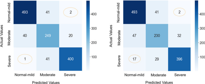
The validation dataset consisted of 1287 breaths including 536 normal-mild (41.7%), 309 moderate (24.0%), and 442 severe Paw deformation (34.4%). The confusion matrix (Fig. 3) shows the breakdown of the classification provided by the machine learning classifiers compared to the human expert labels for the validation phase. The recurrent neural network algorithm accurately classified 92% of normal-mild (493/536), 80.6% of moderate (249/309), and 90.5% of severe (400/442) Paw deformation, and 145 breaths of total validation dataset (11.3%) were misclassified. The recurrent neural network algorithm performed very well at the extremes (severe vs. normal-mild), as it labeled only one severe breath as normal-mild and two normal breaths as severe. Overall, the recurrent neural network performance had 87.9% [87.6–88.3] accuracy, 87.7% [87.5–88.2] precision, 87.9% [87.6–88.3] recall and 87.7% [87.4–88.1] F1 score. The convolutional neural network algorithm accurately classified 92% of normal-mild (493/536), 74.4% of moderate (230/309), and 89.6% of severe (396/442) Paw deformation, and 168 breaths (13.1%) were misclassified. Again, error between the extremes (severe vs. normal-mild) were negligible: 2 normal-mild breaths were classified as severe, and 17 severe breaths were classified as normal-mild. Overall, the convolutional neural network performance was 86.8% [86.6–87.4] accuracy, 87% [86.7–87.3] precision, 86.8% [86.6–87.4] recall and 86.9% [86.6–87.3] F1 score. (Additional file 1: Table E1 in online data supplement shows details of performance metrics obtained during the training and validation process for the 15 times models were trained.)
Fig. 3.
Confusion matrix for the recurrent neural network (RNN) and convolutional neural network (CNN) validation processes, respectively. The implemented models provide a strong performance for normal-mild and severe patterns. The reported performance metrics are the average across the 15 repetitions
Median ventilator inspiratory time, peak inspiratory airflow, respiratory rate, positive end expiratory pressure (PEEP) and expiratory time were similar between the breaths corresponding to the 3 groups of Paw deformation. Tidal volume was lower in the most severe patterns, with no statistically significant differences (Additional file 1: Figure E6 and Table E2 in the online data supplement). Double triggering was only present in breaths with severe Paw deformation (8.8% of breaths with severe deformation).
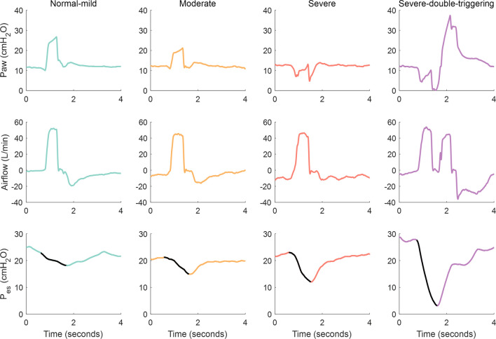
In the secondary analysis of BEARDS patients with esophageal pressure measurements ΔPes was > 8 cmH2O in 2.4%, 35.4%, and 94.8% of breaths with normal-mild, moderate or severe Paw deformation, respectively, whereas ΔPes was > 10 cmH2O in 74.4% of breaths with severe Paw deformation (Additional file 1: Additional information in Table E3 online data supplement). Figure 4 shows representative examples of Paw, airflow and Pes tracings corresponding to breaths of different severity.
Fig. 4.
Representative examples of airway pressure (Paw), airflow and esophageal pressure (Pes) tracings during square-flow assisted control ventilation corresponding to normal-mild breath, moderate breath, severe breath and double triggering, respectively. The esophageal swing is represented by solid black lines on the Pes tracings, which increases in relation to the different patterns (the greater the swing, the greater the inspiratory effort)
Discussion
The main findings of this study are: (1) AI models can detect and classify breath-by-breath Paw deformation patterns with high accuracy; (2) breaths classified as having severe Paw deformation exhibit stronger inspiratory efforts; (3) double triggering only occurs in breaths with severe Paw deformation.
A major goal of IMV is to unload the respiratory muscles to avoid exhaustion while avoiding muscle atrophy [19, 20]. However, during clinical situations of high inspiratory demands or insufficient delivered airflow, patients may develop strong inspiratory efforts [21]. This may be associated with dyspnea and both patient self-induced lung injury and myotrauma [22, 23]. In square-flow volume assist-control ventilation, sometimes the patient triggers the ventilator by slightly lowering Paw, followed by the mechanical insufflation that intends to reduce the work of breathing [20]. The muscular pressure could be estimated by the difference in Paw between passive and active circumstances. The greater drop in the Paw waveform during insufflation, the greater inspiratory effort of the patient [12, 24, 25]. Although the Paw waveform can be quickly examined during square-flow volume assist-control ventilation to identify a significant deformation [26], underdiagnosis is frequent, either because of failure to recognize the deformation or because professionals can only inspect waveforms for short time periods [11].
Convolutional neural network and recurrent neural network models have shown the best results on automatically detecting patient-ventilator asynchronies e.g., double triggering, ineffective effort, delayed cycling and premature cycling [15, 17, 18, 27–30]. Convolutional neural network algorithms detected different types of patient-ventilator asynchronies with an accuracy ranging from 97 to 99% [15, 17, 18, 27–30] whereas recurrent neural networks, in particular long short-term memory, performed slightly lower results between 91 and 98.3% [15]. In the present study, two different neural networks have been implemented, a long short-term memory and a 1D convolutional neural network. Convolutional neural networks are currently considered the most advanced models due to their best results in patient-ventilator asynchronies detection, but in our study, the recurrent neural network model showed similar accuracy. One explanation may be that recurrent neural networks are also suited to handle time-dependent sequences or data [15]. These networks use time series information to identify patterns between input and output. The memory of recurrent neural network algorithms allows them to learn more about the long-term dependencies of the data and understand the full context of the sequence when making the next prediction [15, 31].
Currently, the gold standard for the identification and quantification of strong inspiratory efforts is the measurement of Pes swing. However, it is not commonly used due to its complexity and invasiveness [32–34]. Similarly to our study, Telias et al. [34] have recently developed an automated algorithm based on Pes measurements that accurately generates and quantifies the muscular pressure for synchronous and dyssynchronous inspiratory efforts. They suggest that those patients with strong efforts detected by the algorithm might benefit from Pes monitoring. In recent years, several continuous monitoring systems that integrate signals in real-time have emerged and, through the application of validated algorithms, can automatically and continuously identify asynchronies [13, 35–38]. In the present study, a high percentage of breaths classified as severe exhibit ΔPes > 8 or 10 cmH2O, suggesting that Paw deformation is frequently associated with strong muscular efforts.
Double triggering was present exclusively in breaths with a severe Paw deformation (8.8% of them) [34]. Double triggering is one of the most potentially injurious patient-ventilator asynchronies in assisted volume-controlled ventilation, due to the high Paw and very high tidal volume resulting from the accumulation of two consecutive breaths [39–41]. This can generate higher transpulmonary and transvascular pressure gradients, increasing tissue stress and strain, and resulting in an unequal pressure distribution in lung-dependent areas [42], which can favor ventilator-induced lung injury [43, 44]. Among the factors associated with the development of double triggering, short ventilator inspiratory time and/or low airflow setting have also been recognized as important [41].
Our study make a significant contribution to the field of patient-ventilator asynchrony detection. Firstly, it introduces an innovative solution for classifying flow starvation during square-flow assisted ventilation using convolutional neural network and recurrent neural network models. The majority of existing patient-ventilator asynchrony algorithms [37, 45] primarily focus on identifying common forms of asynchronies such as double triggering, ineffective effort, and short- and prolonged cycling. In contrast to previous studies [27–30] employing a binary classification for asynchrony classification, our work adopts a multiclass approach. This approach enables clinicians to differentiate, for instance, between moderate and severe degrees of Paw deformation. Secondly, our dataset construction strategy, which incorporates waveforms from two different medical centers, allows us to assess the extrapolation capability of deep learning methods. To ensure a balanced representation and prevent overemphasis on specific patients, the number of breaths per patient in each class was capped at a maximum of 350 breaths. Additionally, breaths were selected to create a balanced sample across the initial, intermediate, and final stages of IMV. Thirdly, the architectural design of our implemented models utilizes a single branch corresponding to the inspiratory phase of the Paw waveform, with a fixed size of 80 sample points as input to the tensor. This results in models of lower complexity compared to other studies [27, 30] that employ deep learning approaches for the classification of patient-ventilator asynchronies. Lastly, our work presents an automated algorithm for detecting flow starvation, aiming to improve the underdiagnosis of patient-ventilator asynchronies by visual examination of ventilator waveforms at the bedside [11, 46]. The AI model could provide an accurate classification of breaths with severe Paw deformation, based on the analysis of Paw waveform. Therefore, the continuous assessment of Paw deformation by using AI technologies could alert clinicians about the presence of excessively high inspiratory efforts or associated with insufficient airflow.
This study has limitations. First, the deep learning model was only applied to IMV under square-flow assisted ventilation, but it is one of the most widely used mode of ventilation [47, 48]. Our AI model stands as an initial technological approach that needs further evaluation and implementation with additional data and other ventilator modes to enhance its robustness and generability. Currently the ventilators do not provide alarm systems to notify the presence of abnormal Paw waveforms patterns. From a clinical perspective, computerized systems are needed to connect and agnostically interoperate ventilator waveforms. A continuous analysis of Paw waveforms using AI models could potentially be integrated into ICU mechanical ventilators or monitoring centers, providing valuable support and alert tool for clinicians [49–51]. Second, the recurrent neural network and convolutional neural network models need to be trained with sufficient data [52], and although our sample of about 6500 breaths may appear small, it has yielded very good performance on the training and validation datasets. Higher large-scale labeling efforts are costly and time-consuming, and often require extensive domain knowledge or technical expertise to implement a particular medical task, often resulting in large-scale inefficiencies in clinical AI workflows. Furthermore, these methods can only predict events on which they have been trained, which restricts their widespread applicability. Therefore, these label learning methods may not be as powerful in environments where access to a diverse set of high-quality data is limited [53].
Conclusions
Our study shows that AI, in particular recurrent neural networks, could be an excellent tool to identify airway pressure deformation associated to strong inspiratory efforts during square-flow volume assist-control ventilation, allowing to minimize unrecognized periods of abnormal and potentially injurious patient-ventilator interaction.
Supplementary Information
Additional file 1. Supplementary materials, tables and figures.
Acknowledgements
List of the BEARDS investigators: (1) Saint Michael’s Hospital, Toronto, Ontario, Canada: Laurent Brochard, Irene Telias, Felipe Damiani, Ricard Artigas, Cesar Santis, Tài Pham. (2) Dipartimento di Anestesia, Rianimazione ed Emergenza-Urgenza, Fondazione IRCCS Ca’ Granda Ospedale Maggiore Policlinico, Università degli studi di Milano, Milan, Italy: Tommaso Mauri, Elena Spinelli, Giacomo Grasselli. (3) Department of Morphology, Surgery and Experimental Medicine, Intensive Care Unit University of Ferrara, Sant’Anna Hospital, Ferrara, Italy: Savino Spadaro, Carlo Alberto Volta. (4) Anesthesia and Intensive Care, Fondazione Istituto di Ricovero e Cura a Carattere Scientifico, Policlinico San Matteo, Pavia, Italy: Francesco Mojoli. (5) Department of Intensive Care Medicine, University Hospital of Heraklion and School of Medicine, University of Crete, Heraklion, Crete, Greece: Dimitris Georgopoulos, Eumorfia Kondili, Stella Soundoulounaki. (6) Department of Anesthesiology and Intensive Care Medicine, University Medical Center Schleswig-Holstein, Campus Kiel, Kiel, Germany: Tobias Becher, Norbert Weiler, Dirk Schaedler. (7) Critical Care Department, Vall d’Hebron University Hospital, Vall d’Hebron Research Institute and Ciber Enfermedades Respiratorias, Instituto de Salud Carlos III, Madrid, Spain: Oriol Roca, Manel Santafe. (8) Intensive Care Medicine, Hospital de Sant Pau, Barcelona, Spain: Jordi Mancebo, Nuria Rodríguez. (9) Department of Intensive Care Medicine, Amsterdam UMC, Amsterdam, The Netherlands: Leo Heunks, Heder de Vries. (10) National Cheng Kung University Hospital, College of Medicine, National Cheng-Kung University, Tainan, Taiwan: Chang-Wen Chen. (11) Department of Critical Care Medicine, Beijing Tiantan Hospital, Capital Medical University, Beijing, China: Jian-Xin Zhou周建新, Guang-Qiang Chen 陈光强. (12) Division of Respiratory Diseases and Tuberculosis, Mahidol University Faculty of Medicine Siriraj Hospital, Bangkok, Thailand: Nuttapol Rit-tayamai. (13) Complejo Médico de la Policía Federal Argentina Churruca Visca, Buenos Aires, Argentina: Norberto Tiribelli. (14) Sanatorio de la Trinidad Miter, Buenos Aires, Argentina: Sebastian Fredes. (15) Hospital Clinic, Barcelona, Spain : Ricard Mellado Artigas, Carlos Ferrando Ortolá. (16) Medical Intensive Care Unit, University Hospital of Angers, Angers, France: François Beloncle, Alain Mercat. (17) Service de Réanimation Polyvalente, Hôpital Sainte Musse, Toulon, France: Jean-Michel Arnal. (18) Medical Intensive Care Unit Hôpital Européen Georges Pompidou Assistance Publique-Hôpitaux de Paris, Paris, France: Jean-Luc Diehl. (19) AP-HP, Groupe Hospitalier Pitié-Salpêtrière Charles Foix, Service de Pneumologie, Médecine intensive—Réanimation (Département ’R3S’), Paris, France: Alexandre Demoule, Martin Dres, Quentin Fossé. (20) Groupe Hospital-ier Sud Ile-De-France, Center Hospitalier de Melun, Melun, France: Sébastien Jochmans, Jonathan Chelly. (21) Médecine Intensive Réanimation, C.H.U de Grenoble-Alpes, Grenoble, France: Nicolas Terzi, Claude Guérin. (22) Division of Pulmonary and Critical Care, Beth Israel Deaconess Medical Center and Massachusetts General Hospital, Harvard Medical School, Boston, MA, USA: E Baedorf Kassis. (23) Division of Pulmonary, Allergy, and Critical Care Medicine, Columbia University College of Physicians and Surgeons, NewYork-Presbyte-rian Hospital, New York, New York, USA: Jeremy Beitler. (24) Anesthesia and Intensive Care, Fondazione Istituto di Ricovero e Cura a Carattere Scientifico, Policlinico San Matteo, Pavia, Italy: Davide Chiumello, Erica Ferrari Luca Bol-giaghi. (25) Médecine Intensive Réanimation, Center Hospitalier Universitaire de Poitiers, Poitiers, France : Arnaud W Thille, Rémi Coudroy. (26) Médecine Intensive Réanimation, Hôpital Nord, Hôpitaux de Marseille, Chemin des Bour-rely, 13015, Marseille, France : Laurent Papazian
Lluís Blanch and Leonardo Sarlabous are co-senior authors.
Abbreviations
- AI
Artificial intellligence
- cmH2O
Centimeters of water
- ICU
Intensive Care Unit
- IMV
Invasive mechanical ventilation
- Paw
Airway pressure
- PEEP
Positive end expiratory pressure
- Pes
Esophageal pressure
- Ti
Inspiratory time
Author contributions
CdH, LS and LlB conceived and designed the study. VSP, AXP and LS conducted statistical analysis. CdH, LlB, RF, GMA and GM classified the patterns. CdH, VSP, LS and LlB interpreted the data and drafted the manuscript with input from all authors. IT, RF and LB critically revised the manuscript. CdH, LS and LlB were responsible for the decision to submit the manuscript for publication. All other authors contributed to data acquisition. All the authors reviewed the final draft of the manuscript and agreed on submitting it to the Journal.
Funding
This work was funded by projects RTC-2017-6193-1 (AEI/FEDER EU) and 202118 (413/C/2021) Fundació La Marató de TV3, CIBER-Consorcio Centro de Investigación Biomédica en RED-CB06/06/1097, Instituto de Saludo Carlos III, Ministerio de Ciencia e Innovación and Unión Europea – European Regional Development Fund, CERCA Program/Generalitat de Catalunya and Fundació Institut d’Investigació i Innovació Parc Taulí-I3PT. C. de Haro is granted with a Contrato para la intensificación de la actividad investigadora en el sistema nacional de salud (INT20/00030), AES 2020, by Instituto de Salud Carlos III. L. Sarlabous is supported by Pla Estratègic de Recerca i Innovació en Salut program from the Health Department of Generalitat de Catalunya, Spain.
Availability of data and materials
The datasets used and analyzed during this study are available from the corresponding author on reasonable request.
Declarations
Ethics approval and consent to participate
The institutional review boards of each participating ICU approved the protocol of the study. Patients or their surrogate decision-makers provided informed consent to participate in the study collecting waveforms for processing and analysis.
Consent for publication
Not applicable.
Competing interests
L. Blanch (LlB) is inventor of a US patent owned by Consorci Corporació Sanitària Parc Taulí: “Method and system for managed related patient parameters provided by a monitoring device”, US Patent No. 12/538,940. Gaston Murias and L. Blanch own stock options of BetterCare S.L., a research and development spinoff of Consorci Corporació Sanitària Parc Taulí.
Footnotes
Publisher's Note
Springer Nature remains neutral with regard to jurisdictional claims in published maps and institutional affiliations.
Candelaria de Haro and Verónica Santos-Pulpón equally contributed to the investigation.
Lluís Blanch and Leonardo Sarlabous are co-senior authors.
Contributor Information
Candelaria de Haro, Email: cdeharo@tauli.cat.
the BEARDS study investigators:
Laurent Brochard, Irene Telias, Felipe Damiani, Ricard Artigas, Cesar Santis, Tài Pham, Tommaso Mauri, Elena Spinelli, Giacomo Grasselli, Savino Spadaro, Carlo Alberto Volta, Francesco Mojoli, Dimitris Georgopoulos, Eumorfia Kondili, Stella Soundoulounaki, Tobias Becher, Norbert Weiler, Dirk Schaedler, Oriol Roca, Manel Santafe, Jordi Mancebo, Nuria Rodríguez, Leo Heunks, Heder de Vries, Chang-Wen Chen, Jian-Xin Zhou, Guang-Qiang Chen, Nuttapol Rit-tayamai, Norberto Tiribelli, Sebastian Fredes, Ricard Mellado Artigas, Carlos Ferrando Ortolá, François Beloncle, Alain Mercat, Jean-Michel Arnal, Jean-Luc Diehl, Alexandre Demoule, Martin Dres, Quentin Fossé, Sébastien Jochmans, Jonathan Chelly, Nicolas Terzi, Claude Guérin, E. Baedorf Kassis, Jeremy Beitler, Davide Chiumello, Erica Ferrari Luca Bol-giaghi, Arnaud W. Thille, Rémi Coudroy, and Laurent Papazian
References
- 1.Georgopoulos D, Prinianakis G, Kondili E. Bedside waveforms interpretation as a tool to identify patient-ventilator asynchronies. Intensive Care Med. 2006;32(1):34–47. doi: 10.1007/s00134-005-2828-5. [DOI] [PubMed] [Google Scholar]
- 2.MacIntyre NR, McConnell R, Cheng KCG, Sane A. Patient-ventilator flow dyssynchrony: flow-limited versus pressure- limited breaths. Crit Care Med. 1997;25(10):1671–1677. doi: 10.1097/00003246-199710000-00016. [DOI] [PubMed] [Google Scholar]
- 3.Pham T, Telias I, Piraino T, Yoshida T, Brochard LJ. Asynchrony consequences and management. Crit Care Clin. 2018;34(3):325–341. doi: 10.1016/j.ccc.2018.03.008. [DOI] [PubMed] [Google Scholar]
- 4.Schepens T, Dres M, Heunks L, Goligher EC. Diaphragm-protective mechanical ventilation. Curr Opin Crit Care. 2019;25(1):77–85. doi: 10.1097/MCC.0000000000000578. [DOI] [PubMed] [Google Scholar]
- 5.Bertoni M, Spadaro S, Goligher EC. Monitoring patient respiratory effort during mechanical ventilation: lung and diaphragm-protective ventilation. Crit Care. 2020;24(1):106. doi: 10.1186/s13054-020-2777-y. [DOI] [PMC free article] [PubMed] [Google Scholar]
- 6.Schmidt M, Banzett RB, Raux M, Morélot-Panzini C, Dangers L, Similowski T, et al. Unrecognized suffering in the ICU: Addressing dyspnea in mechanically ventilated patients. Intensive Care Med. 2014;40(1):1–10. doi: 10.1007/s00134-013-3117-3. [DOI] [PMC free article] [PubMed] [Google Scholar]
- 7.Itagaki T, Akimoto Y, Nakano Y, Ueno Y, Ishihara M, Tane N, et al. Relationships between double cycling and inspiratory effort with diaphragm thickness during the early phase of mechanical ventilation: A prospective observational study. PLoS ONE. 2022;17(8):e0273173. doi: 10.1371/journal.pone.0273173. [DOI] [PMC free article] [PubMed] [Google Scholar]
- 8.Hashimoto H, Yoshida T, Firstiogusran AMF, Taenaka H, Nukiwa R, Koyama Y, et al. Asynchrony injures lung and diaphragm in acute respiratory distress syndrome*. Crit Care Med. 2023;51(11):e234–e242. doi: 10.1097/CCM.0000000000005988. [DOI] [PubMed] [Google Scholar]
- 9.Schmidt M, Demoule A, Polito A, Porchet R, Aboab J, Siami S, et al. Dyspnea in mechanically ventilated critically ill patients. Crit Care Med. 2011;39(9):2059–2065. doi: 10.1097/CCM.0b013e31821e8779. [DOI] [PubMed] [Google Scholar]
- 10.Demoule A, Hajage D, Messika J, Jaber S, Diallo H, Coutrot M, et al. Prevalence, intensity, and clinical impact of dyspnea in critically ill patients receiving invasive ventilation. Am J Respir Crit Care Med. 2022;205(8):917–926. doi: 10.1164/rccm.202108-1857OC. [DOI] [PubMed] [Google Scholar]
- 11.Colombo D, Cammarota G, Alemani M, Carenzo L, Barra FL, Vaschetto R, et al. Efficacy of ventilator waveforms observation in detecting patient-ventilator asynchrony. Crit Care Med. 2011;39(11):2452–2457. doi: 10.1097/CCM.0b013e318225753c. [DOI] [PubMed] [Google Scholar]
- 12.Cinnella G, Conti G, Lofaso F, Lorino H, Harf A, Lemaire F, et al. Effects of assisted ventilation on the work of breathing: volume-controlled versus pressure-controlled ventilation. Am J Respir Crit Care Med. 1996;153(3):1025–1033. doi: 10.1164/ajrccm.153.3.8630541. [DOI] [PubMed] [Google Scholar]
- 13.Pham T, Montanya J, Telias I, Piraino T, Magrans R, Coudroy R, et al. Automated detection and quantification of reverse triggering effort under mechanical ventilation. Crit Care. 2021;25(1):60. doi: 10.1186/s13054-020-03387-3. [DOI] [PMC free article] [PubMed] [Google Scholar]
- 14.Sheng VS, Zhang J, Gu B, Wu X. Majority Voting and Pairing with Multiple Noisy Labeling. IEEE Trans Knowl Data Eng. 2019;31(7):1355–1368. doi: 10.1109/TKDE.2017.2659740. [DOI] [Google Scholar]
- 15.Hüsken M, Stagge P. Recurrent neural networks for time series classification. Neurocomputing. 2003;50:223–235. doi: 10.1016/S0925-2312(01)00706-8. [DOI] [Google Scholar]
- 16.Ismail Fawaz H, Forestier G, Weber J, Idoumghar L, Muller P-A. Deep learning for time series classification: a review. Data Min Knowl Discov. 2019;33(4):917–963. doi: 10.1007/s10618-019-00619-1. [DOI] [Google Scholar]
- 17.Du Q, Gu W, Zhang L, Huang S-L. Attention-based LSTM-CNNs For Time-series Classification. In: Proceedings of the 16th ACM Conference on Embedded Networked Sensor Systems. New York, NY, USA: ACM; 2018. p. 410–1. (SenSys ’18).
- 18.Karim F, Majumdar S, Darabi H, Harford S. Multivariate LSTM-FCNs for time series classification. Neural Netw. 2019;116:237–245. doi: 10.1016/j.neunet.2019.04.014. [DOI] [PubMed] [Google Scholar]
- 19.Leung P, Jubran A, Tobin MJ. Comparison of assisted ventilator modes on triggering, patient effort, and dyspnea. Am J Respir Crit Care Med. 1997;155(6):1940–1948. doi: 10.1164/ajrccm.155.6.9196100. [DOI] [PubMed] [Google Scholar]
- 20.Marini JJ, Capps JS, Culver BH. The inspiratory work of breathing during assisted mechanical ventilation. Chest. 1985;87(5):612–618. doi: 10.1378/chest.87.5.612. [DOI] [PubMed] [Google Scholar]
- 21.de Wit M. Monitoring of patient-ventilator interaction at the bedside. Respir Care. 2011;56(1):61–68. doi: 10.4187/respcare.01077. [DOI] [PubMed] [Google Scholar]
- 22.Yoshida T, Uchiyama A, Matsuura N, Mashimo T, Fujino Y. Spontaneous breathing during lung-protective ventilation in an experimental acute lung injury model: high transpulmonary pressure associated with strong spontaneous breathing effort may worsen lung injury. Crit Care Med. 2012;40(5):1578–1585. doi: 10.1097/CCM.0b013e3182451c40. [DOI] [PubMed] [Google Scholar]
- 23.Goligher EC, Fan E, Herridge MS, Murray A, Vorona S, Brace D, et al. Evolution of diaphragm thickness during mechanical ventilation. Impact of inspiratory effort. Am J Respir Crit Care Med. 2015;192(9):1080–1088. doi: 10.1164/rccm.201503-0620OC. [DOI] [PubMed] [Google Scholar]
- 24.Marini JJ, Rodriguez RM, Lamb V. Bedside estimation of the inspiratory work of breathing during mechanical ventilation. Chest. 1986;89(1):56–63. doi: 10.1378/chest.89.1.56. [DOI] [PubMed] [Google Scholar]
- 25.Ward ME, Corbeil C, Gibbons W, Newman S, Macklem PT. Optimization of respiratory muscle relaxation during mechanical ventilation. Anesthesiology. 1988;69(1):29–35. doi: 10.1097/00000542-198807000-00005. [DOI] [PubMed] [Google Scholar]
- 26.Carteaux G, Parfait M, Combet M, Haudebourg A-F, Tuffet S, Mekontso DA. Patient-self inflicted lung injury: a practical review. J Clin Med. 2021;10(12):2738. doi: 10.3390/jcm10122738. [DOI] [PMC free article] [PubMed] [Google Scholar]
- 27.Pan Q, Zhang L, Jia M, Pan J, Gong Q, Lu Y, et al. An interpretable 1D convolutional neural network for detecting patient-ventilator asynchrony in mechanical ventilation. Comput Methods Programs Biomed. 2021;204. [DOI] [PubMed]
- 28.Ge H, Duan K, Wang J, Jiang L, Zhang L, Zhou Y, et al. Risk Factors for Patient–Ventilator Asynchrony and Its Impact on Clinical Outcomes: Analytics Based on Deep Learning Algorithm. Front Med. 2020;7. [DOI] [PMC free article] [PubMed]
- 29.Loo NL, Chiew YS, Tan CP, Mat-Nor MB, Ralib AM. A machine learning approach to assess magnitude of asynchrony breathing. Biomed Signal Process Control. 2021;66:102505. doi: 10.1016/j.bspc.2021.102505. [DOI] [Google Scholar]
- 30.Zhang L, Mao K, Duan K, Fang S, Lu Y, Gong Q, et al. Detection of patient-ventilator asynchrony from mechanical ventilation waveforms using a two-layer long short-term memory neural network. Comput Biol Med. 2020;120. [DOI] [PubMed]
- 31.Fullah Kamara A, Chen E, Liu Q, Pan Z. Combining contextual neural networks for time series classification. Neurocomputing. 2020;384:57–66. doi: 10.1016/j.neucom.2019.10.113. [DOI] [Google Scholar]
- 32.Mauri T, Yoshida T, Bellani G, Goligher EC, Carteaux G, Rittayamai N, et al. Esophageal and transpulmonary pressure in the clinical setting: meaning, usefulness and perspectives. Intensive Care Med. 2016;42(9):1360–1373. doi: 10.1007/s00134-016-4400-x. [DOI] [PubMed] [Google Scholar]
- 33.Akoumianaki E, Maggiore SM, Valenza F, Bellani G, Jubran A, Loring SH, et al. The application of esophageal pressure measurement in patients with respiratory failure. Am J Respir Crit Care Med. 2014;189(5):520–531. doi: 10.1164/rccm.201312-2193CI. [DOI] [PubMed] [Google Scholar]
- 34.Telias I, Madorno M, Pham T, Piraino T, Coudroy R, Sklar MC, et al. Magnitude of synchronous and dyssynchronous inspiratory efforts during mechanical ventilation: a novel method. Am J Respir Crit Care Med. 2023;207(9):1239–1243. doi: 10.1164/rccm.202211-2086LE. [DOI] [PMC free article] [PubMed] [Google Scholar]
- 35.Chen CW, Lin WC, Hsu CH, Cheng KS, Lo CS. Detecting ineffective triggering in the expiratory phase in mechanically ventilated patients based on airway flow and pressure deflection: feasibility of using a computer algorithm. Crit Care Med. 2008;36(2):455–461. doi: 10.1097/01.CCM.0000299734.34469.D9. [DOI] [PubMed] [Google Scholar]
- 36.Blanch L, Sales B, Montanya J, Lucangelo U, Oscar GE, Villagra A, et al. Validation of the Better Care® system to detect ineffective efforts during expiration in mechanically ventilated patients: a pilot study. Intensive Care Med. 2012;38(5):772–780. doi: 10.1007/s00134-012-2493-4. [DOI] [PubMed] [Google Scholar]
- 37.Mulqueeny Q, Ceriana P, Carlucci A, Fanfulla F, Delmastro M, Nava S. Automatic detection of ineffective triggering and double triggering during mechanical ventilation. Intensive Care Med. 2007;33(11):2014–2018. doi: 10.1007/s00134-007-0767-z. [DOI] [PubMed] [Google Scholar]
- 38.Gutierrez G, Ballarino GJ, Turkan H, Abril J, De La Cruz L, Edsall C, et al. Automatic detection of patient-ventilator asynchrony by spectral analysis of airway flow. Crit Care. 2011;15(4):R167. doi: 10.1186/cc10309. [DOI] [PMC free article] [PubMed] [Google Scholar]
- 39.Magrans R, Ferreira F, Sarlabous L, López-Aguilar J, Gomà G, Fernandez-Gonzalo S, et al. The effect of clusters of double triggering and ineffective efforts in critically ill patients. Crit Care Med. 2022;50(7):E619–E629. doi: 10.1097/CCM.0000000000005471. [DOI] [PubMed] [Google Scholar]
- 40.Thille AW, Rodriguez P, Cabello B, Lellouche F, Brochard L. Patient-ventilator asynchrony during assisted mechanical ventilation. Intensive Care Med. 2006;32(10):1515–1522. doi: 10.1007/s00134-006-0301-8. [DOI] [PubMed] [Google Scholar]
- 41.de Haro C, López-Aguilar J, Magrans R, Montanya J, Fernández-Gonzalo S, Turon M, et al. Double cycling during mechanical ventilation: frequency, mechanisms, and physiologic implications. Crit Care Med. 2018;46(9):1385–1392. doi: 10.1097/CCM.0000000000003256. [DOI] [PubMed] [Google Scholar]
- 42.Yoshida T, Fujino Y, Amato MBP, Kavanagh BP. Fifty years of research in ards spontaneous breathing during mechanical ventilation risks, mechanisms, and management. Am J Respir Crit Care Med. 2017;195(8):985–992. doi: 10.1164/rccm.201604-0748CP. [DOI] [PubMed] [Google Scholar]
- 43.Pohlman MC, McCallister KE, Schweickert WD, Pohlman AS, Nigos CP, Krishnan JA, et al. Excessive tidal volume from breath stacking during lung-protective ventilation for acute lung injury. Crit Care Med. 2008;36(11):3019–3023. doi: 10.1097/CCM.0b013e31818b308b. [DOI] [PubMed] [Google Scholar]
- 44.Beitler JR, Sands SA, Loring SH, Owens RL, Malhotra A, Spragg RG, et al. Quantifying unintended exposure to high tidal volumes from breath stacking dyssynchrony in ARDS: the BREATHE criteria. Intensive Care Med. 2016;42(9):1427–1436. doi: 10.1007/s00134-016-4423-3. [DOI] [PMC free article] [PubMed] [Google Scholar]
- 45.Bakkes T, van Diepen A, De Bie A, Montenij L, Mojoli F, Bouwman A, et al. Automated detection and classification of patient–ventilator asynchrony by means of machine learning and simulated data. Comput Methods Programs Biomed. 2023;230:107333. doi: 10.1016/j.cmpb.2022.107333. [DOI] [PubMed] [Google Scholar]
- 46.Ramirez II, Arellano DH, Adasme RS, Landeros JM, Salinas FA, Vargas AG, et al. Ability of ICU health-care professionals to identify patient-ventilator asynchrony using waveform analysis. Respir Care. 2017;62(2):144–149. doi: 10.4187/respcare.04750. [DOI] [PubMed] [Google Scholar]
- 47.Esteban A, Frutos-Vivar F, Muriel A, Ferguson ND, Penuelas O, Abraira V, et al. Evolution of mortality over time in patients receiving mechanical ventilation. Am J Respir Crit Care Med. 2013;188(2):220–230. doi: 10.1164/rccm.201212-2169OC. [DOI] [PubMed] [Google Scholar]
- 48.Jabaley CS, Groff RF, Sharifpour M, Raikhelkar JK, Blum JM. Modes of mechanical ventilation vary between hospitals and intensive care units within a university healthcare system: a retrospective observational study. BMC Res Notes. 2018;11(1):425. doi: 10.1186/s13104-018-3534-z. [DOI] [PMC free article] [PubMed] [Google Scholar]
- 49.de Haro C, Ochagavia A, López-Aguilar J, Fernandez-Gonzalo S, Navarra-Ventura G, Magrans R, et al. Patient-ventilator asynchronies during mechanical ventilation: current knowledge and research priorities. Intensive Care Med Exp. 2019;7(S1):43. doi: 10.1186/s40635-019-0234-5. [DOI] [PMC free article] [PubMed] [Google Scholar]
- 50.Esperanza JA, Sarlabous L, de Haro C, Magrans R, Lopez-Aguilar J, Blanch L. Monitoring asynchrony during invasive mechanical ventilation. Respir Care. 2020;65(6):847–869. doi: 10.4187/respcare.07404. [DOI] [PubMed] [Google Scholar]
- 51.Maslove DM, Tang B, Shankar-Hari M, Lawler PR, Angus DC, Baillie JK, et al. Redefining critical illness. Nat Med. 2022;28(6):1141–1148. doi: 10.1038/s41591-022-01843-x. [DOI] [PubMed] [Google Scholar]
- 52.Fink O, Wang Q, Svensén M, Dersin P, Lee W-J, Ducoffe M. Potential, challenges and future directions for deep learning in prognostics and health management applications. Eng Appl Artif Intell. 2020;92:103678. doi: 10.1016/j.engappai.2020.103678. [DOI] [Google Scholar]
- 53.Tiu E, Talius E, Patel P, Langlotz CP, Ng AY, Rajpurkar P. Expert-level detection of pathologies from unannotated chest X-ray images via self-supervised learning. Nat Biomed Eng. 2022;6(12):1399–1406. doi: 10.1038/s41551-022-00936-9. [DOI] [PMC free article] [PubMed] [Google Scholar]
Associated Data
This section collects any data citations, data availability statements, or supplementary materials included in this article.
Supplementary Materials
Additional file 1. Supplementary materials, tables and figures.
Data Availability Statement
The datasets used and analyzed during this study are available from the corresponding author on reasonable request.


