

Abstract
Introduction and importance
The presentation of spontaneous hematomas remains different between all affected populations, but advanced age and use of anticoagulants are common risk factors in the majority. The progression of the hematoma may require some time to be detected; however, it can prove fatal if it reaches a significant size. A spontaneous calf hematoma can be mistakenly diagnosed as deep vein thrombosis, and the management of both conditions is varying.
Case presentation
A 26-year-old man had recently undergone multiple left lower limb surgeries and was using Rivaroxaban for deep vein thrombosis prophylaxis. He presented with a painful contralateral calf swelling for a duration of one month, which was later diagnosed as a spontaneous chronic calf hematoma. Despite the fact that blood tests were within the normal range, imaging confirmed the diagnosis. After extensive discussion among multidisciplinary teams, a surgical exploration was conducted, resulting in the complete evacuation of the hematoma. Subsequently, a meticulous monitoring of the re-administration of anticoagulant was conducted.
Clinical discussion
spontaneous calf hematoma is not common pathology and affecting elderly and presentation in young is unique and diagnosis will not be reached easily especially in chronic case.
Conclusion
A spontaneous calf hematoma can occur in a young, fit population with coexisting anticoagulant administrations. A thorough history, examination, and imaging must be applied urgently in order to reach a diagnosis.
Keywords: Spontaneous calf hematoma, Case report, Spontaneous hematoma, Leg mass, Muscular hematoma
Highlights
-
•
Spontaneous hematoma has different presentation form and must be carefully diagnosed and managed accordingly.
-
•
Spontaneous hematoma of lower limb may mimic pathologies such as: deep vein thrombosis, cellulitis, traumatic hematoma and sarcoma.
-
•
Doppler ultrasound alongside with computerized tomography useful tools to diagnose spontaneous leg hematoma.
1. Introduction
A spontaneous hematoma is a bizarre condition that is still presented with various atypical features. The literature reports a multitude of presentations, including abdominal, chest, and lower limb hematomas. It is imperative to maintain high suspicion in order to obtain the appropriate diagnosis, particularly in the case of a lower limb hematoma. Almost all patients have acquired coagulopathy induced by blood thinning agents as a major risk factor, along with other comorbidities [[1], [2], [3]].
Muscular hematomas are linked to trauma, inflammatory conditions, such as myositis, and extraordinary activities. Lower limb hematomas mimic deep vein thrombosis, traumatic muscle tear, infection, and even tumors. Computerized tomography angiography (CTA), magnetic resonance imaging (MRI), and ultrasound are all modalities that can be utilized to confirm diagnosis. MRI is a highly effective tool for distinguishing soft tissue tumors and collection, while ultrasound is a more efficient and convenient option, particularly in critical situations [4]. The management protocol is guided by the hemodynamic status of the patient, the expandability of the hematoma, and the facilities. Conservative treatment comes first, followed by surgical intervention or vascular embolization [5].
A spontaneous calf hematoma can occur in a young, fit population with coexisting anticoagulant administrations. The presence of isolated calf swelling without any obvious trauma has prompted suspicion of hematoma, as minor straining may result in the rupture of small vessels, which may later manifest as hematoma [2].
2. Case presentation
A 26-year-old man complained of right painful calf swelling, which increased gradually over a month until became severe. He had a clear medical background, but he underwent external fixation for a left tibial open fracture two months prior to presentation, followed by a rotational flap on different occasions. Both surgeries went uneventfully, except for blood loss, which was corrected by blood transfusion. The initial surgical procedure addressed the left open comminuted tibial fracture and the anterior tibial artery injury. External fixation was utilized to stabilize the limb, and it was processed by anterior tibial artery shunt to reduce the duration of ischaemia (Fig. 1). After skeletal fixation, a formal repair of the anterior tibial artery was performed by utilizing a saphenous vein graft. Following the vascular repair, debridement was attempted, and the wound was covered with an occlusive dressing. He was discharged to the ward, where he received intravenous antibiotics accompanied by anticoagulant (low molecular weight heparin) medication.
Fig. 1.

Left leg X-ray: showing complex tibial bone fracture.
Since then, he has been given Rivaroxaban as a deep vein thrombosis prophylactic therapy, and low molecular weight heparin has been withdrawn. The postoperative period was uneventful (Fig. 2). After three weeks, he underwent debridement and a flap cover procedure. During the operation, the necrotic bone edges were trimmed and all granulation tissue was excised. Subsequently, a fasciocutaneous flap was elevated and rotated to conceal the pre-existing defect. A skin graft with a split thickness was taken from the contralateral thigh. The skin graft was used to cover the flap donor site (Fig. 3). A partial necrosis of the marginal tissue was observed at the tip of the flap, however, it is held by secondary intent (Fig. 4). The skin graft healed perfectly. He resumed the administration of Rivaroxaban once more (Fig. 5).
Fig. 2.

Left leg: post tibia bone fixation and exposed part of tibia bone.
Fig. 3.

Left surgery: rotational flap to cover exposed part of tibia and STSG.
Fig. 4.

Left leg: post-operative complication: marginal necrosis at flap tip.
Fig. 5.

Left leg: after removal of external fixation.
Two months later, he expressed a mild pain that was restricted to the posterior aspect of his right leg and subsided with simple elevation of the limb, whereas swelling was not evident. The Doppler ultrasound revealed a clear appearance of vasculature in the lower limbs and minimal oedema in the soft tissue, leading to the diagnosis of cellulitis. The examination of the contralateral limbs revealed no abnormalities.
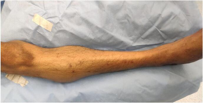
Two weeks later, the swelling and pain were advancing, rendering conventional treatment futile. After a thorough examination, his vital signs (blood pressure, respiratory rate, pulse, and saturation) were within normal values. The examination of the right leg revealed a hot, tender, uniform calf swelling, without any discoloration. Distal pulses were observed, but there was no discernible mass in the remainder of the limb (Fig. 6). The contralateral limb was also examined; however, no abnormalities were detected.
Fig. 6.

Right calves at presentation: showing uniform calf swelling.
Laboratory investigation showed a normal complete blood count (CBC), urea, Creatinine, liver function test (LFT) and bleeding time, but activated Partial Thromboplastin Time (a PTT) showed elevation 41.6 s (Normal lab reference 23- 32 s).
Ultrasound for the right calf showed a well-localized fluid collection at the posterior compartment of the leg beneath the gastrocnemius muscle, and Doppler revealed no vascular abnormalities or leak. The computerized tomography angiography (CTA) of the lower limb was performed, confirming a well-defined heterogeneous lesion measuring 7.2 × 9.7 × 12.5 cm at the posterior aspect of the medial hemisoles. Additionally, contrast leakage was observed at the posteromedial wall of the peroneal artery, resulting in the diagnosis of the right calf chronic haematoma. Following a multidisciplinary decision, the patient underwent surgical evacuation through the medial longitudinal incision (Fig. 7). The dissection proceeded through subcutaneous tissue and fascia, subsequently progressing to a deeper level until the posterior compartment was exposed, wherein clots were identified beneath the gastrocnemius muscle (Fig. 8). The haematoma was evacuated, then space was washed with normal saline (Fig. 9). The pack was inserted for a while, followed by a re-examination of the collection site. There was no bleeding or recollection. The dissection was continued until the peroneal artery was identified, after which it was exploded, and no leakage was detected. In the end, a suction drain was inserted into the cavity, and the wound was closed in layers. The patient was subsequently returned to the room and monitored for the subsequent seven days, which transpired without incident. After the surgical procedure, he was subsequently transferred to Rivaroxaban and thereafter monitored on a weekly basis for a duration of one month until the wound had healed. The follow-up intervals were then extended to every month for six months, with no further complaints.
Fig. 7.
Right lower leg: pre-operative: showing calf mass.
Fig. 8.

Right leg: intra operative medial incision and exploration of posterior compartment.
Fig. 9.

Intra-operative: hematoma after evacuation.
This is report of spontaneous calf hematoma under guide of SCARE guidelines [6].
3. Discussion
A spontaneous calf haematoma remains an unpopular condition, with a list of differential diagnoses must be excluded in order to reach final professional analysis. When the duration of the presentation was short, deep vein thrombosis and muscular tear were prior to other causes, but duration alone will not eliminate rest aetiology. In our patient, recent surgeries were deemed risk factors for DVT, however, the presentation did not meet the criteria for thrombosis. A muscular haematoma is presented by a drop in hemoglobin levels followed by hemodynamic instability and an abnormal coagulation profile, but chronic and minor haematoma can blur the diagnosis. Although spontaneous calf hematomas affected the elderly and middle-aged population, the presentation of the condition in a young previously fit individual was unique [7].
Rivaroxaban is a potent anticoagulant that is utilized for the treatment and prevention of deep vein thrombosis. It induced a state of coagulopathy, but it was not manifested by localized muscular bleeding or haematoma in the majority of patients, although bleeding is recorded side effect. It was found that discontinuation of administration and supportive management, such as fresh frozen plasma, blood transfusion, and vitamin K injection, were the opposite of the Rivaroxaban effect [[8], [9], [10]]. Our case was examined carefully and no signs of bleeding were found, apart from a collection at the right calf, to create a new picture of spontaneous calf haematoma.
The precise pathology of spontaneous haematoma remains uncertain, however, certain hypotheses may provide an explanation for this distinct phenomenon. Myositis and vasculitis can result in vascular wall weakness, and even minimal pressure can result in vessel rupture. This phenomenon is observed in athletes and elderly individuals following prolonged immobilization, and is characterized by sudden muscular pain swelling Furthermore, anticoagulant may cause vascular wall damage and ultimately lead to muscle haematoma [11].
In this particular patient, Magnetic Resonance Imaging (MRI) was not feasible due to the metallic fixation device. However, the utilization of Ultrasound in conjunction with Duplex solved the issue and provided a diagnosis. The confirmation of the diagnosis was confirmed by CTA. Li-ya Su et al. concluded that Doppler ultrasound is a reliable tool to differentiate between calf DVT and haematoma. Although the former technique provides sensitive details about the condition of vascular and soft tissue and is able to categorize space occupying lesions either mass or fluid, CTA provides an accurate detailed diagnosis [12].
After a multidisciplinary assessment, it was determined that surgical intervention was the most effective approach to alleviate compression and conduct a visual assessment of the bleeding source. However, these objectives could not be achieved through ultrasound guided aspiration or embolization. Surgical exploration over radiological coiling and embolization remains debatable, as some literature mentioned muscular ischaemia when embolization was used, but surgical exploration cannot identify minor vascular tear [11]. Furthermore, the cautionary resume of anticoagulants was influenced by intraoperative examination and confirmation of haemostasis, and monitoring by vacuum drain and hemodynamic condition. The post-surgical evacuation is characterized by complications such as recurrence of haematoma, bleeding, and deep vein thrombosis, none of which occurred in our patient. With coexisting anticoagulant administration, a spontaneous calf haematoma can present in the young fit population. The isolated calf pain without obvious trauma raised suspicion of a haematoma [2].
Our case lacks risk factors for spontaneous calf haematoma, apart from anticoagulant use, rendering it an unusual presentation in a previously healthy young individual. Nonetheless, it demonstrates the importance of utilizing a multidisciplinary team approach to effectively manage and develop judiciously drawn management plans.
4. Conclusion
A spontaneous calf hematoma can present in a young fit population, with coexist anticoagulant administration. Thorough history, examination and imaging must be applied urgently to reach a diagnosis.
Ethical approval
Patient confidentiality maintained and data collected after full explanation of research project and aim of it, then written informed consent was obtained and this adherent to national ethical committee (NEC 11/2002)and Military hospital ethical committee, Khartoum, Sudan. 21/7/2023.
Funding
This work received no fund.
Author contribution
I confirm that all the authors have made a significant contribution to this manuscript, have seen and approved the final manuscript and have agreed to its summation. Also I accept full responsibility for the work and the conduct of study, have access to data, and controlling the decision of publishing.
Guarantor
Dr. Momen Mahmoud Ibrahim Mohamed.
Research registration number
No trial or experiment involved human subjects.
Consent
Written consent was obtained by patient to involve in this work, and gives autonomous permission for publication includes photograph and a copy of consent available.
Ethical committee
Patient confidentiality will be maintained and data will be collected after full explanation of purpose of research and obtaining of an informed consent and adherent to National ethical committee (NEC 11/2002).
Conflict of interest statement
No conflict of interest.
References
- 1.Zubaidah Nor Hanipah. Spontaneous calf Haematoma: case report. Med. J. Malaysia February 2014, Vol 69 (1). [PubMed]
- 2.Benbouchta Karima, et al. Spontaneous massive pectoral hematoma induced by vitamin K antagonist therapy: a case report. Pan African Medical Journal. 2021;38(324) doi: 10.11604/pamj.2021.38.324.28454. [DOI] [PMC free article] [PubMed] [Google Scholar]
- 3.Dohan A., et al. Spontaneous soft tissue hematomas. Diagn. Interv. Imaging. 2015 Jul–Aug;96(7–8):789–796. doi: 10.1016/j.diii.2015.03.014. [DOI] [PubMed] [Google Scholar]
- 4.YOSHIHIRO ARAKI,A Traumatic Intermuscular Hematoma Mimicking a Soft-tissue Tumor.. In Vivo Nov 2022, Vol. 36(6) 3018–3022, 10.21873/invivo.13047 ). [DOI] [PMC free article] [PubMed]
- 5.Landolsi Sana, et al. A rare case report: spontaneous rectus sheath and iliopsoas hematomas: clinical presentation, management, and implications. Int. J. Surg. Case Rep. 2023 Sep.;110 doi: 10.1016/j.ijscr.2023.108756. [DOI] [PMC free article] [PubMed] [Google Scholar]
- 6.Sohrabi C, Mathew G, Maria N, Kerwan A, Franchi T, Agha RA. The SCARE HYPERLINK "tel:2023" 2023 guideline: updating consensus Surgical CAse REport (SCARE) guidelines. Int J Surg Lond Engl. HYPERLINK "tel:2023" 2023; HYPERLINK "tel:109" 109 (5): HYPERLINK "tel:1136" 1136). [DOI] [PMC free article] [PubMed]
- 7.Sylvain Lescuyer, A SPONTANEOUS HEMATOMA IN A HEALTHY YOUNG WOMAN.. P108–109, s.l.: INTERNAL MEDICINE FLASHCARD, SEPTEMBER 2020), Vol. 79. [DOI] [PubMed]
- 8.Christopoulou Eliza C. Non-hemorrhage-related adverse effects of rivaroxaban. Arch Med Sci Atheroscler Dis. 2017;2:e108–e112. doi: 10.5114/amsad.2017.72533. [DOI] [PMC free article] [PubMed] [Google Scholar]
- 9.Singh Rajshree. StatPearls Publishing; 2023 Jan. Rivaroxaban. s.l.: Treasure Island (FL): [PubMed] [Google Scholar]
- 10.Javier Ardebol, Mario Cahueque, Carlos Sanchez, Spontaneous rupture and hematoma of the sartorius muscle secondary to rivaroxaban therapy, Journal of Surgical Case Reports, Volume 2020, Issue 4, April 2020, rjaa090, 10.1093/jscr/rjaa090. [DOI] [PMC free article] [PubMed]
- 11.Mendoza Moreno F., Díez Alonso M., Villeta Plaza R., Minaya Bravo A.M., Ovejero Merino E., Córdova García D.M., et al. Hematoma espontáneo del músculo recto anterior del abdomen. Cir. Esp. 2016;94:294–299. doi: 10.1016/j.ciresp.2016.02.012. [DOI] [PubMed] [Google Scholar]
- 12.Li-ya Su, et al. Differential diagnosis of isolated calf muscle vein thrombosis and gastrocnemius hematoma by high-frequency ultrasound. Chin Med J (Engl) 2013 Dec;126(23):4448–4452. [PubMed] [Google Scholar]