Abstract
Background
Postoperative pulmonary complications after esophagectomy still represent a matter of concern. High flow nasal cannula (HFNC) early after major abdominal and thoracic surgery has demonstrated some advantages over conventional oxygen therapy. Data about respiratory effect of HFNC after esophagectomy is scarce. The primary aim of this study is to investigate if the early use of HFNC after esophagectomy could enhance patients’ postoperative respiratory oxygenation (ROX) index and, ultimately, reduce postoperative pneumonia.
Methods
In this single center retrospective study all patients undergoing to esophagectomy for cancer from May 2020 to November 2022 were evaluated. Historical cohort (HC) received postoperative oxygen supplementation with Venturi mask or nasal goggles, and a cohort was put under HFNC (HFNC cohort). ROX index, blood gas analysis, radiological atelectasis score (RAS), post-operative complications’ data and information on hospital stay have been collected and analyzed.
Results
Seventy-one patients were included for the final statistical analysis, 31 in the HFNC and 40 in the HC cohort. Mean age was 64±10 years and body mass index (BMI) was 26 [24–29] kg/m2. ROX index was higher in the HFNC patients than in the HC, 20.8 [16.7–25.9] vs. 14.9 [10.8–18.2] (P<0.0001). In the HFNC cohort patients, pH was higher, 7.42 [7.40–7.44] vs. 7.39 [7.37–7.43] than HC, while PaCO2 was lower in HFNC cohort compared with HC, 39 [36–41] vs. 42 [39–45] mmHg, respectively (P=0.01). RAS was similar between the two cohorts of patients, 1.5±0.98 vs. 1.4±1.04 in the HFNC and the HC cohort, respectively (P=0.611). Lower acute respiratory failure (ARF) rate was recorded among HFNC than HC cohort, 0% vs. 13% respectively, P=0.06. No difference in pneumonia frequency between two cohorts was shown.
Conclusions
HFNC improved the ROX index after esophagectomy through significant respiratory rate reduction. This tool should be considered for early respiratory support after extubation in this category of patients, not only as a rescue therapy for ARF, but also to optimize early postoperative respiratory function. Whether this will improve patients’ outcomes requires further large randomized controlled trials.
Keywords: Pneumonia, esophagectomy, high flow nasal cannula (HFNC), post-operative complications, noninvasive respiratory support
Highlight box.
Key findings
• High flow nasal oxygen after esophagectomy improved blood gas analysis and respiratory oxygenation index, particularly reducing respiratory rate.
What is known and what is new?
• High flow nasal oxygen after cardiac, thoracic or major abdominal surgery reduces the need for reintubation.
• This study highlights the role of high flow nasal cannula (HFNC) after esophagectomy especially as it concerns postoperative respiratory function improvement.
What is the implication, and what should change now?
• Early application of HFNC after esophagectomy ameliorates respiratory parameters. Large randomized trials are warranted to establish if this improvement would translate into a reduced postoperative pulmonary complication rate.
Introduction
The frequency of esophageal cancer has been increasing worldwide, and almost 1 million patients will need esophagectomy by 2040 (1).
These patients are often frail and have suboptimal nutritional status after having undergone neo-adjuvant chemoradiotherapy (CHT-RT) (2). Moreover, they are likely to be exposed to a high rate of perioperative complications (3). Some interventions have been proposed to improve their outcomes, but evidence to support them is still lacking.
On the one hand, according to the Enhanced Recovery After Surgery (ERAS) protocol, a mini-invasive surgical approach with intraoperative protective mechanical ventilation seems to reduce perioperative morbidity (4). On the other hand, postoperative pulmonary complications (PPCs), mainly focusing on pneumonia, represent a significant burden of complications in up to 30% of this population (5). In this regard, noninvasive preventive ventilation (NIV) has been proposed to reduce PPC after extubation, but its role is still being debated (6). Moreover, concerns about possible interference with surgical anastomosis limited the vast NIV application as a standard treatment after extubation (7). Recently, a new noninvasive respiratory device, the high flow nasal cannula (HFNC), has even been widely used in the critical care setting because it increases oxygenation and reduces the need for reintubation in patients considered at high risk (8,9). In addition, it has been shown to produce favorable effects on respiratory function after major abdominal surgery (10). Data on using HFNC after esophagectomy are still scarce (11).
Thus, the primary aim of this pilot study is to investigate if the early use (in the first 24 hours after extubation) of HFNC after esophagectomy could enhance patients’ postoperative respiratory function and, ultimately, reduce postoperative pneumonia. We present this article in accordance with the STROBE reporting checklist (available at https://jtd.amegroups.com/article/view/10.21037/jtd-23-1176/rc).
Methods
Setting and design
We studied all consecutive adult patients from May 2020 to November 2022 who underwent esophagectomy at University Hospital of Udine, a high-volume center in the north-east of Italy in Friuli Venezia Giulia Region. Despite ongoing COVID-19 pandemic, we continued to deliver appropriate care after reorganization the care path at our hospital (12). Notwithstanding, a 20% volume reduction in esophagectomy was noted.
The study was conducted in accordance with the Declaration of Helsinki (as revised in 2013). This observational retrospective study was conducted after it received the Institutional Review Board of the University of Udine’s approval (No. #176-2022). Each patient’s willingness to participate was obtained through a signed general informed consent for research purposes G.E.C.O. European General Data Protection Regulation 2016/679 (G.D.P.R.) was respected. The same standard of care was applied to all patients admitted to the postanesthesia care unit.
Inclusion and exclusion criteria
Inclusion criteria were (I) all adult patients underwent esophagectomy for cancer with radical intent and (II) all were receiving an open or mini-invasive surgical approach extubated within 4 hours after the end of surgery. We excluded patients who (I) underwent esophagectomy combined with other types of surgery (lung or hepatic resection), (II) received esophagectomy with palliative intent, (III) with previous SARS-CoV2 infection, (IV) with incomplete data, and (V) with severe end-stage organ disease (liver cirrhosis, kidney failure under renal replacement treatment or patient awaiting solid organ transplant).
Surgical characteristics
Minimally invasive McKeown esophagectomy was performed with the technique described in our previous publication (13) with patients in a prone position and single-lumen endotracheal intubation. The hybrid approach (laparoscopy/thoracotomy) was the preferred technique for the Ivor-Lewis esophagectomy. In McKeown minimally invasive esophagectomy (MIE), the mediastinal pleura and pulmonary ligament were divided. Moreover, the azygos vein was isolated. Subsequently it was divided at the level of its arc using a vascular stapler. Esophageal dissection with periesophageal tissue and en bloc lymphadenectomy were performed using a coagulating hook caudally to the diaphragmatic hiatus and cranially to the pleural dome. The gastric conduit was created by multiple firings of stapler. The celiac lymph nodes were dissected. Left cervicotomy was performed, and the upper esophagus was isolated and divided. A cervical end-to-side anastomosis was performed using a circular stapler. In Ivor-Lewis procedure, right thoracotomy was performed after celiac lymphadenectomy and gastric conduit creation. The azygos vein was isolated and divided as previously described; esophageal dissection was performed cranially to the azygos arc. The esophagus was then divided and a purse-string was made in the proximal esophageal stump with a Prolene 2/0; the anvil of a circular stapler was introduced into the esophageal stump. An intrathoracic esophago-gastric anastomosis was then performed. After esophagectomy one or two chest tubes were left in the thorax. In all patient we performed an indocyanine green fluorescence near infrared lymphography to obtain the visualization of the thoracic duct (14).
The pleural cavity was drained with a chest tube and trans-hiatal Jackson-Pratt drain.
Preoperative phase
All patients underwent preoperative evaluation following the European Society of Anaesthesiology (ESA)/European Society of Cardiology (ESC) guidelines (15). A dedicated dietician performed the preoperative nutritional plan. Pulmonologists and physiotherapists evaluated patients at least one month before surgery, performing respiratory functionality tests and respiratory prehabilitation with a primarily educational objective.
Intra-operative phase
All patients were monitored with electrocardiography, noninvasive arterial blood pressure, and SpO2.
After anesthesia induction with propofol, fentanyl or remifentanil, and rocuronium, the trachea was intubated with a single or, if needed, a double-lumen tube. Mechanical ventilation was set at 6 mL/kg of tidal volume (TV) to maintain SpO2 ≥92% and end-tidal CO2 (ETCO2) between 35–45 mmHg. Under ultrasound guidance, a radial artery and central venous line were put in place, and a urinary catheter with a temperature probe completed the intraoperative patient’s monitoring.
Anesthesia was maintained with sevoflurane/desflurane or total intravenous anesthesia (TIVA) per the anesthesiologist’s preference and the patient’s history.
After thoracic procedure ended, lung recruitment maneuvers have been performed.
At the end of the surgery, a nasogastric tube was placed with the surgeon’s guidance and left in place until dietary intake was sufficient.
Nonsteroidal anti-inflammatory drugs, opioids, and paracetamol were used for postoperative analgesia.
Postoperative care
Patients with hemodynamic stability, normothermia, complete reversal of neuromuscular block, and standard arterial blood gas analysis were extubated within 4 hours after end of surgery in the operating room or intensive care unit (ICU). Nasogastric tube decompression was maintained until the contrast swallow study on a postoperative day (POD) 7; early mobilization and physiotherapy were started in POD 1; chest tube was removed within 96 hours and when output <100 mL/die. After the swallow study, an oral diet was started.
Chest physiotherapy and oxygen supplementation
On the 1st POD, patients were placed under the care of respiratory physiotherapists. The goals of postsurgical respiratory physiotherapy were to help mucociliary clearance (preventing accumulation and facilitating the removal of excess bronchial secretions), prevent atelectasis, improve the ventilation-to-perfusion ratio, and improve gas exchange. After extubation, patients received oxygen supplementation per our routine practice to maintain SpO2 ≥92%. After a feasibility study to improve the learning curve in using HFNC (AIRVO2, Fisher and Paykel Healthcare, New Zealand) after esophagectomy in surgical wards (16), from May 2021, it has been systematically applied to all patients for 24 hours after extubation. We decided to apply humidified HFNC at a flow rate of 50 L/min when body weight was <80 kg and 60 L/min in case of ≥80 kg of body weight. If not tolerated, we reduced the gas flow by 5 L/min until the patient’s comfort was obtained. Large-bore cannulas were of adequate size based on nostril dimensions. We initially set 34–37 °C of gas temperature according to the patient’s tolerance. Oxygen enrichment was used to obtain SpO2 >92–98%. In contrast, the historical cohort (HC) of patients (May 2020–May 2021) received humidified O2 with a Venturi mask or nasal googles to achieve the same oxygenation targets. In the case of acute respiratory failure (ARF), we used a different form of respiratory support as an escalation approach: conventional oxygen therapy (COT)→HFNC→continuous positive airway pressure (C-PAP)/NIV→endotracheal intubation.
Data collection
Age, gender, body mass index (BMI), American Society of Anesthesiologists (ASA) score, Charlson Comorbidity Index (CCI), Assess Respiratory Risk in Surgical Patients in Catalonia (ARISCAT) score (17), preoperative weight loss, preoperative nutritional support (PreOp NS), preoperative oncological data [type of tumor, site, clinical tumor-node-metastasis (cTNM) VIII edition radiation, chemotherapy (CHT) or CHT-RT] and surgical data [type of surgery (open vs. minimally invasive-MIE), length of surgery] were considered. Intraoperative anesthesiological data included the total amount of fluids, TV during the abdominal and thoracic phase, positive end-expiratory pressure (PEEP), and use of TIVA. Respiratory parameters immediately after extubation included respiratory rate (RR), FiO2, SpO2/FiO2, respiratory oxygenation (ROX) index (18). In addition to the same parameters, 24 hours after extubation, blood gas analysis (pH, pO2, pCO2) were also collected. Data on hospital stay and mortality at 30- and 180-day after surgery were also collected.
Postoperative complications
We defined postoperative complications according to the International Consensus on Standardization of Data Collection for Complications Associated with Esophagectomy published by the Esophagectomy Complications Consensus Group (19).
ARF was defined as PaO2 ≤60 mmHg while breathing room air and/or PaCO2 >50 mmHg in patients with normal preoperative blood gas analysis, with the presence of dyspnoea or tachypnoea (RR >20 apm).
An expert radiologist (L.C.) with over 10 years of experience and dedicated to chest imaging, blinded to patient clinical data, reviewed the chest X-rays for atelectasis between the 2nd and 4th POD according to the score developed by Richter Larsen et al. (20). In detail, the radiological atelectasis score (RAS) is a numerical scale from 0 (clear lung fields) to 4 (bilateral lobar atelectasis) that defines the entity of atelectasis.
Primary outcome
The study’s primary outcome was to assess if the early use of HFNC, compared with COT after esophagectomy improves respiratory gas exchange and ROX index 24 hours after extubation.
Secondary outcome
The secondary outcomes were to evaluate RAS score in the cohorts of patients and observe the frequency of postoperative complications.
Statistical analysis
Continuous data were presented as mean ± standard deviation or median [interquartile range (IQR)] according to the normality of the distribution tested with the Shapiro-Wilk test. Categorical variables were expressed as absolute values and relative frequencies. Comparison between continuous variables was done with t-test or U-Mann-Whitney as appropriate. Chi-square or Fisher’s exact test was applied to examine any differences between categorical variables. The level of statistical significance was set at P<0.05. No imputation was done for missing data. Considering an expected increase in the ROX index by 30% (t-test between two independent means) in the HFNC cohort after our preliminary results (16), with α =0.05 and β =0.90, 27 patients needed to be enrolled per cohort. Statistical analysis was performed with R software and GraphPad Prism v. 9.0.
Results
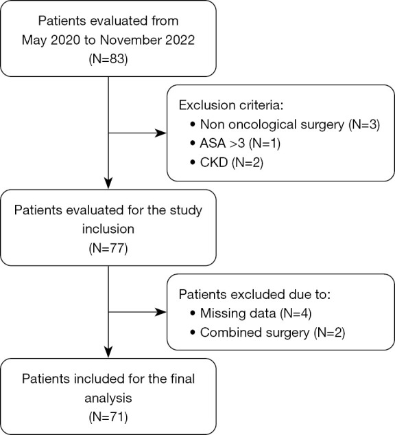
During the study period, 83 esophagectomies were performed in our center. Seventy-one patients were included in the final data analysis, as the study flow chart (Figure 1) shows. Thirty-one patients represent the HFNC cohort, while the remaining 40 patients represent the HC. This last cohort included patients who underwent esophagectomy from May 2020 to May 2021 until the sample size was reached.
Figure 1.

Study flow-chart. ASA, American Society of Anesthesiologists; CKD, chronic kidney disease.
Patients were mainly men (90%) with a mean age of 64±10. Most patients were classified as having an ASA 2 score (54%). All patients can be considered at high risk (42.1% risk) of PPCs according to ARISCAT score.
Comparisons between patients’ baseline characteristics in HFNC and the HC showed no significant differences (Table 1).
Table 1. Baseline characteristics of patients.
| Characteristics | Overall (n=71) | HFNC (n=31) | HC (n=40) | P value |
|---|---|---|---|---|
| Male (%) | 90 | 93 | 88 | 0.65 |
| Age (years) | 64±10 | 63.9±9 | 64.1±11 | 0.71 |
| BMI (kg/m2) | 26 [24–29] | 26 [24–29] | 27.5 [25–30] | 0.16 |
| ASA score | 0.08 | |||
| 1 | 3 [4] | 0 | 3 [8] | |
| 2 | 38 [54] | 14 [45] | 24 [60] | |
| 3 | 30 [42] | 17 [55] | 13 [33] | |
| CCI | 4 [3–5] | 4 [3–6] | 5 [3–6] | 0.80 |
| ARISCAT score | 50 [50–50] | 50 [50–50] | 50 [50–50] | 0.70 |
| SpO2/FiO2 | 461 [447–471] | 461 [447–466] | 462 [461–471] | 0.60 |
| PreOp weight loss (% of BW) | 10 [6–18] | 10 [5–14] | 10 [6–19] | 0.32 |
| PreOp NS | 38 [54] | 16 [52) | 22 [55] | 0.50 |
Data are expressed as mean ± standard deviation, percentage/absolute frequency and percentage [%] or as median and interquartile ranges [25–75]. HFNC, high flow nasal cannula cohort; HC, historical cohort; BMI, body mass index; ASA, American Society of Anesthesiologists; CCI, Charlson’s Comorbidity Index; ARISCAT, Assess Respiratory Risk in Surgical Patients in Catalonia; PreOP, preoperative; BW, body weight; NS, nutritional support.
Adenocarcinoma was the most frequent histological type of cancer (72%) in the lower third part of the esophagus (85%), as shown in Table 2.
Table 2. Pre-operative oncological data.
| Pre-operative data | Overall (n=71) | HFNC (n=31) | HC (n=40) | P value |
|---|---|---|---|---|
| Type of tumor, n [%] | 0.43 | |||
| ADK | 51 [72] | 24 [77] | 27 [68] | |
| SCC | 20 [28] | 7 [23] | 13 [33] | |
| Site, n [%] | 0.42 | |||
| Middle | 11 [15] | 6 [19] | 5 [13] | |
| Lower | 60 [85] | 25 [81] | 35 [88] | |
| cTNM VIII edition, n [%] | 0.36 | |||
| I | 3 [4] | 2 [6] | 1 [3] | |
| II | 6 [8] | 3 [10] | 3 [8] | |
| III | 60 [85] | 25 [81] | 35 [88] | |
| IV | 2 [3] | 1 [3] | 1 [3] | |
| CHT neo, n [%] | 21 [30] | 10 [32] | 11 [28] | 0.79 |
| CHT-RT neo, n [%] | 39 [55] | 20 [65] | 19 [48] | 0.15 |
HFNC, high flow nasal cannula cohort; HC, historical cohort; ADK, adenocarcinoma; SCC, squamous carcinoma; cTNM, clinical tumor-node-metastasis; CHT, chemotherapy; neo, neoadjuvant; CHT-RT, chemoradiotherapy.
Fifty-three patients (75%) underwent the open Ivor-Lewis approach (Table 3).
Table 3. Main surgical and anesthesiological intraoperative data.
| Intraoperative data | Overall (n=71) | HFNC (n=31) | HC (n=40) | P value |
|---|---|---|---|---|
| Type of surgery | 0.72 | |||
| Open Ivor-Lewis | 53 [75] | 23 [74] | 30 [75] | |
| MIE Ivor-Lewis | 2 [3] | 0 | 2 [5] | |
| Open McKeown | 3 [4] | 2 [6] | 1 [3] | |
| MIE McKeown | 13 [18] | 6 [19] | 7 [18] | |
| Length of surgery (min) | 305 [240–350] | 330 [280–370] | 267 [230–341] | <0.01 |
| Total IOP fluids (mL) | 3,500 [3,000–4,460] | 3,500 [2,300–4,500] | 3,200 [2,550–4,000] | 0.28 |
| IOP fluid balance (mL) | 1,550 [725–2,238] | 1,850 [1,050–2,438] | 1,500 [625–2,150] | 0.21 |
| TVABD (mL/kg IBW) | 7.6 [7.2–8.1] | 7.6 [7–8] | 7.6 [7.4–8.4] | 0.27 |
| TVTHOR (mL/kg IBW) | 5.5 [4.8–6.2] | 5.5 [4.6–6.1] | 5.6 [5–6.2] | 0.13 |
| PEEPABD (cmH2O) | 5 [5–5] | 5 [5–6] | 5 [5–5] | 0.84 |
| PEEPTHOR (cmH2O) | 5 [4–6] | 5 [3–5] | 5 [5–7] | 0.04 |
| TIVA | 6 [8] | 3 [10] | 3 [8] | 0.74 |
Data are expressed as absolute frequency and percentage [%] or as median and interquartile ranges [25–75]. HFNC, high flow nasal cannula cohort; HC, historical cohort; MIE, minimally invasive esophagectomy; IOP, intraoperative; TV, tidal volume; ABD, abdominal phase; IBW, ideal body weight; THOR, thoracic phase; PEEP, positive end-expiratory pressure; TIVA, total intravenous anesthesia.
The HC showed a shorter duration of surgery (P<0.01). No significative difference among intraoperative anesthesiological data was revealed when comparing the two cohorts except regarding the lower PEEP level in the control than in the HFNC cohort, P=0.04 (Table 3).
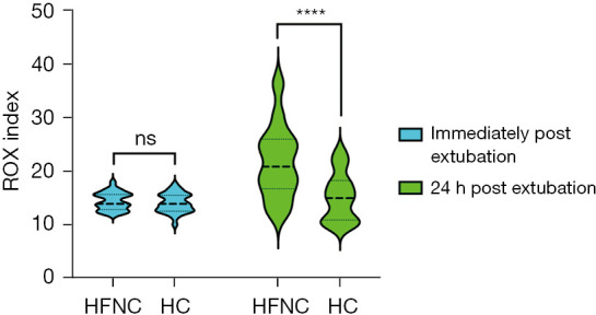
ROX index was similar just after extubation in the two cohorts, 13.8 [IQR, 12.8–15.6] vs. 13.7 [IQR, 12–15.4] in HFNC and HC cohort respectively (P=0.547).
Higher ROX index value was recorded 24 hours after extubation in HFNC than HC cohort, 20.8 [IQR, 16.7–25.9] vs. 14.9 [IQR, 10.8–18.2] respectively (P<0.0001) as shown in Figure 2.
Figure 2.

ROX index immediately and 24 hours after extubation. In the left part (light blue box violin plot), ROX index was similar just after extubation in the two cohorts. On the opposite, 24 hours after extubation (light green box violin plot) ROX index was higher in the HFNC than HC cohort. ns, not significant; ****, P<0.0001. HFNC, high flow nasal cannula cohort; HC, historical cohort; ROX, respiratory oxygenation.
RR was significantly lower 24 hours after extubation in HFNC than HC cohort, 15 [IQR, 13–18] vs. 21 [IQR, 18–23] apm (P<0.0001) respectively.
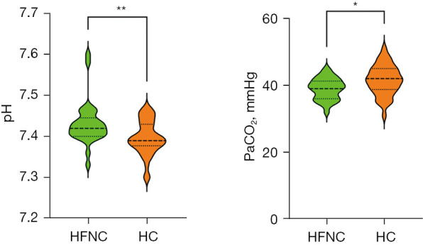
Regarding blood gas analysis, in the HFNC cohort patients’ pH was higher, 7.42 [IQR, 7.40–7.44] vs. 7.39 [IQR, 7.37–7.43] (P<0.001), than HC, and PaCO2 was lower in HFNC cohort compared with HC, 39 [IQR, 36–41] vs. 42 [IQR, 39–45] mmHg, respectively (P=0.01), as shown in Figure 3.
Figure 3.

Blood gas analysis at 24 hours after extubation. pH was higher and PaCO2 lower in HFNC cohort than HC cohort. *, P=0.01; **, P<0.001. HFNC, high flow nasal cannula cohort; HC, historical cohort.
The mean flow delivered through HFNC was 47±6 L/min at a median temperature of 34 [IQR, 31–37] °C. In most cases (65%), patients tolerated the prescribed gas flow rate. In the remaining (35%), gas flow or temperature was reduced to achieve the patients’ comfort.
RAS was similar between the two cohorts of patients, 1.5±0.98 vs. 1.4±1.04 in the HFNC and the HC cohort, respectively (P=0.611).
A lower postoperative respiratory complication was found in the HFNC patients than in the HC patients regarding pneumonia and anastomotic leak, even though it did not reach statistical significance (Table 4).
Table 4. Post-operative complications.
| Complications | Overall (n=71) | HFNC (n=31) | HC (n=40) | P value |
|---|---|---|---|---|
| Overall, n [%] | 35 [49] | 15 [48] | 20 [50] | 0.89 |
| Pulmonary†, n [%] | 18 [25] | 8 [26] | 10 [25] | 0.93 |
| Pneumonia | 15 [21] | 6 [19] | 9 [23] | 0.74 |
| Drained pleural effusion | 3 [4] | 1 [3] | 2 [5] | 0.73 |
| PNX requiring drainage | 2 [3] | 1 [3] | 1 [3] | 0.86 |
| ARF | 5 [7] | – | 5 [13] | 0.06 |
| Cardiovascular, n [%] | 13 [18] | 6 [19] | 7 [18] | 0.84 |
| AF | 11 [15] | 5 [16] | 6 [15] | 0.90 |
| CA | 1 [1] | 1 [3] | – | – |
| PE | 1 [1] | – | 1 [3] | – |
| Surgical, n [%] | 8 [11] | 3 [10] | 5 [13] | 0.71 |
| Anastomotic leak | 7 [10] | 2 [6] | 5 [13] | 0.40 |
| Vocal cord paralysis | 1 [1] | 1 [3] | – | – |
| Infectious, n [%] | 7 [10] | 2 [6] | 5 [13] | 0.72 |
†, the number of pulmonary complications represents each single patient with at least one pulmonary complication. However, some patients experienced more than one complication. For this reason, while listing all complication, the sum does not equal to the number reported in pulmonary. HFNC, high flow nasal cannula cohort; HC, historical cohort; PNX, pneumothorax; ARF, acute respiratory failure; AF, atrial fibrillation; CA, cardiac arrest; PE, pulmonary embolism.
A higher rate of ARF was registered among the HC cohort than in the HFNC, 13% vs. 0% (P=0.06), see Table 5. The five patients to whom ARF was diagnosed developed low oxygen saturation (SpO2 <92%) after 46 [IQR, 40–54] hours from extubation. Bacterial pneumonia was diagnosed in three of them, and antibiotic treatment improved the condition. For the remaining two patients, a negative computed tomography (CT) scan ruled out pulmonary embolism but revealed large right atelectasis, so fiberoptic bronchoscopy revealed bronchial obstruction due to secretions with subsequent improvement after their removal.
Table 5. Post-operative and hospital data.
| Hospital data | Overall (n=71) | HFNC (n=31) | HC (n=40) | P value |
|---|---|---|---|---|
| LOSICU (days) | 1 [1–2] | 1 [1–2] | 1 [1–3] | 0.40 |
| LOSHOSP (days) | 15 [12–20] | 15 [13–19] | 15 [12–22] | 0.97 |
| Food intake (days) | 10 [8–14] | 11 [9–14] | 10 [8–13] | 0.38 |
| 30-day mortality | 0 | 0 | 0 | – |
| 180-day mortality | 0 | 0 | 0 | – |
Data are expressed as median and interquartile ranges [25–75] or percentage (%). HFNC, high flow nasal cannula cohort; HC, historical cohort; LOSICU, length of ICU stay; ICU, intensive care unit; LOSHOSP, length of hospital stay.
One required reintubation and mechanical ventilation for 7 days, and four cases were treated with HFNC lasting 3 [IQR, 2–4] days.
Median length of hospital stay (LOSHOSP) was 15 [IQR, 12–20] days in both groups. No death was registered at the 180-day follow-up (Table 5).
Discussion
The main finding of this study is that early application of HFNC after esophagectomy significantly improved blood gas analysis and ROX index. At the same time, the effect of HFNC on postoperative atelectasis reduction is negligible.
Esophagectomy still carries a high rate of postoperative complications, and early extubation after esophagectomy remains a matter of debate (21). Although prolonged invasive mechanical ventilation can lead to higher rates of pneumonia and barotrauma, early ARF after extubation with reintubation significantly worsens patients’ outcomes (22). Moreover, patients who underwent esophagectomy are considered at high risk of reintubation (23). A supportive tool that mediates between these opposite situations is consequently advocated. Evidence from large randomized trials in major abdominal, thoracic, and cardiac surgery has highlighted the possible benefits from early post-extubation application of HFNC in terms of reduced need for reintubation and LOSHOSP (24). However, substantial evidence in the specific setting of esophageal surgery needs to be better documented. Xia and colleagues analyzed the effect of HFNC in the setting of post-esophagectomy ARF. In their retrospective study they found that HFNC improved hypoxemia, increased the flow sputum and reduced post-operative pulmonary complications (25).
From a pathophysiological point of view, the anticipated benefits of the application of HFNC are (I) delivery of heated and humified gas with improved secretions clearance and (II) increased dead space washout and support of a low level of PEEP (26). The net effect of these mechanisms of action should sustain improvements in some critical patients, which could decrease atelectasis, increase oxygenation, improve CO2 elimination with reduced RR, and reduce inspiratory effort and work of breathing, finally reducing dyspnea and ameliorating clinical outcomes (27). All these benefits are desirable after esophagectomy, but they need to be proven. We found that in patients treated with HFNC, the level of PaCO2 was significantly lower than in the HC group, with a consequently higher pH value. Moreover, patients demonstrated a significantly lower RR in the HFNC cohort. As a final result, the ROX index was considerably higher in the HFNC cohort (20.8 vs. 14.9, P<0.0001) than the HC one. Our results support the HFNC effect of more efficient dead space washout (26).
In a recent physiologic study, Mauri et al. demonstrated that ROX index increase depends on the flow rate set with HFNC, highlighting how higher values were reached at higher flow rates (28). For practical reasons, we chose to deliver 50 or 60 lt/min according to the patient’s body weight. At this point, we should consider that HFNC often requires temperature and flow rate adjustments to achieve the patient’s comfort and tolerance. In our study, 65% of patients tolerated the set flow rate, while the remaining required lower than predefined values due to poor tolerability. Higher flow rates are probably better tolerated by ARF patients, such as the critically ill, who immediately feel the benefit. In contrast, the lower tolerance observed in this study could reflect the better oxygenation of this postoperative category of patients. Although the ROX index has been validated for reintubation risk prediction after the institution of HFNC, it can be considered a global compound of respiratory function (29).
Postoperative patients with less RR and better oxygenation, i.e., lower work of breathing, could perform physiotherapy and mobilization sooner and better, speeding up the recovery phase.
A recent meta-analysis demonstrated that postoperative rehabilitation resulted in a lower incidence of pneumonia, a shorter LOSHOSP, and better health related quality of life scores for dyspnea and physical functioning (30).
However, this was not the main focus of this study and was not properly investigated.
Pulmonary complications after esophagectomy are supposed to be caused by atelectasis (31). Many factors contribute to developing atelectasis after esophagectomy: intraoperative one-lung ventilation, pneumoperitoneum or induced pneumothorax, mediastinal dissection, and the necessity of lung retraction to optimize esophageal exposure. Indeed, some postoperative factors also increase the risk of their formation, such as diaphragmatic dysfunction with consequent impaired cough, pain, and reduced ability to clear tracheobronchial secretions. In addition, esophagectomy comprehends abdominal and thoracic cavity access, with all the consequent complications of both these types of major surgery. Atelectasis occurs 24–72 h after surgery with various degrees of clinical signs, from mild to severe symptoms of respiratory failure requiring endotracheal intubation and mechanical ventilation (32). Consequently, applying HFNC theoretically should reduce atelectasis formation through the delivery of low levels of PEEP. However, we were not able to demonstrate this in our study. RAS score was similar in the two cohorts of patients. We acknowledge that the RAS score has limitations, and a CT scan would be the examination of choice. However, a chest X-ray is sufficiently useful after esophagectomy unless a clinical scenario requires other image modalities. Moreover, it implies lower radiation exposure than a CT scan. This finding carries two considerations: atelectasis probably is not the sole hit that leads to PPCs, and, secondly, PEEP generated by HFNC could not be sufficient to reopen closed alveoli within atelectasis (33). This last point is a matter of debate. If some evidence supports that HFNC, especially at high flow rates, produces PEEP near 5 cmH2O, simply opening the mouth decreases the PEEP level at lower values (about 1 cmH2O), making improbable a net effect or alveolar recruitment (34). Moreover, it cannot be excluded that besides recruitment in dependent lung regions, HFNC could also overdistend the nondependent parts without oxygenation improvement (35). More evidence is needed in this regard. Evidence highlights that postoperative complication worsens patient outcomes (36).
But it is not surprising that the overall complication rate reached 49%. In a recent benchmarking study from the Esophageal Complications Consensus Group (ECCG)’s large database (ESODATA), 59% of patients developed postoperative complications, with PPC the most represented group (27.8% in 1,595 patients) (37,38).
Patients undergoing esophagectomy have many concomitant risk factors for postoperative complications. Pre-operative ones such as nutritional status or neoadjuvant CHT-RT, intraoperative factors like fluid administration and mechanical ventilation, and postoperative ones such as adequate mobilization can all contribute to the patient’s outcome (39). Moreover, it is difficult to extrapolate a single factor that, per se, could influence a patient’s outcome. In this regard, HFNC is part of a continuum of care that has been demonstrated to improve postoperative pulmonary function in this study. Even though we did not record significant differences in terms of PPC between groups, it is worth noting that no reintubation within 7 days nor ARF has been observed in the HFNC cohort of patients, while 5 (13%) patients in the HC had an ARF (P=0.06). Whether the systematic application of HFNC early after esophagectomy will improve outcomes still needs to be determined. In this regard, an ongoing randomized multicenter study (NCT05718284) will produce more data and evidence on this important topic with a strong physiological basis.
This study has some limitations. First, its retrospective design limits some argumentations, and the lack of randomization carries selection biases. Second, this is a single-center study, so generalizability could be questioned. Third, we did not record the tolerability of HFNC after 24 hours, but this was not the topic of this preliminary study because it was already explored in a pilot study, as was previously mentioned. Consequently, we cannot exclude that if higher rates had been delivered, some additional benefit could have further ameliorated postoperative outcomes.
Conclusions
In conclusion, HFNC improved the oxygenation index after esophagectomy. This tool should be considered for early respiratory support after extubation in this category of patients, not only as a rescue therapy for ARF, but also to optimize early postoperative respiratory function. Whether this will improve patients’ outcomes requires further large randomized controlled trials.
Supplementary
The article’s supplementary files as
Acknowledgments
Funding: None.
Ethical Statement: The authors are accountable for all aspects of the work in ensuring that questions related to the accuracy or integrity of any part of the work are appropriately investigated and resolved. The study was conducted in accordance with the Declaration of Helsinki (as revised in 2013). This observational retrospective study was conducted after it received the Institutional Review Board of the University of Udine’s approval (No. #176-2022). Each patient’s willingness to participate was obtained through a signed general informed consent for research purposes G.E.C.O. and the European General Data Protection Regulation 2016/679 (G.D.P.R.) was respected.
Footnotes
Reporting Checklist: The authors have completed the STROBE reporting checklist. Available at https://jtd.amegroups.com/article/view/10.21037/jtd-23-1176/rc
Data Sharing Statement: Available at https://jtd.amegroups.com/article/view/10.21037/jtd-23-1176/dss
Peer Review File: Available at https://jtd.amegroups.com/article/view/10.21037/jtd-23-1176/prf
Conflicts of Interest: All authors have completed the ICMJE uniform disclosure form (available at https://jtd.amegroups.com/article/view/10.21037/jtd-23-1176/coif). The authors have no conflicts of interest to declare.
References
- 1.International Agency for Research on Cancer, World Health Organization. (Accessed on 10th July 2023). Available online: https://gco.iarc.fr/tomorrow/en/dataviz/isotype?cancers=6&single_unit=50000
- 2.Movahed S, Norouzy A, Ghanbari-Motlagh A, et al. Nutritional Status in Patients with Esophageal Cancer Receiving Chemoradiation and Assessing the Efficacy of Usual Care for Nutritional Managements. Asian Pac J Cancer Prev 2020;21:2315-23. 10.31557/APJCP.2020.21.8.2315 [DOI] [PMC free article] [PubMed] [Google Scholar]
- 3.Bartels K, Fiegel M, Stevens Q, et al. Approaches to perioperative care for esophagectomy. J Cardiothorac Vasc Anesth 2015;29:472-80. 10.1053/j.jvca.2014.10.029 [DOI] [PubMed] [Google Scholar]
- 4.Low DE, Allum W, De Manzoni G, et al. Guidelines for Perioperative Care in Esophagectomy: Enhanced Recovery After Surgery (ERAS®) Society Recommendations. World J Surg 2019;43:299-330. 10.1007/s00268-018-4786-4 [DOI] [PubMed] [Google Scholar]
- 5.Deana C, Vetrugno L, Bignami E, et al. Peri-operative approach to esophagectomy: a narrative review from the anesthesiological standpoint. J Thorac Dis 2021;13:6037-51. 10.21037/jtd-21-940 [DOI] [PMC free article] [PubMed] [Google Scholar]
- 6.Yu KY, Zhao L, Chen Z, et al. Noninvasive positive pressure ventilation for the treatment of acute respiratory distress syndrome following esophagectomy for esophageal cancer: a clinical comparative study. J Thorac Dis 2013;5:777-82. 10.3978/j.issn.2072-1439.2013.09.09 [DOI] [PMC free article] [PubMed] [Google Scholar]
- 7.Charlesworth M, Lawton T, Fletcher S. Noninvasive positive pressure ventilation for acute respiratory failure following oesophagectomy: Is it safe? A systematic review of the literature. J Intensive Care Soc 2015;16:215-21. 10.1177/1751143715571698 [DOI] [PMC free article] [PubMed] [Google Scholar]
- 8.Frat JP, Thille AW, Mercat A, et al. High-flow oxygen through nasal cannula in acute hypoxemic respiratory failure. N Engl J Med 2015;372:2185-96. 10.1056/NEJMoa1503326 [DOI] [PubMed] [Google Scholar]
- 9.Rochwerg B, Granton D, Wang DX, et al. High flow nasal cannula compared with conventional oxygen therapy for acute hypoxemic respiratory failure: a systematic review and meta-analysis. Intensive Care Med 2019;45:563-72. 10.1007/s00134-019-05590-5 [DOI] [PubMed] [Google Scholar]
- 10.Chaudhuri D, Granton D, Wang DX, et al. High-Flow Nasal Cannula in the Immediate Postoperative Period: A Systematic Review and Meta-analysis. Chest 2020;158:1934-46. 10.1016/j.chest.2020.06.038 [DOI] [PubMed] [Google Scholar]
- 11.Hsiao WL, Hung WT, Yang CH, et al. Effects of high flow nasal cannula following minimally invasive esophagectomy in ICU patients: A prospective pre-post study. J Formos Med Assoc 2023;122:1247-54. 10.1016/j.jfma.2023.05.016 [DOI] [PubMed] [Google Scholar]
- 12.Deana C, Rovida S, Orso D, et al. Learning from the Italian experience during COVID-19 pandemic waves: be prepared and mind some crucial aspects. Acta Biomed 2021;92:e2021097. 10.23750/abm.v92i2.11159 [DOI] [PMC free article] [PubMed] [Google Scholar]
- 13.Petri R, Zuccolo M, Brizzolari M, et al. Minimally invasive esophagectomy: thoracoscopic esophageal mobilization for esophageal cancer with the patient in prone position. Surg Endosc 2012;26:1102-7. 10.1007/s00464-011-2006-5 [DOI] [PubMed] [Google Scholar]
- 14.Vecchiato M, Martino A, Sponza M, et al. Thoracic duct identification with indocyanine green fluorescence during minimally invasive esophagectomy with patient in prone position. Dis Esophagus 2020;33:doaa030. 10.1093/dote/doaa030 [DOI] [PMC free article] [PubMed] [Google Scholar]
- 15.Kristensen SD, Knuuti J, Saraste A, et al. 2014 ESC/ESA Guidelines on non-cardiac surgery: cardiovascular assessment and management: The Joint Task Force on non-cardiac surgery: cardiovascular assessment and management of the European Society of Cardiology (ESC) and the European Society of Anaesthesiology (ESA). Eur J Anaesthesiol 2014;31:517-73. 10.1097/EJA.0000000000000150 [DOI] [PubMed] [Google Scholar]
- 16.Vecchiato M, Deana C, Ziccarelli A, et al. 263. Effects of prehabilitation and post-operative high-flow nasal cannula after open esophagectomy on pulmonary complications: a feasibility study. Diseases of the Esophagus 2022;35:doac051.263.
- 17.Canet J, Gallart L, Gomar C, et al. Prediction of postoperative pulmonary complications in a population-based surgical cohort. Anesthesiology 2010;113:1338-50. 10.1097/ALN.0b013e3181fc6e0a [DOI] [PubMed] [Google Scholar]
- 18.Roca O, Caralt B, Messika J, et al. An Index Combining Respiratory Rate and Oxygenation to Predict Outcome of Nasal High-Flow Therapy. Am J Respir Crit Care Med 2019;199:1368-76. 10.1164/rccm.201803-0589OC [DOI] [PubMed] [Google Scholar]
- 19.Low DE, Alderson D, Cecconello I, et al. International Consensus on Standardization of Data Collection for Complications Associated With Esophagectomy: Esophagectomy Complications Consensus Group (ECCG). Ann Surg 2015;262:286-94. 10.1097/SLA.0000000000001098 [DOI] [PubMed] [Google Scholar]
- 20.Richter Larsen K, Ingwersen U, Thode S, et al. Mask physiotherapy in patients after heart surgery: a controlled study. Intensive Care Med 1995;21:469-74. 10.1007/BF01706199 [DOI] [PubMed] [Google Scholar]
- 21.Serafim MCA, Orlandini MF, Datrino LN, et al. Is early extubation after esophagectomy safe? A systematic review and meta-analysis. J Surg Oncol 2022;126:68-75. 10.1002/jso.26821 [DOI] [PubMed] [Google Scholar]
- 22.Whitmore D, Mahambray T. Reintubation following planned extubation: incidence, mortality and risk factors. Intensive Care Med Exp 2015;3:A684. [Google Scholar]
- 23.Liu JL, Jin JW, Lin LL, et al. Emergency tracheal intubation peri-operative risk factors and prognostic impact after esophagectomy. BMC Anesthesiol 2022;22:367. 10.1186/s12871-022-01918-9 [DOI] [PMC free article] [PubMed] [Google Scholar]
- 24.Lu Z, Chang W, Meng S, et al. The Effect of High-Flow Nasal Oxygen Therapy on Postoperative Pulmonary Complications and Hospital Length of Stay in Postoperative Patients: A Systematic Review and Meta-Analysis. J Intensive Care Med 2020;35:1129-40. 10.1177/0885066618817718 [DOI] [PubMed] [Google Scholar]
- 25.Xia M, Li W, Yao J, et al. A postoperative comparison of high-flow nasal cannula therapy and conventional oxygen therapy for esophageal cancer patients. Ann Palliat Med 2021;10:2530-9. 10.21037/apm-20-1539 [DOI] [PubMed] [Google Scholar]
- 26.Rochwerg B, Einav S, Chaudhuri D, et al. The role for high flow nasal cannula as a respiratory support strategy in adults: a clinical practice guideline. Intensive Care Med 2020;46:2226-37. 10.1007/s00134-020-06312-y [DOI] [PMC free article] [PubMed] [Google Scholar]
- 27.Goligher EC, Slutsky AS. Not Just Oxygen? Mechanisms of Benefit from High-Flow Nasal Cannula in Hypoxemic Respiratory Failure. Am J Respir Crit Care Med 2017;195:1128-31. 10.1164/rccm.201701-0006ED [DOI] [PubMed] [Google Scholar]
- 28.Mauri T, Carlesso E, Spinelli E, et al. Increasing support by nasal high flow acutely modifies the ROX index in hypoxemic patients: A physiologic study. J Crit Care 2019;53:183-5. 10.1016/j.jcrc.2019.06.020 [DOI] [PubMed] [Google Scholar]
- 29.Gallardo A, Zamarrón-López E, Deloya-Tomas E, et al. Advantages and limitations of the ROX index. Pulmonology 2022;28:320-1. 10.1016/j.pulmoe.2022.02.008 [DOI] [PMC free article] [PubMed] [Google Scholar]
- 30.Tukanova KH, Chidambaram S, Guidozzi N, et al. Physiotherapy Regimens in Esophagectomy and Gastrectomy: a Systematic Review and Meta-Analysis. Ann Surg Oncol 2022;29:3148-67. 10.1245/s10434-021-11122-7 [DOI] [PMC free article] [PubMed] [Google Scholar]
- 31.Hua R, Jiang H, Sun Y, et al. Postoperative complications and management of minimally invasive esophagectomy. Shanghai Chest 2018;2:57. [Google Scholar]
- 32.Lagier D, Zeng C, Fernandez-Bustamante A, et al. Perioperative Pulmonary Atelectasis: Part II. Clinical Implications. Anesthesiology 2022;136:206-36. 10.1097/ALN.0000000000004009 [DOI] [PMC free article] [PubMed] [Google Scholar]
- 33.Chertoff J. High-Flow Oxygen, Positive End-Expiratory Pressure, and the Berlin Definition of Acute Respiratory Distress Syndrome: Are They Mutually Exclusive? Am J Respir Crit Care Med 2017;196:396-7. 10.1164/rccm.201701-0005LE [DOI] [PubMed] [Google Scholar]
- 34.Ritchie JE, Williams AB, Gerard C, et al. Evaluation of a humidified nasal high-flow oxygen system, using oxygraphy, capnography and measurement of upper airway pressures. Anaesth Intensive Care 2011;39:1103-10. 10.1177/0310057X1103900620 [DOI] [PubMed] [Google Scholar]
- 35.Li J, Albuainain FA, Tan W, et al. The effects of flow settings during high-flow nasal cannula support for adult subjects: a systematic review. Crit Care 2023;27:78. 10.1186/s13054-023-04361-5 [DOI] [PMC free article] [PubMed] [Google Scholar]
- 36.Deana C, Vetrugno L, Stefani F, et al. Postoperative complications after minimally invasive esophagectomy in the prone position: any anesthesia-related factor? Tumori 2021;107:525-35. 10.1177/0300891620979358 [DOI] [PubMed] [Google Scholar]
- 37.Low DE, Kuppusamy MK, Alderson D, et al. Benchmarking Complications Associated with Esophagectomy. Ann Surg 2019;269:291-8. 10.1097/SLA.0000000000003382 [DOI] [PubMed] [Google Scholar]
- 38.Xu SJ, Lin LQ, Chen C, et al. Textbook outcome after minimally invasive esophagectomy is an important prognostic indicator for predicting long-term oncological outcomes with locally advanced esophageal squamous cell carcinoma. Ann Transl Med 2022;10:161. 10.21037/atm-22-506 [DOI] [PMC free article] [PubMed] [Google Scholar]
- 39.Della Rocca G, Vetrugno L, Coccia C, et al. Preoperative Evaluation of Patients Undergoing Lung Resection Surgery: Defining the Role of the Anesthesiologist on a Multidisciplinary Team. J Cardiothorac Vasc Anesth 2016;30:530-8. 10.1053/j.jvca.2015.11.018 [DOI] [PubMed] [Google Scholar]
Associated Data
This section collects any data citations, data availability statements, or supplementary materials included in this article.
Supplementary Materials
The article’s supplementary files as


