Abstract
Introduction
Mycobacterium simiae is a slow-growing non-tuberculous mycobacterium that can cause non-tuberculous mycobacterium (NTM) pulmonary disease and extrapulmonary infections. Until now, detailed genomic and clinical characteristics, as well as possible transmission routes of this rare pathogen remain largely unknown.
Methods
We conducted whole genome sequencing of available M. simiae isolates collected at a tertiary care centre in Central Germany from 2006 to 2020 and set them into context with publicly available M. simiae complex sequences through phylogenetic analysis. Resistance, virulence and stress genes, as well as known Mycobacteriaceae plasmid sequences were detected in whole genome raw reads. Clinical data and course were retrieved and correlated with genomic data.
Results
We included 33 M. simiae sensu stricto isolates from seven patients. M. simiae showed low clinical relevance with only two patients fulfilling American Thoracic Society (ATS) criteria in our cohort and three receiving NTM-effective therapy. The bacterial populations were highly stable over time periods of up to 14 years, and no instances of mixed or re-infections with other strains of M. simiae were observed. Clustering with <12 single nucleotide polymorphisms distance was evident among isolates from different patients; however, proof for human-to-human transmission could not be established from epidemiological data.
Conclusion
Overall, the available sequence data for M. simiae complex was significantly extended and new insights into its pathogenomic traits were obtained. We demonstrate high longitudinal genomic stability within single patients. Although we cannot exclude human-to-human transmission, we consider it unlikely in the light of available epidemiological data.
Shareable abstract
Mycobacterium simiae is a rare cause of NTM pulmonary disease. This study demonstrates high longitudinal genomic stability of the pathogen, as well as low clinical relevance in this cohort. https://bit.ly/3SFl9kL
Introduction
Mycobacterium simiae is a slow-growing non-tuberculous mycobacterium (NTM) first isolated from a rhesus macaque (Macaca mulatta) in 1965 [1]. It belongs to the M. simiae complex, which currently comprises 20 species (M. simiae, M. genavense, Mycobacterium lentiflavum and others) [2, 3]. The bacterium has been isolated from humans and animals, but also environmental sources such as patient shower, source and tap water [4]. In humans, M. simiae can lead to various extrapulmonary manifestations, such as lymphadenitis in children, disseminated disease in immunocompromised hosts and NTM pulmonary disease (NTM-PD) [5]. NTM-PD is characterised by symptoms such as severe cough, increased sputum production, haemoptysis and chest pain [5]. However, the clinical relevance of isolation from pulmonary sources has been rated low in comparison to other NTM species, as the diagnostic criteria of the American Thoracic Society (ATS) have been shown to be only fulfilled in 21% of cases in a European setting [6, 7].
M. simiae exhibits a differential geographical distribution [8] being more common in the Middle East and Central Asia [9]. In Germany, it has been described to be rarely isolated from patients with cystic fibrosis (CF) and patients with non-CF bronchiectasis [10]. A Dutch case series comprised just 28 nationwide cases for the whole country during a 7-year period [6]. Optimal treatment of infection due to M. simiae has not yet been established, but the current consensus guideline for the treatment of less common NTM infections recommends a regimen consisting of a minimum of three drugs (out of a macrolide, amikacin, moxifloxacin, clofazimine and cotrimoxazole) [11].
Genomic data for M. simiae first became available with sequencing of the type strain DSM 44165 in 2013, which has a high GC content (65.2%) and a length of ∼5.8 megabase pairs [12]. Since then, other mycobacteria have been classified into the M. simiae complex based on 16S ribosomal DNA sequence identity [2]. Still, sequencing data of M. simiae complex members remains scarce.
NTMs are readily found in the environment as well as in household, recreational area and hospital settings [13]. They can be transmitted to humans through activities such as gardening [14], showering [15] or swimming [16] and via contaminated drinking water sources [17, 18]. NTM infections have also been frequently related to medical procedures [19], while human-to-human transmission is considered rare [20, 21]. Transmission routes and reservoirs are largely unknown for M. simiae strains.
For M. abscessus, one of the most clinically relevant NTMs, clusters of genetically closely related isolates, so-called dominant circulating clones, have been demonstrated in a large population of CF and non-CF patients [20, 22]. In M. avium complex, the picture is more diverse, although clustering could be observed as well [23]. However, for less common NTM species, similar data are lacking. Furthermore, whether M. simiae isolates in a single patient remain stable with regard to their genome over a certain period of time or re-infection takes place, as is frequently described in M. avium complex [24], is not known.
To get more insight into the global population structure of M. simiae complex, we analysed publicly available genomic data of M. simiae complex isolates (n=91) together with 36 new genomes sequenced within the scope of this study. In addition, we analysed possible in-hospital transmission and the clinical characteristics and course of M. simiae infection or colonisation.
Methods
Clinical and radiological assessment
Patients with M. simiae isolates collected between 2006 and 2020 were identified retrospectively at University Hospital Frankfurt. We retrieved clinical data, such as age, sex, disposition (CF, malignoma, HIV, structural lung disease), immunosuppressive therapies (such as steroids, mycophenolatemofetil, ciclosporin, tacrolimus, azathioprine, methothrexate, tumour necrosis factor-α inhibitors), NTM therapy, clinical outcome and lung function during the course of time by chart review. Diagnostic criteria for relevant NTM pulmonary disease were applied according to the ATS [5]. For patients with pulmonary isolates and available computed tomography scans of the lung, the radiological score by Song et al. [25] was applied by a radiologist to all images that were made during the period of positive cultures of M. simiae. This study was approved by our local ethics committee under file number 2022–672.
Culture and sequencing
M. simiae isolates were cultivated on Middlebrook Agar 7H10 at 37°C until visible growth was detected. DNA extraction was performed with the cetyltrimethylammonium bromide (CTAB) protocol as previously described [21]. Next-generation sequencing libraries were generated from extracted genomic DNA, using a modified Illumina Nextera library kit protocol [26]. Libraries were sequenced in a 150-bp paired-end run on an Illumina NextSeq 500 or 2000 instrument (Illumina, San Diego, CA, USA). All sequence reads generated can be accessed under project number PRJEB66336 at the European Nucleotide Archive.
Bioinformatical analysis
We downloaded all available whole genome sequence data of the M. simiae complex available on NCBI on 31 December 2022 including the reference genomes of M. simiae, M. sherrisii, M. genavense, M. ahvazicum, M. lentiflavum, M. montefiorense, M. kubicae, M. triplex, M. florentinum, M. stomatepiae, M. shigaense, M. palustre, M. heidelbergense, M. parascrofulaceum, M. saskawatchewanense, M. europeaum, M. interjectum, M. intermedium, M. paraense and M. parmense, as well as available sequence data of non-reference strains within the M. simiae complex (table S1). Available metadata of these sequences were recorded. For isolates with only genomic assemblies available, raw reads were simulated using dwgsim v. 0.1.12–13 [27]. Sequence quality of all read data was evaluated with fastqc v. 0.11.9 [28], multiqc v. 1.13 [29] and kraken2 v. 2.1.2 [30]. Fastp v.0.23.2 with --cut_tail and –cut_tail_window_size 1 was used to remove low-quality bases and adapters [31]. De novo assemblies for all genomes were generated using shovill v.1.1.0 [32]. The respective species was initially differentiated using NTM-Profiler 0.2.0 (starting from fastQ) [33] and type strain genome server (TYGS) (starting from fastA) [34]. Contaminated (>2% reads belonging to a genus other than Mycobacterium) and low-quality samples (>300 contigs, assembly failed, or estimated coverage <30) were excluded (n=43). The corresponding paired-end fastQ files were applied to the MTBseq pipeline with default settings and M. simiae JCM12377 (Accession code: DRR161197) as a reference genome [35]. A phylogenetic tree was constructed from 112.261 concatenated SNP positions (1.9% of the reference genome) with RaxML v.8.2.12 with 500 bootstraps and GTRGAMMA as a substitution model [36]. This tree was then visualised in R using the ggtree package [37]. For identified clusters within M. simiae with <12 SNPs, further in-depth analysis was performed: if the cluster contained more than one isolate of a single patient, the sequence of the first isolate was used to generate a de novo genome assembly with shovill 1.1.0. This assembly was subsequently used as a reference genome in MTBseq for another joint analysis including only isolates from the respective cluster. Re-infection or mixed infection was defined as the subsequent occurrence or co-occurrence of different strains from the M. simiae complex within the same patient, respectively.
Average nucleotide identities were calculated using fastani for every genome-to-genome combination resulting in a distance matrix [38]. Resistance, virulence and stress genes were identified with AMRfinder plus v.3.11.2 with database v. 2023-07-13.2 [39], using the assemblies as input. Known Mycobacteriaceae plasmids (n=152) downloaded from the curated plasmid database PLSDB v. 2021-06-23-v2 [40] were identified directly from short sequencing reads using SRST v.0.2.0 with default settings [41]. The 152 PLSDB plasmids were classified into 87 groups based on their mash distances (maximum mash distance between plasmids in one group = 0.05).
Network analysis and statistics
All data were analysed within R Version 4.2.2 (www.r-project.org) using tidyverse packages [42]. Concomitant hospital visits and hospitalisations in all patient-to-patient combinations were identified using R. Social interaction was defined as “no contact”, “living in the same city”, “same day at the hospital” and “same day at the same ward/outpatient clinic”. Categorical variables are depicted as frequencies with numerator and denominator, and continuous variables as mean with range for normally distributed data and median with interquartile range for non-normally distributed data. For all statistical tests a significance level of α=0.05 was used.
Results
Phylogenetic relations and taxonomy of the M. simiae complex
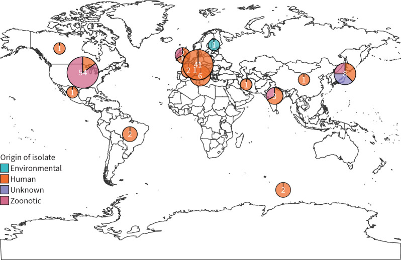
To investigate the taxonomy and global population structure of the M. simiae complex we included 33 M. simiae sensu stricto isolates, two M. interjectum isolates, one M. palustre isolate from our centre and 91 publicly available genome sequences from different geographical origins, resulting in a final dataset of 127 sequences (figures 1 and 2, supplementary table S1). The majority of isolates were of human origin (n=75), while 51 isolates were isolated from animals (mainly birds) and only one from the environment.
FIGURE 1.
Map of included Mycobacterium simiae complex isolates (public and from this study) and their respective sampling origin (environmental, human, zoonotic).
FIGURE 2.
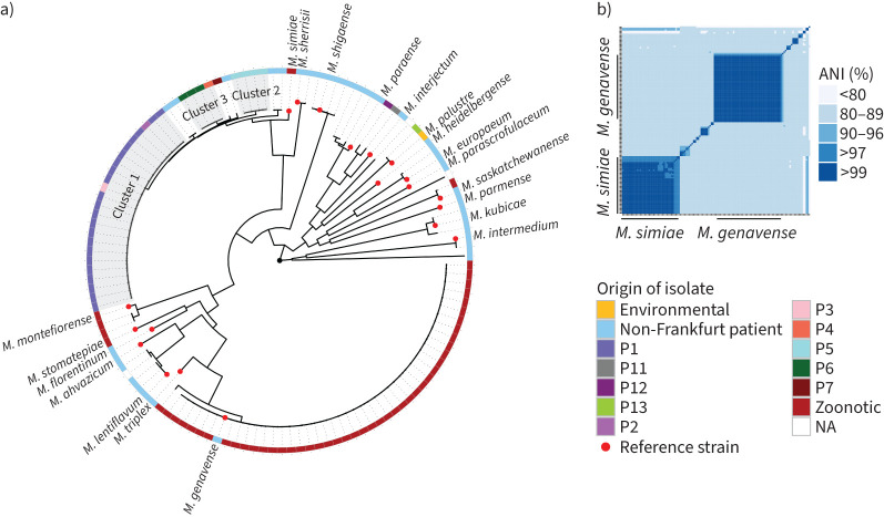
a) Phylogenetic tree of publicly available sequences and isolates from included patients attending the University Hospital Frankfurt from 2006 to 2020 and b) heat map of average nucleotide identities of included isolates. NA: not applicable; P: patient; ANI: average nucleotide identity.
While the NTM-specific tool NTMprofiler could assign species only for 63 out of 127 isolates, TYGS identified 124 out of 127 isolates up to species level. DRR171302, DRR016032 and GCA_008370645.1 are labelled as M. interjectum, M. triplex and M. simiae, respectively, in the public database NCBI, but were classified as potential new species by TYGS. All M. simiae sensu stricto isolates from our study clustered with the reference genome M. simiae JCM12377 with <500 SNPs distance (figure 2a). Average nucleotide identity (ANI) of the included sequences ranged from 79.0% to 100.00% (figure 2b). Excluding the potential new species, all intra-species comparisons had ANI comparisons above 97% and all inter-species comparisons had ANI above 79.58% with the maximum ANI being 91.74%.
Plasmids, resistance and virulence factors in M. simiae complex
Previously known Mycobacteriaceae plasmid sequences were found in 13 out of 127 isolates belonging to six M. simiae complex species (M. simiae, M. interjectum, M. kubicae, M. parascrofulaceum, M. europaeum and M. lentiflavum) (supplementary figure S1). Five out of 13 strains with plasmid sequences were isolated from three patients at our centre (all available isolates from patients P6 (n=3), P11 (n=1) and P4 (n=1)). The identified plasmid sequences belonged to eight plasmid groups (intra-group mash distance <0.05) and had lengths between 16.5 and 147 kbp. Four isolates harboured sequences belonging to more than one plasmid group, indicating the presence of multiple plasmids within one strain.
Antibiotic resistance genes were highly prevalent and found in 125 out of 127 M. simiae complex isolates. The only isolate from M. parascrofulaceum and one out of two M. europaeum isolates contained no known resistance markers. The most common resistance-associated genes were acc(2′)-Ic (coding for low-level aminoglycoside resistance) in 121 isolates, rgt in 114 isolates (rifamycin resistance), abaF (fosfomycin resistance) in 114 isolates, arr (rifamycin resistance) in 109 isolates, bla (β-lactam resistance) in 61 isolates and dfrA3 (trimethoprim) in 49 isolates (supplementary table S2 and figure S2). In total, 124 M. simiae complex isolates were predicted to be resistant against aminoglycosides and 119 against rifamycin. All M. simiae complex isolates had at least one stress response gene with arsC (in 126 isolates) and acr3 (in 89 isolates) being most prevalent (supplementary table S2). Only two known virulence genes were identified: katP, coding for a catalase/peroxidase, in all 127 M. simiae complex isolates; and bpsD, coding for a sugar transferase, in 19 isolates. Longitudinal analysis in patients with multiple serial isolates did not show any change in pathogenomic traits of the respective strains. Additionally, the subset of detected resistance genes was identical for all isolates within a respective species (supplementary figure S2).
Patient characteristics, clinical course and intra-individual genetic stability of M. simiae infection or colonisation
Overall, 33 M. simiae sensu stricto isolates from seven patients attending the University Hospital Frankfurt between 2006 and 2020 were included (tables 1 and 2). Five patients were male and two female. Median age at time point of first isolation of M. simiae was 41 years (IQR 36.5–66.5). Three patients were suffering from CF, two from malignoma and two from structural lung disease (non-CF bronchiectasis). One patient was suffering from HIV/AIDS. This patient was the only one receiving an immunosuppressive therapy during the observation period (prednisone) and presented with disseminated disease and isolation of M. simiae from a blood sample. All other patients had pulmonary isolation of M. simiae only. ATS criteria were fulfilled in two out of six patients with pulmonary isolates. Three out of seven patients received NTM-effective therapy during the observation period (including two patients fulfilling ATS criteria and one patient with disseminated disease, table 2). Therapies consisted of macrolides, fluoroquinolones, clofazimine, cotrimoxazole, ethambutol or rifamycins. Culture conversion was observed for all three patients receiving therapy as well as for two patients without therapy. No patient died during the observation period. In three patients, other NTM were also isolated.
TABLE 1.
General characteristics of patients with isolation of Mycobacterium simiae
| Sex | |
| Male | 5/7 |
| Female | 2/7 |
| Disposition | |
| CF | 3/7 |
| Non-CF | 4/7 |
| HIV | 1/7 |
| Malignoma | 2/7 |
| SLD | 2/7 |
| Clinical manifestation | |
| Isolated pulmonary | 6/7 |
| Isolated extrapulmonary | 0/7 |
| Disseminated | 1/7 |
| ATS criteria | 2/6 |
| Pulmonary symptoms and radiology | 2/6 |
| Exclusion other diagnosis | 2/6 |
| Two culture-positive sputa | 3/6 |
| Positive culture in BALF | 3/6 |
| Biopsy and culture | 1/6 |
| NTM therapy | 3/7 |
| Macrolide | 3/3 |
| Ethambutol | 1/3 |
| Rifamycin | 1/3 |
| Fluoroquinolone | 3/3 |
| Clofazimin | 1/3 |
| Cotrimoxazol | 2/3 |
| Linezolid | 1/3 |
| Deceased | 0/7 |
| Culture conversion | 5/7 |
| Other NTM isolated during the observation period | |
| M. abscessus | 2/7 |
| M. chimaera | 1/7 |
| M. intracellulare | 1/7 |
| M. chelonae | 1/7 |
CF: cystic fibrosis; SLD: structural lung disease; BALF: bronchoalveolar lavage fluid; NTM: non-tuberculous mycobacterium.
TABLE 2.
Clinical course of patients with isolation of Mycobacterium simiae
| Patient | Sex | Age years | Disposition | Clinical manifestation | Pulmonary radiology | ATS criteria fulfilled | Other NTM species | NTM therapy | Outcome |
| P1 | m | 37 | CF | Pulmonary | Bronchiectasis, nodular changes | y | M. chimaera | Clarithromycin, moxifloxacin, clofazimine, cotrimoxazole | Culture conversion, alive |
| P2 | m | 70 | Malignoma | Pulmonary | Bronchiectasis, malignoma | n | NA | n | Culture conversion, alive |
| P3 | m | 78 | Malignoma | Pulmonary | Infiltration, pleural effusion | n | NA | n | Alive |
| P4 | m | 63 | SLD (emphysema, bronchiectasis) | Pulmonary | Bronchiectasis, carnificating pneumonia | n | NA | n | Alive |
| P5 | f | 36 | CF | Pulmonary | Bronchiectasis | y | M. abscessus, M. intracellulare, M. chelonae | Clarithromycin, levofloxacin, linezolid | Culture conversion, alive |
| P6 | m | 36 | CF | Pulmonary | Bronchiectasis, infiltration | n | M. abscessus | n | Culture conversion, alive |
| P7 | f | 41 | HIV CDC Stadium C3 | Disseminated | Slight emphysema | NA | NA | Clarithromycin/azithromycin, ethambutol, rifabutin, levofloxacin, cotrimoxazole | Culture conversion, alive |
NTM: non-tuberculous mycobacterium; P: patient; CF: cystic fibrosis; SLD: structural lung disease; CDC: Centers for Disease Control and Prevention; NA: not applicable; m: male; f: female; y: yes; n: no.
Within patients with serial M. simiae isolates (P1, P5 and P6) a high intra-individual genetic stability of the bacterial isolates was observed. SNP distances were at a median of 0 SNPs over a 14-year period in patient 1, at 0 SNPs over an 18-month period in patient 5 and at 1 SNP over a 15-month period in patient 6. These SNP distances did not change substantially in the cluster-specific analyses including only sequences from a respective cluster mapped against an assembly of their primary isolate (supplementary table S3).
Two of these three patients received antimycobacterial therapy, but all of them achieved culture conversion (table 2). Two exhibited subsequent isolation of another NTM (M. chimaera and M. abscessus, respectively). The patient with the longest period of M. simiae isolation showed only slight changes in clinical presentation: vital capacity went down from 3.47 L in 2011 to 2.57 L in 2020, forced expiratory volume in 1 s from 1.67 L to 1.41 L. The CT score increased from 10 to 11 between 2015 and 2021, but was slightly improved after the initiation of NTM active therapy (supplementary figure S3).
Cluster and social network analysis
Interestingly, isolates of two patients (P2 and P3) clustered with a 0 SNP distance with isolates from patient P1, indicating two possible transmission events (cluster 1, figure 2). However, the isolate of patient P3 was taken on the same day as one of the isolates from patient P1, and the isolate of patient P2 in very close temporal vicinity. Both patients never produced any other specimen positive for M. simiae afterwards, and both did not fulfill ATS criteria. Cluster 2 consisted of isolates from a single patient. Finally, cluster 3 contained isolates from patients P4 and P6 that were not taken on the same day. One patient was suffering from CF, the other was a non-CF patient. SNP distances did not change substantially after assembling a sequence within the cluster and mapping all other reads against it, respectively (supplementary table S3).
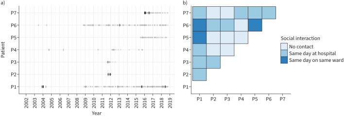
However, social network analysis demonstrated that patient P1 was in the hospital on the same day as all other patients on at least one occasion but not on the same ward (figure 3). Only three patient-to-patient combinations showed occasions on which patients were on the same ward on the same day. However, none of these patient isolates clustered together.
FIGURE 3.
a) Timeline of patients’ visits to the hospital (grey dots depict singular visits or dates, black bars represent in-hospital stays) and b) heatmap of patient interaction. P: patient.
Discussion
To our knowledge, this study is the largest genomic analysis of M. simiae complex strains hitherto performed. We present comprehensive data on 91 publicly available genomic sequences supplemented with an additional set of 36 sequences from Germany. In addition, this data is set into clinical context based on seven patients from Germany.
In line with previous reports [6, 7], clinical relevance of the isolation of M. simiae was relatively low with only two out of six patients fulfilling ATS criteria and only one patient with disseminated disease. Patients with longitudinal isolates showed very stable bacterial populations of M. simiae with one patient exhibiting nearly identical isolates even over a 14-year period culminating in eradication of the pathogen by antimycobacterial therapy. However, changes in lung function and CT score are always difficult to attribute to the isolation of NTM, especially when underlying conditions such as CF are present and have to be interpreted in the context of co-cultivated pathogens and the natural course of comorbidities.
Interestingly, even in this relatively small dataset of seven patients with M. simiae isolates, we found a genomic cluster with <12 SNPs distance and almost identical isolates. However, all isolates occurring in cluster 1 were taken in very close temporal vicinity, two of them even on the same day. A mislabeling of the samples or lab contamination can therefore not be excluded. This hypothesis is underlined by the fact that two of the three patients within the cluster never exhibited any M. simiae isolates afterwards and did not fulfill ATS criteria. We consider human-to-human transmission improbable for two reasons: 1) patients within clusters never had contact on the same wards; and 2) social network analysis showed the possibility of in-hospital transmission between patient P1 and all other patients at the hospital on the same day or even on the same ward, but no clustering with isolates from other patients occurred. However, hospital outbreaks caused by contaminated water have been described in the past [4], and we cannot entirely exclude this possibility. Finally, the presence of global lineages or clones such as in M. abscessus might be another explanation for this effect [22]. However, we could not detect any clustering between isolates from different countries.
Plasmids might play a role in transmitting virulence factors and resistance genes, but studies on these genetic elements in NTM species are scarce. Unfortunately, de novo plasmid reconstruction from short read sequencing data is still challenging [43]. Therefore, we limited our plasmid analysis to the detection of known Mycobacteriaceae plasmids included in the curated plasmid database PLSDB. We found such known plasmid sequences in 13 out of 127 M. simiae isolates. Most of these plasmids were originally isolated from non-simiae complex members: M. avium, M. intracellulare, M. paragordonae and M. kansasii, strengthening previous observations that transfer is possible across species barriers [44, 45].
We also found putative resistance genes for rifamycin and aminoglycoside antibiotics in most M. simiae complex isolates. For rifampicin, this prediction is in line with results of phenotypic drug susceptibility testing [46]. However, for aminoglycosides, data are conflicting: the predicted presence of the enzyme aac(2′)-Ic did not translate into phenotypic resistance in studies with other mycobacteria, and the substance is part of the current recommendation for the treatment of M. simiae pulmonary disease [11, 47–49]. As we cannot correlate our results to phenotypic drug susceptibility data, we feel that this should be further investigated, and no treatment recommendations should be deduced from our results for the time being.
The main limitation of this study is its retrospective character. Case numbers are relatively low but have to be interpreted in the light of the low overall prevalence of M. simiae lung disease. Although questionable isolates were sequenced in duplicate to confirm the results of our phylogenetic analysis, we cannot rule out mislabeling or contamination at the time of sample isolation, either at the lab or on hospital wards. Secondly, we could not provide phenotypic drug susceptibility data nor did we look at mutational resistance mechanisms. This will be the aim of future studies.
Conclusion
M. simiae is a rare cause of NTM pulmonary disease, and clinical relevance seems to be low. Patients exhibit highly stable bacterial populations over the course of time. We cannot exclude human-to-human transmission but consider it improbable in the light of underlying epidemiological data.
Supplementary material
Please note: supplementary material is not edited by the Editorial Office, and is uploaded as it has been supplied by the author.
Supplementary material 00773-2023.SUPPLEMENT (1.3MB, pdf)
Acknowledgement
We thank Denia Frank, Kuflom Gebreamlack and Nina Avemaria (Goethe University Frankfurt, University Hospital, Institute of Medical Microbiology and Infection Control, Frankfurt am Main, Germany) for excellent technical support.
Provenance: Submitted article, peer reviewed.
Conflict of interest: N. Wetzstein, M. Diricks, S. Andres, M. Kuhns, L. Marschall, T. Biciusca, C. Smaczny, I. Friesen and S. Niemann have nothing to disclose.
Conflict of interest: T.A. Wichelhaus reports research grants from BMBF, JPIAMR, Deutsche Krebshilfe and MSD, and speaker fees and consulting honoraria from Insmed and Osartis, all outside the submitted work.
Support statement: This study was funded by the German Center of Infection Research (Deutsches Zentrum für Infektionsforschung). Funding information for this article has been deposited with the Crossref Funder Registry.
Ethics statement: Ethical consent was given by our local ethics committee.
References
- 1.Karassova V, Weissfeiler J, Krasznay E. Occurrence of atypical mycobacteria in Macacus rhesus. Acta Microbiol Acad Sci Hung 1965; 12: 275–282. [PubMed] [Google Scholar]
- 2.Bouam A, Heidarieh P, Hashemi Shahraki A, et al. Mycobacterium ahvazicum sp. nov., the nineteenth species of the Mycobacterium simiae complex. Sci Rep 2018; 8: 4138. doi: 10.1038/s41598-018-22526-z [DOI] [PMC free article] [PubMed] [Google Scholar]
- 3.NIH/NCBI/NLM. Mycobacterium simiae complex. Date last accessed: 11 August 2023. www.ncbi.nlm.nih.gov/datasets/taxonomy/2249310/
- 4.Conger NG, O'Connell RJ, Laurel VL, et al. Mycobacterium simiae outbreak associated with a hospital water supply. Infect Control Hosp Epidemiol 2004; 25: 1050–1055. doi: 10.1086/502342 [DOI] [PubMed] [Google Scholar]
- 5.Griffith DE, Aksamit T, Brown-Elliott BA, et al. An Official ATS/IDSA statement: diagnosis, treatment, and prevention of nontuberculous mycobacterial diseases. Am J Respir Crit Care Med 2007; 175: 367–416. doi: 10.1164/rccm.200604-571ST [DOI] [PubMed] [Google Scholar]
- 6.van Ingen J, Boeree MJ, Dekhuijzen PNR, et al. Clinical relevance of Mycobacterium simiae in pulmonary samples. Eur Respir J 2008; 31: 106–109. doi: 10.1183/09031936.00076107 [DOI] [PubMed] [Google Scholar]
- 7.Coolen-Allou N, Touron T, Belmonte O, et al. Clinical, radiological, and microbiological characteristics of Mycobacterium simiae infection in 97 patients. Antimicrob Agents Chemother 2018; 62: e00395-18. doi: 10.1128/AAC.00395-18 [DOI] [PMC free article] [PubMed] [Google Scholar]
- 8.Honda J, Virdi R, Chan ED. Global environmental nontuberculous mycobacteria and their contemporaneous man-made and natural niches. Front Microbiol 2018; 9: 2029. doi: 10.3389/fmicb.2018.02029 [DOI] [PMC free article] [PubMed] [Google Scholar]
- 9.Nasiri MJ, Heidary M, Azimi T, et al. Mycobacterium simiae pulmonary disease in Iran: systematic review and meta-analysis. New Microbes New Infect 2018; 26: 118–123. doi: 10.1016/j.nmni.2018.09.008 [DOI] [PMC free article] [PubMed] [Google Scholar]
- 10.Wetzstein N, Diricks M, Kohl TA, et al. Molecular epidemiology of Mycobacterium abscessus isolates recovered from German cystic fibrosis patients. Microbiol Spectr 2022; 10: e0171422. doi: 10.1128/spectrum.01714-22 [DOI] [PMC free article] [PubMed] [Google Scholar]
- 11.Lange C, Böttger EC, Cambau E, et al. Consensus management recommendations for less common non-tuberculous mycobacterial pulmonary diseases. Lancet Infect Dis 2022; 22: e178–e190. doi: 10.1016/S1473-3099(21)00586-7 [DOI] [PubMed] [Google Scholar]
- 12.Sassi M, Robert C, Raoult D, et al. Non-contiguous genome sequence of Mycobacterium simiae strain DSM 44165(T.). Stand Genomic Sci 2013; 8: 306–317. doi: 10.4056/sigs.3707349 [DOI] [PMC free article] [PubMed] [Google Scholar]
- 13.Jeon D. Infection source and epidemiology of nontuberculous mycobacterial lung disease. Tuberc Respir Dis (Seoul) 2019; 82: 94–101. doi: 10.4046/trd.2018.0026 [DOI] [PMC free article] [PubMed] [Google Scholar]
- 14.De Groote MA, Pace NR, Fulton K, et al. Relationships between Mycobacterium isolates from patients with pulmonary mycobacterial infection and potting soils. Appl Environ Microbiol 2006; 72: 7602–7606. doi: 10.1128/AEM.00930-06 [DOI] [PMC free article] [PubMed] [Google Scholar]
- 15.Feazel LM, Baumgartner LK, Peterson KL, et al. Opportunistic pathogens enriched in showerhead biofilms. Proc Natl Acad Sci U S A 2009; 106: 16393–16399. doi: 10.1073/pnas.0908446106 [DOI] [PMC free article] [PubMed] [Google Scholar]
- 16.Prevots DR, Adjagingn J, Fernandez AG, et al. Environmental risks for nontuberculous mycobacteria. Individual exposures and climatic factors in the cystic fibrosis population. Ann Am Thorac Soc 2014; 11: 1032–1038. doi: 10.1513/AnnalsATS.201404-184OC [DOI] [PMC free article] [PubMed] [Google Scholar]
- 17.Thomson R, Tolson C, Carter R, et al. Isolation of nontuberculous mycobacteria (NTM) from household water and shower aerosols in patients with pulmonary disease caused by NTM. J Clin Microbiol 2013; 51: 3006–3011. doi: 10.1128/JCM.00899-13 [DOI] [PMC free article] [PubMed] [Google Scholar]
- 18.Thomson RM, Carter R, Tolson C, et al. Factors associated with the isolation of Nontuberculous mycobacteria (NTM) from a large municipal water system in Brisbane, Australia. BMC Microbiol 2013; 13: 89. doi: 10.1186/1471-2180-13-89 [DOI] [PMC free article] [PubMed] [Google Scholar]
- 19.Wetzstein N, Kohl TA, Diricks M, et al. Clinical characteristics and outcome of Mycobacterium chimaera infections after cardiac surgery: systematic review and meta-analysis of 180 heater-cooler unit associated cases. Clin Microbiol Infect 2023; 29: 1008–1014. doi: 10.1016/j.cmi.2023.03.005 [DOI] [PubMed] [Google Scholar]
- 20.Bryant JM, Grogono DM, Greaves D, et al. Whole-genome sequencing to identify transmission of Mycobacterium abscessus between patients with cystic fibrosis: a retrospective cohort study. Lancet 2013; 381: 1551–1560. doi: 10.1016/S0140-6736(13)60632-7 [DOI] [PMC free article] [PubMed] [Google Scholar]
- 21.Wetzstein N, Kohl TA, Schultze TG, et al. Antimicrobial susceptibility and phylogenetic relations in a German cohort infected with Mycobacterium abscessus. J Clin Microbiol 2020; 58: e01813-20. doi: 10.1128/jcm.01813-20 [DOI] [PMC free article] [PubMed] [Google Scholar]
- 22.Ruis C, Bryant JM, Bell SC, et al. Dissemination of Mycobacterium abscessus via global transmission networks. Nat Microbiol 2021; 6: 1279–1288. doi: 10.1038/s41564-021-00963-3 [DOI] [PMC free article] [PubMed] [Google Scholar]
- 23.van Tonder AJ, Ellis HC, Churchward Cp, et al. Mycobacterium avium complex (MAC) genomics and transmission in a London hospital. Eur Respir J 2022; 61: 2201237. doi: 10.1183/13993003.01237-2022 [DOI] [PMC free article] [PubMed] [Google Scholar]
- 24.Boyle DP, Zembower TR, Qi C. Relapse versus reinfection of Mycobacterium avium complex pulmonary disease. Patient characteristics and macrolide susceptibility. Ann Am Thorac Soc 2016; 13: 1956–1961. doi: 10.1513/AnnalsATS.201605-344BC [DOI] [PubMed] [Google Scholar]
- 25.Song JW, Koh W-J, Lee KS, et al. High-resolution CT findings of Mycobacterium avium-intracellulare complex pulmonary disease: correlation with pulmonary function test results. Am J Roentgenol 2008; 191: W160–W166. doi: 10.2214/AJR.07.3505 [DOI] [PubMed] [Google Scholar]
- 26.Baym M, Kryazhimskiy S, Lieberman TD, et al. Inexpensive multiplexed library preparation for megabase-sized genomes. PLoS One 2015; 10: e0128036. doi: 10.1371/journal.pone.0128036 [DOI] [PMC free article] [PubMed] [Google Scholar]
- 27.GitHub. nh13/DWGSIM: Whole Genome Simulator for Next-Generation Sequencing. Date last accessed: 11 August 2023. https://github.com/nh13/DWGSIM
- 28.GitHub. s-andrews/FastQC: A quality control analysis tool for high throughput sequencing data. Date last accessed: 11 August 2023. https://github.com/s-andrews/FastQC
- 29.Ewels P, Magnusson M, Lundin S, et al. MultiQC: summarize analysis results for multiple tools and samples in a single report. Bioinformatics 2016; 32: 3047–3048. doi: 10.1093/bioinformatics/btw354 [DOI] [PMC free article] [PubMed] [Google Scholar]
- 30.Wood DE, Lu J, Langmead B. Improved metagenomic analysis with Kraken 2. Genome Biol 2019; 20: 257. doi: 10.1186/s13059-019-1891-0 [DOI] [PMC free article] [PubMed] [Google Scholar]
- 31.Chen S. Ultrafast one-pass FASTQ data preprocessing, quality control, and deduplication using fastp. iMeta 2023; 2: e107. doi: 10.1002/imt2.107 [DOI] [PMC free article] [PubMed] [Google Scholar]
- 32.GitHub. tseemann/shovill: Assemble bacterial isolate genomes from Illumina paired-end reads. Date last accessed: 11 August 2023. https://github.com/tseemann/shovill
- 33.GitHub. jodyphelan/NTM-Profiler: Profiling NTM WGS data. Date last accessed: 11 August 2023. https://github.com/jodyphelan/NTM-Profiler
- 34.Meier-Kolthoff JP, Göker M. TYGS is an automated high-throughput platform for state-of-the-art genome-based taxonomy. Nat Commun 2019; 10: 2182. doi: 10.1038/s41467-018-07882-8 [DOI] [PMC free article] [PubMed] [Google Scholar]
- 35.Kohl TA, Utpatel C, Schleusener V, et al. MTBseq: a comprehensive pipeline for whole genome sequence analysis of Mycobacterium tuberculosis complex isolates. PeerJ 2018; 6: e5895. doi: 10.7717/peerj.5895 [DOI] [PMC free article] [PubMed] [Google Scholar]
- 36.Stamatakis A. RAxML version 8: a tool for phylogenetic analysis and post-analysis of large phylogenies. Bioinformatics 2014; 30: 1312–1313. doi: 10.1093/bioinformatics/btu033 [DOI] [PMC free article] [PubMed] [Google Scholar]
- 37.Yu G, Smith D, Zhu H, et al. ggtree: an R package for visualization and annotation of phylogenetic trees with their covariates and other associated data. Methods Ecol Evol 2017; 8: 28–36. doi: 10.1111/2041-210X.12628 [DOI] [Google Scholar]
- 38.GitHub. ParBLiSS/FastANI: Fast Whole-Genome Similarity (ANI) Estimation. Date last accessed: 11 August 2023. https://github.com/ParBLiSS/FastANI
- 39.Feldgarden M, Brover V, Gonzalez-Escalona N, et al. AMRFinderPlus and the reference Gene Catalog facilitate examination of the genomic links among antimicrobial resistance, stress response, and virulence. Sci Rep 2021; 11: 12728. doi: 10.1038/s41598-021-91456-0 [DOI] [PMC free article] [PubMed] [Google Scholar]
- 40.Schmartz GP, Hartung A, Hirsch P, et al. PLSDB: advancing a comprehensive database of bacterial plasmids. Nucleic Acids Res 2022; 50: D273–D278. doi: 10.1093/nar/gkab1111 [DOI] [PMC free article] [PubMed] [Google Scholar]
- 41.Inouye M, Dashnow H, Raven LA, et al. SRST2: rapid genomic surveillance for public health and hospital microbiology labs. Genome Med 2014; 6: 90. doi: 10.1186/s13073-014-0090-6 [DOI] [PMC free article] [PubMed] [Google Scholar]
- 42.Wickham H, Averick M, Bryan J, et al. Welcome to the {tidyverse}. J Open Source Softw 2019; 4: 1686. doi: 10.21105/joss.01686 [DOI] [Google Scholar]
- 43.Arredondo-Alonso S, Willems RJ, van Schaik W, et al. On the (im)possibility of reconstructing plasmids from whole-genome short-read sequencing data. Microb Genomics 2017; 3: e000128. doi: 10.1099/mgen.0.000128 [DOI] [PMC free article] [PubMed] [Google Scholar]
- 44.Reva O, Korotetskiy I, Ilin A. Role of the horizontal gene exchange in evolution of pathogenic Mycobacteria. BMC Evol Biol 2015; 15: Suppl 1, S2. doi: 10.1186/1471-2148-15-S1-S2 [DOI] [PMC free article] [PubMed] [Google Scholar]
- 45.Rabello MdS, Matsumoto CK, de Almeida LGP, et al. First description of natural and experimental conjugation between Mycobacteria mediated by a linear plasmid. PLoS One 2012; 7: e29884. doi: 10.1371/journal.pone.0029884 [DOI] [PMC free article] [PubMed] [Google Scholar]
- 46.van Ingen J, Totten SE, Heifets LB, et al. Drug susceptibility testing and pharmacokinetics question current treatment regimens in Mycobacterium simiae complex disease. Int J Antimicrob Agents 2012; 39: 173–176. doi: 10.1016/j.ijantimicag.2011.09.019 [DOI] [PubMed] [Google Scholar]
- 47.Cowman S, Burns K, Benson S, et al. The antimicrobial susceptibility of non-tuberculous mycobacteria. J Infect 2016; 72: 324–331. doi: 10.1016/j.jinf.2015.12.007 [DOI] [PubMed] [Google Scholar]
- 48.van Ingen J, van der Laan T, Dekhuijzen R, et al. In vitro drug susceptibility of 2275 clinical non-tuberculous Mycobacterium isolates of 49 species in The Netherlands. Int J Antimicrob Agents 2010; 35: 169–173. doi: 10.1016/j.ijantimicag.2009.09.023 [DOI] [PubMed] [Google Scholar]
- 49.Maurer FP, Bruderer VL, Castelberg C, et al. Aminoglycoside-modifying enzymes determine the innate susceptibility to aminoglycoside antibiotics in rapidly growing mycobacteria. J Antimicrob Chemother 2015; 70: 1412–1419. doi: 10.1093/jac/dku550 [DOI] [PubMed] [Google Scholar]
Associated Data
This section collects any data citations, data availability statements, or supplementary materials included in this article.
Supplementary Materials
Please note: supplementary material is not edited by the Editorial Office, and is uploaded as it has been supplied by the author.
Supplementary material 00773-2023.SUPPLEMENT (1.3MB, pdf)


