. 2024 Jan 31;12:RP86577. doi: 10.7554/eLife.86577

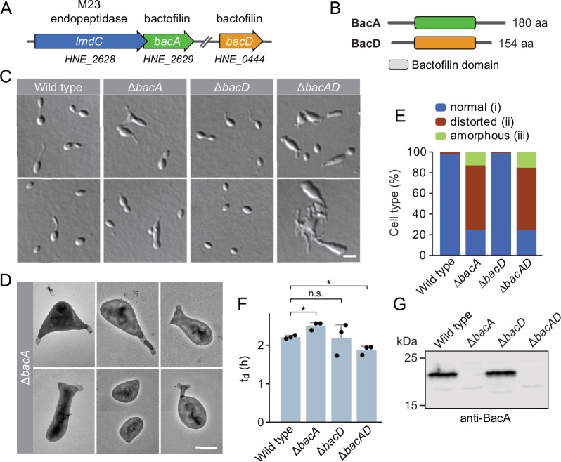
Figure 1. The bactofilin homolog BacA is required for proper cell morphology in H. neptunium.

(A) Schematic representation of the two bactofilin genes present in the H. neptunium genome. bacA lies adjacent to the M23 peptidase gene lmdC. Arrows indicate the direction of transcription. (B) Domain organization of BacA and BacD from H. neptunium. The bactofilin polymerization domain (colored boxes) is flanked by non-structured N- and C-terminal regions. (C) Morphology of H. neptunium bactofilin mutants. Shown are representative cells of strains LE670 (wild type), EC28 (ΔbacA), EC23 (ΔbacD), and EC33 (ΔbacAD), imaged by differential interference contrast (DIC) microscopy. Bar: 4 µm. (D) Transmission electron micrographs of representative ΔbacA cells at the early stalked-cell stage. Bar: 1 µm. (E) Quantification of the proportion of phenotypically abnormal stalked and budding cells in cultures of the strains analyzed in panel C (n=100 cells per strain). (F) Doubling times of the indicated H. neptunium strains. Bars represent the mean (± SD) of three independent experiments (dots). Statistical significance was determined using an unpaired two-sided Welch’s t-test (*p<0.05; n.s., not significant). (G) Immunoblot analysis of the strains shown in panel C, performed using anti-BacA antibodies.

Figure 1—source data 1. Raw images for the immunoblot analysis in Figure 1G.

Figure 1.

Figure 1—figure supplement 1. The dimorphic lifecycle of H. neptunium.
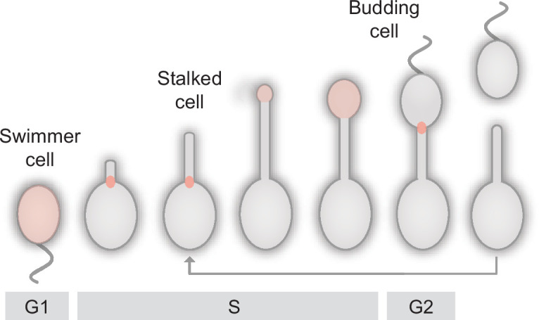
Figure 1—figure supplement 1.

A motile, flagellated swimmer cell sheds its flagellum and forms a stalk at the opposite cell pole. At a defined point in the cell cycle, the terminal segment of the stalk dilates and develops into a new swimmer cell. After cell division, the newborn swimmer cell first needs to differentiate into a stalked cell to initiate a new round of cell division, whereas the stalked mother cell immediately enters the next replication cycle. The predominant growth zones (Cserti et al., 2017) are indicated in red.
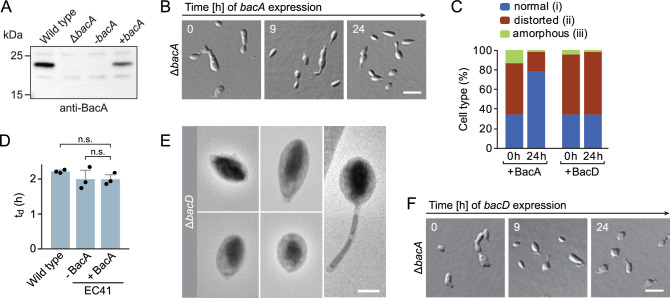
Figure 1—figure supplement 2. Phenotypic analysis of H.neptunium bactofilin mutants.

Figure 1—figure supplement 2.

(A) Immunoblot analysis of strains LE670 (wild-type), EC28 (ΔbacA), and EC41 (ΔbacA PCu::PCu‐bacA) grown in the absence (-bacA) and presence (+bacA) of 0.5 mM CuSO4, performed with an anti-BacA antibody. (B) Rescue of the phenotype of a ΔbacA mutant by ectopic expression of bacA. Cells of strain EC41 (ΔbacA PCu::PCu‐bacA) were grown in copper-free medium, induced by the addition of copper, and imaged after the indicated time incubation times. The images show representative cells. Bar: 3 µm. (C) Quantification of the proportion of phenotypically abnormal stalked and budding cells in the cultures of strains EC41 (ΔbacA PCu::PCu‐bacA) and EC43 (ΔbacA PZn::PZn‐bacD) analyzed in panels B and F before (t=0 hr) and 24 hr after induction (n=100 cells per time point). (D) Doubling times of the indicated H. neptunium strains. Strain EC41 (ΔbacA PCu::PCu‐bacA) as analyzed in the absence (-) and presence (+) of copper. Strain LE670 (wild-type) grown in copper-free medium is shown as a control. Bars represent the mean (± SD) of three independent experiments (dots). Statistical significance was determined using an unpaired two-sided Welch’s t-test (n.s., not significant). (E) Transmission electron micrographs of representative ΔbacD cells at the swimmer and stalked cell stage. Bar: 1 µm. (F) Representative DIC images of a ΔbacA mutant overproducing BacD from a zinc-inducible promoter. Cells of strain EC43 (ΔbacA PZn::PZn‐bacD) were grown in inducer-free medium, induced by the addition of 0.5 mM ZnSO4, and imaged after the indicated incubation times. Bar: 3 µm.
Figure 1—figure supplement 2—source data 1. Raw images for the immunoblot analysis in Figure 1—figure supplement 2A.