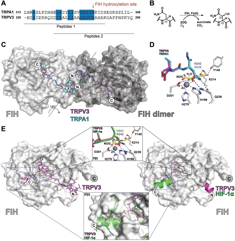
Figure 3.
Views of crystal structures of FIH in complex with TRP channel‐derived peptides. A) Sequence alignment of TRP channel FIH substrates used for crystallography. B) Scheme for FIH‐mediated Asn‐residue hydroxylation reactions. C) Overlay of crystal structure views of FIH in complex with TRPV3 (220–246) and TRPA1 (313–339) peptides. The FIH dimer is in dark grey. D) Overlaying views from structures of FIH complexed with TRPA1 (220–246) and TRPV3 (220–246) peptides reveals near‐identical binding modes at the active site. E) Comparison of a structure of FIH complexed with TRPV3 (229–255) with a crystal structure of FIH in complex with HIF‐1α (PDB ID: 1H2K) [29] indicating differences in their crystallographically observed binding modes.

