

Abstract
Background
Probiotic strains have the potential to modulate immune responses, reduce intestinal inflammation, normalize intestinal mucosal function and decrease allergic reactions.
Objective
This study aimed to investigate the effect of oral probiotic supplements containing Bacillus subtilis and Bacillus coagulans spores on clinical symptoms, haematological factors and immune responses to allergic contact dermatitis in dogs induced by dinitrochlorobenzene (DNCB).
Methods
DNCB was injected subcutaneously into the scapular region of 20 healthy adult dogs of both sexes, divided into four groups, to induce experimental allergic contact dermatitis. Dogs in Group 1 received food without probiotics or medication. Oral prednisolone was administered to Group 2 for 30 days at a dosage of 0.25 mg/kg every other day. The dogs in Group 3 were treated with a combination of oral prednisolone and probiotics. The dogs in Group 4 were fed daily with a mixture of 109 B. subtilis and B. coagulans bacteria for 30 days. The immune system responses and related gene expression were analysed in the treated animals.
Results
The administration of probiotics for 30 days resulted in a reduction in clinical symptoms and duration of wound repair. The probiotics treatment also significantly increased the serum bactericidal effects against Staphylococcus aureus and Escherichia coli. It enhanced both the classic and alternative activity of the complement, as well as lysozyme activity. Additionally, the probiotics led to higher total immunoglobulin levels and significant reductions in anti‐trypsin and C‐reactive protein levels. Furthermore, the expression of IgE, induction of interferon‐gamma and IL‐4 genes were also reduced.
Conclusions
According to the results, B. subtilis and B. coagulans can be further investigated as a viable alternative to corticosteroids in treating allergic contact dermatitis in dogs.
Keywords: Bacillus coagulans, Bacillus subtilis, dog, dermatitis, dinitrochlorobenzene, probiotic
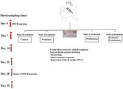
Graphical Abstract: Dinitrochlorobenzene (DNCB) was injected subcutaneously into 20 healthy adult dogs to induce allergic contact dermatitis. Dogs were divided into four groups including control, probiotics, prednisolone and both prednisolone and probiotics. The administration of probiotics resulted in a reduction in clinical symptoms, an increase in most of the innate immune elements, total immunoglobulin levels and significant reductions in anti‐trypsin and CRP levels. Furthermore, it reduced the expression of IgE, IFN‐γ and IL‐4 genes.
1. INTRODUCTION
Probiotics are non‐pathogenic living microorganisms that remain viable in adequate numbers in the gastrointestinal tract and produce various beneficial health effects in the host species (Collado et al., 2009; Ozdemir, 2010). Certain probiotic strains have the potential to modulate the gastrointestinal microbiome, enhance immune responses, reduce intestinal inflammation, normalize intestinal mucosal function and decrease allergic reactions in humans and animals (Collado et al., 2009; Sivamaruthi et al., 2021). Bacillus subtilis is a non‐pathogenic, gram‐positive bacterium. It has been used as an additive to improve animal growth performance. This bacterium can regulate the intestinal microbiota, improve intestinal digestion and enhance host immunity in dogs (Yang et al., 2023). In addition, it induces immune responses such as an increase in IgG and IgA levels, regulation of the balance between Th1 and Th2 pathways, induction of interferon‐gamma (IFN‐γ) and Th1 cytokines, as well as the pro‐inflammatory tumour necrosis factor beta cytokine. It is also used in vaccine formulation as an adjuvant (Amuguni & Tzipori, 2012; de Almeida et al., 2022).
Bacillus coagulans is a gram‐positive, aerobic or facultative anaerobic spore‐forming bacterium. These spores protect the cell wall against heat and pressure and maintain stability during passage through the digestive system (Zhou et al., 2020). The main effects of these bacteria include the stimulation of toll‐like receptors 2, induction of costimulatory molecules and the activation of dendritic cells. B. coagulans spores exert specific immunosuppressive effects by decreasing interleukin 8 (IL‐8) and increasing the secretion of IL‐10 from a human cell line (Shinde et al., 2019). The bacterium could induce a significant increase in inflammatory cytokines such as IL‐1 alpha, IL‐6, IL‐17A, tumour necrosis factor alpha and IFN‐γ cytokines or anti‐inflammatory cytokines as IL‐1 receptor antagonist and IL‐10 in human or animal species (Jensen et al., 2017; Zhou et al., 2020). Allergic reactions often occur due to genetic predisposition and environmental factors. Recently, it is confirmed that human bacterial flora, particularly the intestinal microbiota, plays a crucial role in the development of mucosal immune tolerance during the allergy process; the bacteria interact with innate immune cells like macrophages and dendritic cells and contribute to the maturation of the immune organs and differentiation of Th cells (Crovesy et al., 2017).
Canine contact dermatitis is characterized by mild‐to‐severe local or generalized skin damage of thinly haired or glabrous areas, depending on the causative agent. As a result, the skin keratinocyte undergoes denaturation due to the induction of the local inflammatory responses (Ho et al., 2015). Two types of contact dermatitis are irritant contact dermatitis and allergic contact dermatitis (Ho et al., 2015). Allergic contact dermatitis causes symptoms such as hives on the skin, along with redness, oedema, erythema and papules, and chronic lesions include scaling, fissuring and lichenification. These symptoms occur after skin contact with a hapten, which is a small chemical compound that is too small to elicit an allergic reaction on its own. However, they covalently bind to host self‐proteins, creating neo‐antigens that can induce type 4 hypersensitivity reactions. Examples include chemical elements, topical medications like neomycin or skin products, compounds like benzoic acid, paints, carpets and woods (Ho et al., 2015; Olivry et al., 1990).
This study aimed to investigate the effect of B. subtilis and B. coagulans on clinical symptoms, haematological factors and immune responses of dogs to experimentally induced allergic contact dermatitis. Dinitrochlorobenzene (DNCB) was selected for the experimental induction of an allergic reaction in dogs. The contact sensitivity caused by DNCB is frequently utilized in animal studies, particularly with mice, to investigate the development of contact dermatitis (Tuckermann et al., 2007). If these probiotics are able to modulate allergic responses by affecting Th1 differentiation, the required dose or duration of corticosteroid therapy in dogs with allergic contact dermatitis could be reduced. This study investigated alternative treatments using probiotics and combination therapy involving probiotics and prednisolone to assist veterinarians in small animal practice in managing dogs suffering from allergic contact dermatitis.
2. MATERIALS AND METHODS
2.1. Animals
This study included 20 clinically healthy, intact adult male and female dogs of mixed breed, aged 1–1.5 years and weighing 18.5 ± 2.5 kg, from Khuzestan province, Iran. Before the experiments, the animals were housed in separate cages for 2 weeks in the animal housing area. Twice‐daily feeding with a homemade diet of rice and chicken, along with a single dose of Caniverm forte tablets (Bioveta) as an anti‐parasitic treatment, were conducted as usual. This was done to enable the animals to adapt to their environmental conditions. All of the experiments were carried out in accordance with the animal protection guidelines and research ethics of the Faculty of Veterinary Medicine at Shahid Chamran University of Ahvaz, with the accepted code of EE/1400.3.02, 7810/scu.ac.ir.
2.2. Preparation of probiotics
Separate probiotic powders, including unformulated B. subtilis and B. coagulans spores, were prepared by Pardis Roshd Mehrgan, Shiraz, Iran (ParsiLact, formulated for cats and dogs). The commercial probiotic powder was cultured in nutrient and blood agar media. The identification of the bacteria was determined through biochemical and molecular tests, which are described below. The biochemical tests performed included catalase, oxidase, casein hydrolysis, indole production and consumption of sugars such as mannitol, salicin and inulin, nitrate resuscitation, gelatin hydrolysis, glycogen consumption and evaluation of lecithinase production after culturing bacteria in agar medium containing egg yolk.
2.3. Molecular confirmation of the bacteria
After phenotypic and biochemical evaluation of the studied bacteria, purified colonies were suspended in a microtube containing 100 µL of sterile distilled water. The microtube was incubated in boiling water for 20 min and then centrifuged at 1000 rpm for 10 min. The supernatant was used as a source of DNA for the polymerase chain reaction (PCR) test.
16S ribosomal RNA nucleotide sequencing was used to confirm the presence of these bacteria. The amplification of the 16S ribosomal RNA was conducted using the universal primer pair 27F (5′‐AGAGTTTGATCCTGGCTCAG‐3′) and 1492R (5′‐GGTTACCTTGTTACGACTT‐3′) (Ansari et al., 2019). The reactive mixture was prepared in a 25 µL volume, which included 12.5 µL of master mix (Ampliqon), forward primer 1 µL, reverse primers 1 µL (CinnaGen), 4 µL of DNA sample and 6.5 µL of distilled water. PCR thermal cycles (Eppendorf) were adjusted as follows: initial denaturing at 95°C for 10 min, 30 cycles of denaturing at 95°C for 30 s, annealing at 60°C for 30 s and extension at 72°C for 45 s; the final extension stage was achieved at 72°C for 10 min. The amplified products were electrophoresed on a 1.5% agarose gel containing a safe stain (CinnaGen). After evaluating the amplified product, 30 µL of the PCR products were sent for sequencing to Gene Fanavaran Company. The sequencing results were reviewed using BioEdit software and aligned with the nucleotide sequences from the NCBI database (http://blast.ncbi.nlm.nih.gov/Blast.cgi).
A bacterial spore weighing 0.5 g was suspended in 4.5 mL of sterile distilled water. Then, 10 serial 1:10 dilutions were prepared. A volume of 50 µL of the dilutions numbered 6, 7, 8, 9 and 10 were poured into the nutrient agar medium and incubated at 37°C for 24 h. After that, the bacteria were counted based on CFU/gr. B. coagulans and B. subtilis spores, with a count of 109 CFU, then were combined and poured into capsules (Mounika et al., 2019).
2.4. Induction of dermatitis
Based on a previous study (Zayerzadeh et al., 2020), a solution of 2% DNCB was prepared using a mixture of DSMO, olive oil and ethanol in a ratio of 1:1:3. Subsequently, 200 µL of the solution was injected subcutaneously at five points on the scapular skin of the enrolled dogs. Dogs were periodically examined for general health. After ensuring their well‐being, they were randomly divided into the following four groups. The groups were similar regarding even distribution of gender, age and weight.
The control group was given the usual diet, without probiotics or medication.
Oral prednisolone was administered to the prednisolone group at a dose of 0.25 mg/kg every other day for 30 days.
The probiotic and prednisolone combination group was treated orally with a mixture of 109 spores of each bacterium including B. subtilis and B. coagulans and oral prednisolone 0.25 mg/kg every other day for 30 days.
The probiotic group was orally treated with 109 spores of each bacterium including B. subtilis and B. coagulans for a period of 30 days (Mounika et al., 2019).
Prior to the DNCB injection, blood samples were collected on Day 0. The blood samples were collected again after 7 days. At this time point, the animals started treatment with the specified medication based on their assigned groups. Three weeks after the initial DNCB injection, another 200 µL of the DNCB solution was injected, and blood samples were collected twice at 7‐day intervals. The general health status of the animals was examined daily, and the clinical symptoms of dermatitis were documented weekly by two observers according to the Canine Atopic Dermatitis Lesion Index (CADLE), scoring 0–5 based (0: none; 1: mild, 3: moderate, 4: severe and 5: extensive) on the severity of short‐term lesions, including depth and length of excoriation, erythema and erosion and long‐lasting symptoms of lichenification, discoloration and hyperpigmentation of the lesions during this period (Plant et al., 2012). The healing symptoms of the dogs were scored based on the following criteria: paling of the lesion from red to bright red, pink, pale pink, yellow to white colour, reduction of swelling, shrinking margin of the wound, knitting over the wounds and restoration to normal tissue. Haematology, innate immunity and humoral immunity were assessed on predetermined days, including Days 0, 7, 21, 28 and 35.
2.5. Haematology
The blood samples were collected in microtubes containing heparin (LEO Pharma). Haematology parameters, including haematocrit, red blood cell (RBC) count, white blood cell (WBC) count and platelet count, were evaluated on heparinized blood using an automatic impedance cell counter (Mindray 2800‐vet). The total protein and albumin levels were assessed in serum samples using a biochemical auto‐analyser, following the reference laboratory method (BT‐TARGA‐3000 model).
2.6. Innate immune system evaluation
Innate immunity was assessed by evaluating complement activity, lysozyme, serum bactericidal and myeloperoxidase (MPO) activity on Days 0, 7, 21, 28 and 35.
2.6.1. Serum bactericidal effects
The antibacterial effects of animal sera were evaluated using the microdilution broth method in a sterile 96‐well microplate against Staphylococcus aureus and Escherichia coli. The bacterial strains were cultured in blood agar and MacConkey medium, respectively. After the bacterial growth, the resulting bacterial colonies were transferred into a nutrient broth media and incubated at 37°C until they reached a turbidity equivalent to the 0.5 McFarland standard. The serum samples (25 µL) were mixed with an equal volume of brain and heart infusion medium in specified microplate wells. The bacteria were added to the wells, 25 µL in total. After measuring the optical density (O.D) at 600 nm using a spectrophotometer, the plates were incubated for 24 h at 37°C, and the O.D was detected as before. The results were interpreted by calculating the bacterial growth in the test and control wells (Markey et al., 2013).
2.6.2. The serum lysozyme activity
The activity of serum lysozyme was determined using the turbidimetry assay. The bacterial suspension was prepared by adding 20 mg of Micrococcus lysodeikticus to 100 mL of acetate buffer (0.02 M, pH 5.5). The bacterial suspension of 150 µL was mixed with 15 µL of the serum samples in 96 microplate wells and incubated at room temperature. The O.D was detected at a wavelength of 600 nm after 15 min. A decreased O.D equal to 0.001 per minute was taken as one unit of lysozyme activity (Saidana et al., 2006).
2.6.3. Serum myeloperoxidase activity
The activity of the serum MPO was determined by measuring the oxidation of 3,3′,5,5′‐tetramethylbenzidine (TMB) by MPO. Serum samples (10 µL) were mixed with 90 µL of Hanks balanced salt solution without Ca2+ or Mg2+ in 96‐well plates. Then, 35 µL of 20 mM TMB and 5 mM H2O2 were added to each well. The reaction was stopped after 2 min by adding 35 µL of 4 M sulphuric acid. The enzyme activity was determined calorimetrically using a plate reader at a wavelength of 450 nm, and the results were expressed as O.D (Malle et al., 2007).
2.6.4. Evaluation of complement activity
Activity of the classic complement pathway was analysed by preparing sensitized sheep RBCs (SRBCs). An SRBC 10% solution, with a volume of 10 mL, was mixed with 1 mL of rat anti‐sheep RBC antibody at a concentration of 50 µg/mL for 12 h. After centrifugation at 2000 rpm for 5 min, the sensitized cells were suspended in 10 mL of phosphate‐buffered saline (PBS). Then, 25 µL of each serum sample, 375 µL of PBS containing Ca+ and Mg2+ and 100 µL of sensitized SRBC were mixed in microtubes. The samples were incubated in 37°C incubators for 45 min and then centrifuged for 5 min at 3000 revolutions per minute (rpm). A volume of 100 µL of the supernatant from the sample was removed, and the optical densities were measured at a wavelength of 490 nm. Activity of the alternative complement pathway was evaluated using the same method, except that unsensitized SRBCs were mixed with the serum samples (Inglis et al., 2008).
2.6.5. Evaluation of serum antiprotease activity
Trypsin 5% solution (20 µL) was mixed with 100 µL of Tris–HCl solution (50 mM, pH 8.2). Then, 10 µL of the serum samples were added to this mixture in the microplate wells. The positive control samples contained all of these elements, except for the serum samples. The plate was incubated at room temperature for 1 h. Nα‐Benzoyl‐dl‐arginine p‐nitroanilide hydrochloride substrate (0.1 mM) was prepared in Tris–HCl (50 mM) solution containing calcium chloride (20 mM) and added to the wells. The O.D was measured at 450 nm wavelength at times 0 and after 15 min incubation at room temperature. The anti‐trypsin activity was defined by calculating the difference in wavelength of the mean positive control samples to each sample, divided into the mean positive control samples (Altshuler et al., 2012).
2.7. Measurement of total serum immunoglobulin levels
Total serum immunoglobulin was assessed using the zinc sulphate precipitation method. A 0.7 mM zinc sulphate buffer was prepared and adjusted to a pH of 5.8. The serum samples (12.5 µL each) were poured separately into sterile microtubes and mixed with 850 µL of zinc sulphate. Four consecutive dilutions of the standard serum were prepared as samples. After 2 h of incubation at room temperature, 100 µL of the solution was poured into the 96‐well microplate, and the O.D was detected at a wavelength of 600 nm (Sedlinska et al., 2005). According to the standard curve formula, the total amount of immunoglobulin was calculated for each sample. The total immunoglobulin concentration in the standard was 7.5 mg/mL.
2.8. Evaluation of the C‐reactive protein
The C‐reactive protein (CRP) level was evaluated using the Enison CRP agglutination kit. According to the severity and reaction time within 0–4 min, the results were reported semi‐quantitatively after mixing 30 µL of serum with 30 µL of latex solution. The grading scale ranged from 0 to 4.
2.9. Evaluation of immune system‐related gene expression
The effect of administering B. coagulans and B. subtilis on the expression levels of IFN‐γ, IL‐4 and IgE genes in treated dogs was investigated using the real‐time PCR method. The GAPDH gene was used as the housekeeping control gene. The sequences of the used primers are mentioned in Table 1.
TABLE 1.
Primer sequences used for real‐time polymerase chain reaction (PCR) analysis.
| Primer name | Sequence 5′ → 3′ | Gene accession number | Product length | References |
|---|---|---|---|---|
| IL‐4 (F) | CGAGAAACGACTCGTGCATGG | AF054833 | 83 | blast.ncbi.nlm.nih.gov |
| IL‐4 (R) | GCGAGAAACGACTCGTGC | |||
| IgE (F) | CTCATGCAGCCTCTCACACA | L36872.1 | 97 | blast.ncbi.nlm.nih.gov |
| IgE (R) | CGCCTTGTGGACATACAGGT | |||
| IFN‐γ (F) | GCGCAAGGCGATAAATGAAC | AF126247 | 82 | Majewska et al. (2016) |
| IFN‐γ (R) | CTGACTCCTTTTCCGCTTCCT | |||
| GAPDH (F) | GGAGAAAGCTGCCAAATATG | NM_001003142.1 | 100 | Majewska et al. (2016) |
| GAPDH (R) | ACCAGGAAATGAGCTTGACA |
Abbreviations: IFN‐γ, induction of interferon‐gamma; IL‐4, interleukin‐4.
Blood samples were collected from the cephalic vein using heparin as an anticoagulant. The buffy coats were separated by centrifugation at 1200 g for 15 min. The RBC lysis buffer, containing 0.15 M ammonium chloride, 10 mM potassium bicarbonate and 0.1 mM EDTA, was added to the microtubes. After 5 min, the microtubes were centrifuged as previously described. The supernatants were discarded, and 200 µL of RNX‐Plus (Sinacolon Company; EX6101) was added to each buffy coat sample. The samples were then stored in a −70°C freezer. The RNA extraction, DNase treatment and construction of the cDNA samples were performed according to the kit's instructions. The prepared cDNA was stored at −70°C until the real‐time PCR tests. The elements were mixed in the following order and with the specified values for the real‐time PCR reactions, with a total volume of 20 µL: 6 µL of dH2O, 10 µL of Taq Man real‐time Master Mix, 2 µL of cDNA sample, 1 µL of forward primer and 1 µL of reverse primer. The evaluated cytokines included IFN‐γ, IL‐4 and IgE, along with the housekeeping GAPDH gene. They reacted to annealing at 60°C. A total of 40 cycles were considered for gene amplification.
2.10. Statistical evaluation
The gene expression data were evaluated using the StepOne software with the ΔΔCT comparative method. The results were reported based on the general formula of ΔΔCT as [ΔΔCT = (Ct target − Ct β2M) sample − (Ct target − Ct β2M)]. Finally, the data were compared among the groups using SPSS software version 22 and one‐way ANOVA statistical tests. A p < 0.05 value was considered statistically significant. Data are presented as mean ± standard deviations.
3. RESULTS
3.1. Evaluation of probiotic bacteria
3.1.1. Biochemical tests
Identification of the provided probiotic bacteria, based on biochemical tests, showed that the characteristics of the bacteria were consistent with the expected results. The results of the tests performed on the bacteria are shown in Table 2. Results of biochemical tests identified the bacteria as B. subtilis NATO strain and B. coagulans strain KM‐1.
TABLE 2.
Biochemical tests for the identification of Bacillus subtilis and Bacillus coagulans.
| B. subtilis | B. coagulans | B. S. | B. C. | |
|---|---|---|---|---|
| Anaerobic growth | − | + | ||
| Casein hydrolysis | + | − | + | |
| Glycogen | + | − | ||
| Inulin | (+) | − | + | − |
| Mannitol | + | − | + | − |
| Salicin | + | V | − | − |
| Egg | − | + | − | + |
| SIM (indole) | − | − | − | − |
| Nitrate | + | V | + | + |
| Gelatin | + | V | + | + |
Note: B. S and B. C are shown the biochemical results of the used B. subtilis and B. coagulans strains, respectively.
3.1.2. Genomic identification of the bacteria
The bacteria identification was done using PCR. Amplification of a 1510 bp fragment of the ribosomal RNA 16S gene was confirmed through 1% agarose gel electrophoresis and sequencing of the nucleotide fragments. The consistency of the B. subtilis and B. coagulans sequences with the sequences in the NCBI genomic database showed a 100% and 99.2% identity, respectively, to the total length of the fragment (JX569800; MT271920).
3.2. Clinical symptoms of the treated animals
The control groups exhibited clinical manifestations such as oedema, erythema and even necrosis at the injection sites (Figure 1). Based on CADLE score, these manifestations were significantly more severe compared to the other three groups on Days 21 and 35 (Figure 2). Additionally, following the second injection of DNCB, the severity of the lesions seemed to progress to a greater extent than in the other groups. Furthermore, the rate of symptom resolution and improvement in the control group was lower compared to the other groups. Clinical symptoms were observed in the prednisolone group at the injection site, including erythema, oedema, hyperpigmentation and necrosis. The probiotic group exhibited significantly lower severity and incidence of symptoms 21 days after treatment compared to the control group (p = 0.01, 0.021 and 0.017 for Days 21, 28 and 35, respectively). Figure 1 displays the clinical symptoms observed in the studied groups on different days. Although the treated groups did not show a significant difference, the symptoms of dermatitis were less severe, and the rate of improvement was higher in the probiotic plus prednisolone group compared to the other groups (see Figures 1 and 2).
FIGURE 1.
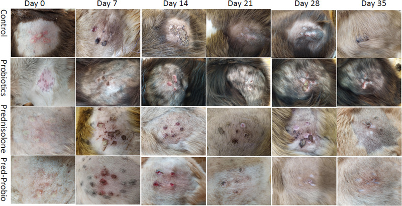
Clinical manifestations in dogs with experimentally induced allergic contact dermatitis.
FIGURE 2.
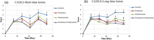
The Canine Atopic Dermatitis Lesion Index (CADLE) score in treated dogs with experimentally induced allergic contact dermatitis: (a) short‐term lesions, including erythema, excoriation and lesions; (b) long‐term lesion, including lichenification, discoloration and hyperpigmentation. The significant difference of the treated groups compare to the control group was indicated by a star on different days.
3.3. Evaluation albumin and total protein level of serum
The serum albumin values showed no significant difference among the studied groups. The prednisolone group had significantly higher serum albumin levels (p = 0.042) on Day 35 compared to Day 0. Figure 3 shows the level of serum albumin and serum total protein in the evaluated groups. The serum total protein level was not significantly different between the evaluated times within each group or between the studied groups.
FIGURE 3.
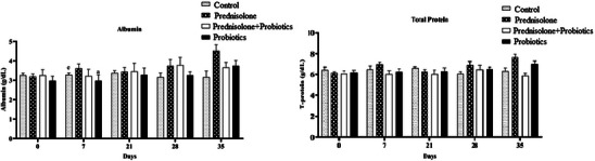
Evaluation of serum albumin and protein levels in dogs with experimentally induced dermatitis using dinitrochlorobenzene (DNCB). The evaluated animals were randomly divided into four groups: Group 1 control group without probiotics and medication; Group 2 prednisolone‐treated animals; Group 3 prednisolone plus probiotic‐treated animals and Group 4 probiotic‐treated animals. The significant difference is indicated by the use of different letters for special days: a for Day 0, b for Day 7, c for Day 21, d for Day 28 and e for Day 35.
3.4. Haematology evaluation
On Day 7 of the study, the WBC levels of the probiotic group (p = 0.001), the probiotic plus prednisolone group (p = 0.001) and the prednisolone group (p = 0.003) were significantly reduced compared to the control group. In addition, the control group had significantly higher WBC levels on Day 7 compared to the other evaluated time points; Days 35 (p = 0.003), 28 (p = 0.001) and 21 (p = 0.005) (Figure 4).
FIGURE 4.
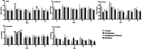
Evaluation of white blood cell counts, including lymphocytes, monocytes, neutrophils and eosinophils, in dogs with experimentally induced dermatitis using dinitrochlorobenzene (DNCB). The animals under evaluation were randomly divided into four groups. Group 1 consisted of control animals without probiotics and medication, Group 2 included animals treated with prednisolone, Group 3 included animals treated with both prednisolone and probiotic and Group 4 included animals treated with probiotics only. The significant differences were indicated by letters on various days: (a) Day 0, (b) Day 7, (c) Day 21, (d) Day 28 and (e) Day 35.
On Day 21 of the study, the WBC level in the probiotic group (p = 0.001) and the probiotic plus prednisolone group (p = 0.007) was lower than that in the prednisolone group. Additionally, the prednisolone group had a higher WBC level than the control group (p = 0.003). In the probiotic group, the WBC level was significantly lower on Day 21 compared to Day 7 (p = 0.006) and Day 0 (p = 0.022). However, the WBC level on Day 28 was higher than on Day 21 (p = 0.027). On Day 35, there was a significant decrease in WBC levels compared to Day 28 (p = 0.035). In the probiotic plus prednisolone group, the WBC levels on Day 28 were significantly higher compared to Days 21 (p = 0.001), 7 (p = 0.009) and 0 (p = 0.003). Additionally, the WBC level on Day 35 was significantly lower than on Day 28 (p = 0.002). There were no significant changes in serum lymphocyte levels at different times and between the groups (Figure 4).
There were no significant changes in serum monocyte, neutrophil and eosinophil levels between the groups. However, monocyte levels decreased in the control group on Days 21 and 28 compared to Day 7. Additionally, all groups showed significantly higher neutrophil levels on Day 7 compared to the other evaluated days. The serum levels of eosinophils in the control group were lower on Day 21 (p = 0.004) compared to Day 0. There were no significant changes in the number of RBC, haemoglobin, haematocrit, Mean Corpuscular Volume (MCV), Mean Cell Hemoglobin (MCH), Mean Corpuscular Hemoglobin Concentration (MCHC) and Red cell Distribution Width (RDW) levels at different times and between the groups (Figure 4).
3.5. Assessment of the innate immune system
3.5.1. Evaluation of myeloperoxidase activity
A comparison of the measured times among groups showed that MPO activity was not significantly different. In the probiotic group, MPO activity was significantly higher 12 h after re‐injection (Day 21 of the study) compared to Day 0 (p = 0.013) and 7 days after the initial injection (p = 0.012). Figure 5 depicts the assessment of MPO activity in the various groups.
FIGURE 5.
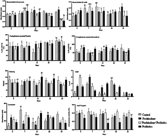
Evaluation of innate immune responses in dogs with dinitrochlorobenzene (DNCB)‐induced dermatitis. The animals under evaluation were randomly divided into four groups. Group 1 consisted of control animals without probiotics and medication, Group 2 included animals treated with prednisolone, Group 3 comprised animals treated with prednisolone plus probiotics and Group 4 consisted of animals treated with probiotics alone. The significant differences were indicated by letters on various days: (a) Day 0, (b) Day 7, (c) Day 21, (d) Day 28 and (e) Day 35.
3.5.2. Serum bactericidal activity
On Day 35 of the study, both the probiotic and probiotic plus prednisolone groups exhibited significantly higher serum bactericidal activity against S. aureus compared to the control group (p = 0.001 and p = 0.001, respectively) and the prednisolone group (p = 0.01 and p = 0.001, respectively). On Day 35 of the study, the bactericidal activity against E. coli in the prednisolone group was significantly lower than in the probiotic plus prednisolone group (p = 0.004) and the probiotic group (p = 0.005). Figure 5 shows the evaluation of serum bactericidal activity against S. aureus and E. coli.
3.5.3. Evaluation of complement activity
The activity of the classic complement pathway showed no significant difference among the evaluated groups at all of the times. In the control group, the activity of this pathway on Day 35 was significantly higher than on Day 21 (p = 0.032). In the probiotic plus prednisolone group, the activity on Day 35 was significantly higher than on other days (Figure 4). On Day 21, the activity of the alternative complement pathway in the probiotic group was significantly higher than in the control group (p = 0.001) and the prednisolone group (p = 0.015). Additionally, the probiotic plus prednisolone group had higher activity than the control group (p = 0.017). On Day 35, the activity in the probiotic group was higher than in the probiotic plus prednisolone and prednisolone group (p = 0.004). Additionally, on Day 35, the probiotic plus prednisolone groups exhibited the lowest lysis activity of the alternative complement pathway compared to the other groups (p = 0.005) (Figure 5).
3.5.4. Anti‐trypsin activity
On Day 28 of the study, the anti‐trypsin activity in the probiotic group was significantly lower than in the probiotic plus prednisolone group (p = 0.003). In the probiotic group, the anti‐trypsin activity significantly increased on Day 35 compared to Days 21 (p = 0.001) and 28 (p = 0.001). Additionally, the anti‐trypsin activity of the probiotic group was significantly decreased on Day 21 compared to Days 7 (p = 0.024) and 0 (p = 0.035) (Figure 5).
3.5.5. Evaluation of lysozyme activity
On Day 21 of the study, the probiotic group exhibited significantly higher lysozyme activity compared to the control group (p = 0.012) and the prednisolone group (p = 0.003). On Day 35, the lysozyme activity in the probiotic group (p = 0.004) and the prednisolone group (p = 0.047) was significantly lower than that in the control group. Serum lysozyme activity in the control group was significantly higher on Day 7 compared to Day 21 and 0. In the prednisolone group, the serum lysozyme activity on Day 28 was significantly higher than Days 7 (p = 0.019) and 21 (p = 0.022) (Figure 5).
3.6. CRP Rate
The CRP level was not significantly different among the evaluated groups. In the control group, the CRP level on Day 28 was significantly higher than on Days 35 (p = 0.024), 0 (p = 0.025) and 21 (p = 0.025). In the probiotic plus prednisolone group, CRP levels were significantly lower on Days 35 and 21 compared to Day 0 (p = 0.005 and 0.008, respectively) (Figure 5).
3.7. Total serum immunoglobulin levels
On Days 21 and 28 of the study, the probiotic group had significantly higher levels of total immunoglobulin than the prednisolone group (p = 0.001 and p = 0.011, respectively) and the probiotic plus prednisolone group (p = 0.01 and p = 0.013, respectively). Serum immunoglobulin levels in the control group decreased significantly on Day 7 of the study compared to Day 0 (p = 0.03). In contrast, the immunoglobulin level in the probiotic group significantly increased on Days 28 and 21 of the study compared to Day 7 and 0 (p = 0.024) (Figure 5).
3.8. Analysis of immune genes expression
The expression of the IgE gene in the prednisolone group (p = 0.01) was significantly higher than in the control group (p = 0.01) on Days 7 and 35 of the study. The probiotic group (p = 0.04) and the probiotic plus prednisolone group (p = 0.03) had lower levels of IgE gene expression compared to the control group on Days 7, 28 and 35 of the study. The expressions of the IgE gene in the control and prednisolone groups were at the highest level on Day 7.
The level of IFN‐γ gene expression in the probiotic and probiotic plus prednisolone groups on Days 7 (p = 0.001 and p = 0.001, respectively), 28 (p = 0.001 and p = 0.002, respectively) and 35 (p = 0.001 and p = 0.001, respectively) was significantly lower than in the prednisolone (p = 0.001) groups. The prednisolone and control groups had the highest IFN‐γ gene expression on Day 7 of the study (p = 0.001) (Figure 6).
FIGURE 6.
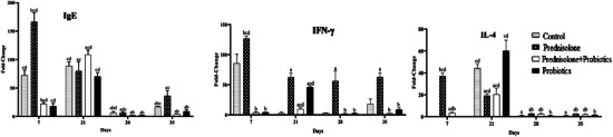
Evaluation of the expression of IgE, induction of interferon‐gamma (IFN‐γ) and IL‐4 genes in dogs with dinitrochlorobenzene (DNCB)‐induced dermatitis. The animals under evaluation were randomly divided into four groups. Group 1 consisted of control animals without probiotics and medication, Group 2 included animals treated with prednisolone, Group 3 included animals treated with prednisolone plus probiotics and Group 4 included animals treated with probiotics alone. Statistical differences are denoted by different letters for special days: (a) Day 0, (b) Day 7, (c) Day 21, (d) Day 28 and (e) Day 35.
The prednisolone group had a significantly lower level of IL‐4 gene expression in comparison to the control and probiotic groups on Day 21 of the study. On Day 35 of the study, both the prednisolone group (p = 0.001) and the probiotic plus prednisolone group (p = 0.04) exhibited significantly higher levels of IL‐4 compared to the control group. The control, probiotic and prednisolone plus probiotic groups exhibited higher levels of IL‐4 gene expression on Day 21 of the study (Figure 6).
4. DISCUSSION
Allergic contact dermatitis is a multifactorial inflammatory skin disease in both humans and dogs. The involved factors that contribute to the condition include genetics, environment, pharmaceutical agents, epidermal skin and immunological factors (Ho et al., 2015; Martins et al., 2016). Deviation of Th cell balance and harmonizing regulatory T cells are the main mechanisms through which probiotics affect immune responses (De Roock et al., 2010). The benefits of using probiotics in dogs include protection against infections caused by intestinal bacteria, treatment of enteropathies, increased weight gain, immunity and control of allergic contact dermatitis (Sivamaruthi et al., 2021; Xu et al., 2019). Based on the current study, DNCB 2% in DMSO, ethanol and olive oil in a ratio of 1:3:1, respectively, induces similar lesions to those seen in canine contact dermatitis such as erythema, oedema, epidermal thickening and vesiculation. As there were few studies on the development of contact dermatitis in dogs, these reactions were optimized in mice and dogs (Zayerzadeh et al., 2020). The 5% and 10% DNCB solutions caused more severe lesions and are not suitable for inducing contact dermatitis‐like lesions in dogs (Krawiec & Ghaafar, 1975; Kim et al., 2012).
In the current study, the administration of prednisolone tablets to dogs at a dose of 0.25 mg/kg every other day for 30 days reduced the severity, number and size of inflamed areas containing erythema. The symptoms of dogs receiving probiotics, similar to those in the prednisolone plus probiotic group, decreased more quickly and favourably compared to the control group. Various studies investigated the effects of probiotics on the treatment of dermatitis (Kiecka et al., 2023; Lolou & Panayiotidis, 2019). Previously, Lactobacillus casei (Chapat et al., 2004), Lactobacillus acidophilus (Shah et al., 2012) and Bifidobacterium longum 51A (Ribeiro et al., 2021) were reported as effective probiotics that could regulate the mice immune responses and used in the management of allergic contact dermatitis.
Goto et al. (2011) developed an experimental dermatitis in mice using Dermatophagoides antigens. The affected mice were treated with 1.2 × 10⁷ CFU of B. subtilis probiotics to reduce the appearance of skin symptoms such as erythema and oedema after 4 weeks due to the limited mast cell proliferation and secretion. Haematological studies showed similar results to the previous studies (Stromfoa et al., 2012). In the current study, the various haemogram indices indicate that there were no significant changes in dogs with DNCB‐induced dermatitis after probiotics administration.
The impact of various probiotics on MPO levels has been examined in several studies, such as Kim et al. (2013) and Sharma et al. (2014). They showed that Lactobacillus lactis and Lactobacillus fermentum significantly increased MPO activity in flounder olive fish and mice, respectively. Our study showed that MPO activity increased in all groups, but this increase was only significant in the probiotic group. Inflammation, as a result of dermatitis, can be attributed to elevated activity of this enzyme in all of the groups. B. coagulans was used in two grouper and zebrafish infected with Vibrio vulnificus bacteria. They demonstrated that B. coagulans possesses antimicrobial properties against S. aureus and E. coli, and it is capable of inhibiting the growth of these two bacteria (Pan et al., 2013).
Sun et al. (2010) evaluated the effects of the B. pumilus and B. clausii bacteria on the complement activity of grouper fish. Contrary to the classical pathway, the activity of the alternative complement pathway was significantly increased in both groups compared to the control group. Cerezuela et al. (2012), Liu et al. (2012) and Galagarza et al. (2018) reported a significant elevation in complement activity in Sparus aurata, grouper and tilapia fish treated with B. subtilis, respectively. Altogether, the effects of B. subtilis and B. coagulans on the complement activity of dogs with experimental dermatitis were consistent with the findings of the mentioned studies in fish species. These results indicate an increase in the alternative complement pathway and an insignificant effect on the classical pathway activity. Considering the various sources of C3, the stimulation of complement element‐producing cells, such as skin keratinocytes, which are the source of C3 and factor B (Pasch et al., 2000), can be considered the cause of the elevation of the alternative complement pathway.
A study conducted by Viljanen et al. (2005) found that Lactobacillus rhamnosus significantly reduced the levels of anti‐trypsin in human infants with dermatitis. In the present study, the anti‐trypsin activity was consistently lower in the probiotic group compared to the other groups. Serum anti‐trypsin can be reduced by reacting with probiotic bacteria. Liu et al. (2012) and Galagarza et al. (2018) found that B. subtilis can increase the plasma concentration of lysozyme activity in fish. In line with the mentioned results, there was a significant increase in lysozyme activity on Day 21 of the present study. This increase could be attributed to the stimulation of neutrophils by Bacillus bacteria, leading to an increase in the production of this enzyme. The CRP level decreased on Day 35 compared to Days 7 (initial injection of DNCB) and 21 (re‐injection of DNCB). This decrease was higher in the group receiving both probiotic and prednisolone compared to the other groups. This finding is consistent with the clinical symptoms and the expected therapeutic effect of prednisolone and probiotic treatment. The indirect effect of probiotics on the secretion of stimulatory cytokines by B lymphocytes can be considered the cause of the probiotics’ impact on serum total immunoglobulin levels.
The probiotics L. casei, L. rhamnosus, Lactobacillus plantarum and Bifidobacterium lactis, both individually and in combination, were found to reduce serum IgE and IL‐4 levels and improve symptoms of experimentally induced allergic dermatitis in mice by DNCB (Seo et al., 2011). The current study showed that after DNCB injection on Day 0, IgE expression increased after 1 week. This increase continued until Day 21 in all of the treated groups. The expression of the IgE gene reached its maximum level on Day 21 compared to other time points. Re‐injection of DNCB on Day 21 increased IgE levels again on Day 35 of the study. A delayed and reduced increase in IgE levels during the secondary response (14 days after injection) compared to the initial response (7 days after injection) may be attributed to the lower dosage administered and the neutralization of a portion of the injected DNCB by antibodies from the first injection. According to the levels of IgE and cytokines, probiotics may have an effect on related mechanisms such as the ratio of Th1/Th2 cells, suppression of Th2 responses, elevation of T regulatory subtype, mast cell infiltration or degranulation (Lim et al., 2017). These effects prompt a different pattern of immune responses against DNCB compared to the control group, leading to a decrease in the clinical manifestations associated with dermatitis in dogs.
Increased levels of IFN‐γ were reported in mice after induction of dermatitis using DNCB (Hayashi et al., 2009). The oral administration of Lactobacillus brevis reduces IFN‐γ production in mice affected by experimentally induced dermatitis with picryl chloride (Segawa et al., 2008). Kim et al. (2012) administered poly‐gamma glutamic acid obtained from B. subtilis to dogs with DNCB‐induced dermatitis. This treatment significantly reduced IFN‐γ‐producing cells in the skin. The results of the DNCB effect on IFN‐γ gene expression show that IFN‐γ expression decreased in the control group. This decrease was even higher after the re‐injection of DNCB on Day 21. Reduction in IFN‐γ gene expression in the control group could be attributed to a decrease in Th1/Th2 cells or immune response deviations as a consequence of treatment with DNCB. IFN‐γ expression decreased in the prednisolone group; this drug inhibits the t‐bet transcription factor in Th‐1 cells, thereby reducing the expression of the IFN‐γ gene. In the current study, an increase in IL‐4 gene expression was detected in all groups after DNCB re‐injection. Two weeks after treatment, the groups treated with prednisolone and the combination of probiotic plus prednisolone showed lower IL‐4 gene expression compared to the groups treated with probiotic alone and the control group. Furthermore, on Days 28 and 35, a considerable decrease in IL‐4 gene expression was observed in all groups compared to Day 21, even after a long period of re‐injection.
5. CONCLUSION
B. subtilis and B. coagulans improve the clinical symptoms of allergic contact dermatitis induced by DNCB in dogs. Probiotics have more favourable effects compared to prednisolone alone or the combination of probiotics with prednisolone. The increasing anti‐trypsin activity from Day 21 to 35, along with the decreased activity of lysosomes, MPO and the alternative and classic complement pathways, as well as the CRP level during this period, corresponded with the clinical observations in the probiotics group. In addition, unlike the other groups, the probiotics group received positive feedback on specific aspects of innate and acquired immune responses. Considering the limitations of the current study, such as the small size of the groups and the induction of experimentally induced contact dermatitis in animals rather than naturally affected ones, it has been concluded that two probiotics, B. subtilis and B. coagulans, can be further investigated separately or in combination with prednisolone for treatment or maintenance therapy in a larger group of animals or clinical cases of allergic contact dermatitis in dogs.
AUTHOR CONTRIBUTIONS
Conceptualization; data curation; formal analysis; funding acquisition; investigation; methodology; project administration; resources; software; supervision; validation; visualization; writing — original draft; writing — review and editing: Mohammad Khosravi. Conceptualization; data curation; formal analysis; funding acquisition; investigation; methodology; project administration; resources; software; supervision; validation; visualization; writing — review and editing: Reza Avizeh. Data curation; formal analysis; investigation; validation; visualization: Akram Zayerzadeh. Conceptualization; data curation; formal analysis; investigation; methodology; project administration; validation; visualization: Darioush Gharibi. Data curation; formal analysis; investigation; methodology; project administration; visualization: Mohammad Razijalali.
ETHICS STATEMENT
The enrolled animals were 20 adult male and female clinically healthy dogs with mixed breed aged 1–1.5 years. All of the experiments were carried out according to animal protection and guidelines of research ethics committee of Shahid Chamran University of Ahvaz with verification code of scu.ac.ir /1400.3.02.7810/EE.
PEER REVIEW
The peer review history for this article is available at https://publons.com/publon/10.1002/vms3.1410.
ACKNOWLEDGEMENTS
This study was supported by Shahid Chamran University of Ahvaz, (Grant Number: SCU.VP1402.12470.)
Khosravi, M. , Avizeh, R. , Zayerzadeh, A. , Gharibi, D. , & Razijalali, M. (2024). Effect of Bacillus subtilis and Bacillus coagulans spores on induced allergic contact dermatitis in dogs. Veterinary Medicine and Science, e1410. 10.1002/vms3.1410
DATA AVAILABILITY STATEMENT
The datasets used and/or analysed during the current study are available from the corresponding author upon request.
REFERENCES
- Altshuler, A. E. , Penn, A. H. , Yang, J. A. , Kim, G. R. , & Schmid‐Schonbein, G. W. (2012). Protease activity increases in plasma, peritoneal fluid, and vital organs after hemorrhagic shock in rats. PLoS ONE, 7(3), e32672. 10.1371/journal.pone.0032672 [DOI] [PMC free article] [PubMed] [Google Scholar]
- Amuguni, H. , & Tzipori, S. (2012). Bacillus subtilis: A temperature resistant and needle free delivery system of immunogens. Human Vaccines & Immunotherapeutics, 8(7), 979–986. [DOI] [PMC free article] [PubMed] [Google Scholar]
- Ansari, J. M. , Colasacco, C. , Emmanouil, E. , Kohlhepp, S. , & Harriott, O. (2019). Strain‐level diversity of commercial probiotic isolates of Bacillus, Lactobacillus, and Saccharomyces species illustrated by molecular identification and phenotypic profiling. PloS ONE, 14(3), e0213841. [DOI] [PMC free article] [PubMed] [Google Scholar]
- Cerezuela, R. , Guardiola, F. A. , Gonzalez, P. , Meseguer, J. , & Esteban, M. A. (2012). Effects of dietary Bacillus subtilis, Tetraselmis chuii, and Phaeodactylum tricornutum, singularly or in combination, on the immune response and disease resistance of sea bream (Sparus aurata L.). Fish and Shellfish Immunology, 33(2), 342–349. [DOI] [PubMed] [Google Scholar]
- Chapat, L. , Chemin, K. , Dubois, B. , Bourdet‐Sicard, R. , & Kaiserlian, D. (2004). Lactobacillus casei reduces CD8+ T cell‐mediated skin inflammation. European Journal of Immunology, 34(9), 2520–2528. [DOI] [PubMed] [Google Scholar]
- Collado, M. C. , Isolauri, E. , Salminen, S. , & Sanz, Y. (2009). The impact of probiotic on gut health. Current Drug Metabolism, 10(1), 68–78. [DOI] [PubMed] [Google Scholar]
- Crovesy, L. , Gongalves, D. C. , & Trigo, E. L. (2017). Probiotics in allergy treatment: A literature review. Revista Española de Nutrición Humana y Dietética, 21(3), 293–299. [Google Scholar]
- de Almeida, M. E. M. , Alves, K. C. S. , de Vasconcelos, M. G. S. , Pinto, T. S. , Gloria, J. C. , Chaves, Y. O. , Neves, W. L. L. , Tarrago, A. M. , de Souza Neto, J. N. , Astolfi‐Filho, S. , & Pontes, G. S. (2022). Bacillus subtilis spores as delivery system for nasal Plasmodium falciparum circumsporozoite surface protein immunization in a murine model. Scientific Reports, 12(1), 1531. [DOI] [PMC free article] [PubMed] [Google Scholar]
- De Roock, S. , Van Elk, M. , Van Dijk, M. E. , Timmerman, H. M. , Rijkers, G. T. , Prakken, B. J. , Hoekstra, M. O. , & De Kleer, I. M. (2010). Lactic acid bacteria differ in their ability to induce functional regulatory T cells in humans. Clinical and Experimental Allergy, 40(1), 103–110. [DOI] [PubMed] [Google Scholar]
- Galagarza, O. A. , Smith, S. A. , Drahos, D. J. , Eifert, J. D. , Williams, R. C. , & Kuhn, D. D. (2018). Modulation of innate immunity in Nile tilapia (Oreochromisniloticus) by dietary supplementation of Bacillus subtilis endospores. Fish and Shellfish Immunology, 83, 171–179. [DOI] [PubMed] [Google Scholar]
- Goto, K. , Iwasawa, D. , Kamimura, Y. , Yasuda, M. , Matsumura, M. , & Shimada, T. (2011). Clinical and histopathological evaluation of Dermatophagoides farinae‐induced dermatitis in NC/Nga mice orally administered Bacillus subtilis . Journal of Veterinary Medical Science, 73(5), 649–654. [DOI] [PubMed] [Google Scholar]
- Hayashi, T. , Hara, S. , & Hasegawa, K. (2009). Enhanced contact hypersensitivity by delayed T‐helper 2 response in BALB/c mice. Allergy and Asthma Proceedings, 30(4), 440–457. [DOI] [PubMed] [Google Scholar]
- Ho, K. K. , Campbell, K. L. , & Lavergne, S. N. (2015). Contact dermatitis: A comparative and translational review of the literature. Veterinary Dermatology, 26(5), 314–327. [DOI] [PubMed] [Google Scholar]
- Inglis, J. E. , Radziwon, K. A. , & Maniero, G. D. (2008). The serum complement system: A simplified laboratory exercise to measure the activity of an important component of the immune system. Advances in Physiology Education, 32(4), 317–321. 10.1152/advan.00061.2007 [DOI] [PubMed] [Google Scholar]
- Jensen, G. S. , Cash, H. A. , Farmer, S. , & Keller, D. (2017). Inactivated probiotic Bacillus coagulans GBI‐30 induces complex immune activating, anti‐inflammatory, and regenerative markers in vitro. The Journal of Inflammation Research, 10, 107–117. [DOI] [PMC free article] [PubMed] [Google Scholar]
- Kiecka, A. , Macura, B. , & Szczepanik, M. (2023). Modulation of allergic contact dermatitis via gut microbiota modified by diet, vitamins, probiotics, prebiotics, and antibiotics. Pharmacological Reports, 75(2), 236–248. [DOI] [PMC free article] [PubMed] [Google Scholar]
- Kim, D. , Beck, B. R. , Heo, S. B. , Kim, J. , Kim, H. D. , Lee, S. M. , Kim, Y. , Oh, S. Y. , Lee, K. , Do, H. , & Lee, K. (2013). Lactococcus lactis BFE920 activates the innate immune system of olive flounder (Paralichthys olivaceus), resulting in protection against Streptococcus iniae infection and enhancing feed efficiency and weight gain in large‐scale field studies. Fish and Shellfish Immunology, 35(5), 1585–1590. [DOI] [PubMed] [Google Scholar]
- Kim, H. G. , Kim, K. S. , & Oh, T. H. (2012). Anti‐inflammatory effects of poly‐gamma glutamic acid on DNCB‐induced allergic contact dermatitis in dogs. Journal of Veterinary Clinics, 29(4), 283–296. [Google Scholar]
- Krawiec, D. R. , & Gaafar, S. M. (1975). A comparative study of allergic and primary irritant contact dermatitis with dinitrochlorobenzene (DNCB) in dogs. Journal of Investigative Dermatology, 65(2), 248–251. [DOI] [PubMed] [Google Scholar]
- Lim, S. K. , Kwon, M. S. , Lee, J. , Oh, Y. J. , Jang, J. Y. , Lee, J. H. , Park, H. W. , Nam, Y. D. , Seo, M. J. , Roh, S. W. , & Choi, H. J. (2017). Weissella cibaria WIKIM28 ameliorates atopic dermatitis‐like skin lesions by inducing tolerogenic dendritic cells and regulatory T cells in BALB/c mice. Scientific Reports, 7(1), 40040. [DOI] [PMC free article] [PubMed] [Google Scholar]
- Liu, C. H. , Chiu, C. H. , Wang, S. W. , & Cheng, W. (2012). Dietary administration of the probiotic, Bacillus subtilis E20, enhances the growth, innate immune responses, and disease resistance of the grouper, Epinephelus coioides . Fish and Shellfish Immunology, 33(4), 699–706. [DOI] [PubMed] [Google Scholar]
- Lolou, V. , & Panayiotidis, M. I. (2019). Functional role of probiotics and prebiotics on skin health and disease. Fermentation, 5(2), 41. [Google Scholar]
- Majewska, A. , Gajewska, M. , Dembele, K. , Maciejewski, H. , Prostek, A. , & Jank, M. (2016). Lymphocytic, cytokine and transcriptomic profiles in peripheral blood of dogs with atopic dermatitis. BMC Veterinary Research, 12(1), 1–4. [DOI] [PMC free article] [PubMed] [Google Scholar]
- Malle, E. , Furtmuller, P. G. , Sattler, W. , & Obinger, C. (2007). Myeloperoxidase: A target for new drug development? British Journal of Pharmacology, 152(6), 838–854. 10.1038/sj.bjp.0707358 [DOI] [PMC free article] [PubMed] [Google Scholar]
- Markey, B. , Leonard, F. , Archambault, M. , Cullinane, A. , & Maguire, D. (2013). Clinical veterinary microbiology (Vol. 30). Elsevier Health Sciences. 10.1111/j.2042-3306.1995.tb03032.x [DOI] [Google Scholar]
- Martins, L. L. , Bento, O. P. , & Inacio, F. F. (2016). Veterinary allergy diagnosis: Past, present and future perspectives. Allergo Journal International, 25, 20–32. [Google Scholar]
- Mounika, B. , Kumar, B. A. , Reddy, A. G. , Kumar, D. A. , & Madhuri, G. (2019). Effect of probiotic formulation containing Bacillus spp. on diarrhoea in dogs. Journal of Pharmaceutical Innovation, 8(6), 81–85. [Google Scholar]
- Olivry, T. , Prelaud, P. , Heripret, D. , & Atlee, B. A. (1990). Allergic contact dermatitis in the dog: Principles and diagnosis. Veterinary Clinics of North America: Small Animal Practice, 20(6), 1443–1456. [DOI] [PubMed] [Google Scholar]
- Ozdemir, O. (2010). Various effects of different probiotic strains in allergic disorders: An update from laboratory and clinical data. Clinical and Experimental Immunology, 160(3), 295–304. [DOI] [PMC free article] [PubMed] [Google Scholar]
- Pan, C. Y. , Wang, Y. D. , & Chen, J. Y. (2013). Immunomodulatory effects of dietary Bacillus coagulans in grouper (Epinephelus coioides) and zebrafish (Danio rerio) infected with Vibrio vulnificus . Aquaculture International, 21, 1155–1168. [Google Scholar]
- Pasch, M. C. , van den Bosch, N. H. , Bos, J. D. , Asghar, S. S. , & Daha, M. R. (2000). Synthesis of complement components C3 and factor B in human keratinocytes is differentially regulated by cytokines. Journal of Investigative Dermatology, 114(1), 78–82. [DOI] [PubMed] [Google Scholar]
- Plant, J. D. , Gortel, K. , Kovalik, M. , Polissar, N. L. , & Neradilek, M. B. (2012). Development and validation of the canine atopic dermatitis lesion index, a scale for the rapid scoring of lesion severity in canine atopic dermatitis. Veterinary Dermatology, 23(6), 515e103. [DOI] [PubMed] [Google Scholar]
- Ribeiro, W. R. , Queiroz, A. G. , Mendes, E. , Casaro, M. B. , Nascimento, C. M. , Coelho, L. S. S. F. , Martins, F. S. , Leite‐Silva, V. R. , & Ferreira, C. M. (2021). Preventive oral supplementation with Bifidobacterium longum 51A alleviates oxazolone‐induced allergic contact dermatitis‐like skin inflammation in mice. Beneficial Microbes, 12(2), 199–209. [DOI] [PubMed] [Google Scholar]
- Saidana, D. , Mahjoub, M. A. , Boussaada, O. , Chriaa, J. , Cheraif, I. , Daami, M. , Mighri, Z. , & Helal, A. N. (2006). Chemical composition and antimicrobial activity of volatile compounds of Tamarix boveana (Tamaricaceae). Microbiological Research, 163(4), 445–455. 10.1016/j.micres.2006.07.009 [DOI] [PubMed] [Google Scholar]
- Sedlinska, M. , Krejci, J. , & Vyskocil, M. (2005). Evaluation of field methods for determining immunoglobulin in sucking foals. Acta Veterinaria Brno, 74(1), 51–58. [Google Scholar]
- Segawa, S. , Wakita, Y. , Hirata, H. , & Watari, J. (2008). Oral administration of heat‐killed Lactobacillus brevis SBC8803 ameliorates alcoholic liver disease in ethanol‐containing diet‐fed C57BL/6N mice. International Journal of Food Microbiology, 128(2), 371–377. [DOI] [PubMed] [Google Scholar]
- Seo, J. G. , Chung, M. J. , & Lee, H. G. (2011). Alleviation of atopic dermatitis through probiotic and mixed‐probiotic treatments in an atopic dermatitis model. Food Science of Animal Resources, 31(3), 420–427. [Google Scholar]
- Shah, M. M. , Saio, M. , Yamashita, H. , Tanaka, H. , Takami, T. , Ezaki, T. , & Inagaki, N. (2012). Lactobacillus acidophilus strain L‐92 induces CD4+ CD25+ Foxp3+ regulatory T cells and suppresses allergic contact dermatitis. Biological and Pharmaceutical Bulletin, 35(4), 612–616. [DOI] [PubMed] [Google Scholar]
- Sharma, R. , Kapila, R. , Kapasiya, M. , Saliganti, V. , Dass, G. , & Kapila, S. (2014). Dietary supplementation of milk fermented with probiotic Lactobacillus fermentum enhances systemic immune response and antioxidant capacity in aging mice. Nutrition Research, 34(11), 968–981. [DOI] [PubMed] [Google Scholar]
- Sivamaruthi, B. S. , Kesika, P. , & Chaiyasut, C. (2021). Influence of probiotic supplementation on health status of the dogs: A review. Applied Sciences, 11(23), 11384. [Google Scholar]
- Shinde, T. , Vemuri, R. , Shastri, M. D. , Perera, A. P. , Tristram, S. , Stanley, R. , & Eri, R. (2019). Probiotic Bacillus coagulans MTCC 5856 spores exhibit excellent in‐vitro functional efficacy in simulated gastric survival, mucosal adhesion and immunomodulation. Journal of Functional Foods, 52, 100–108. [Google Scholar]
- Strompfova, V. , Placha, I. , Cobanova, K. , Gancarcikova, S. , Mudronova, D. , & Laukova, A. (2012). Experimental addition of Eleutherococcus senticosus and probiotic to the canine diet. Central European Journal of Biology, 7, 436–447. [Google Scholar]
- Sun, Y. Z. , Yang, H. L. , Ma, R. L. , & Lin, W. Y. (2010). Probiotic applications of two dominant gut Bacillus strains with antagonistic activity improved the growth performance and immune responses of grouper Epinephelus coioides . Fish and Shellfish Immunology, 29(5), 803–809. [DOI] [PubMed] [Google Scholar]
- Tuckermann, J. P. , Kleiman, A. , Moriggl, R. , Spanbroek, R. , Neumann, A. , Illing, A. , Clausen, B. E. , Stride, B. , Forster, I. , Habenicht, A. J. , & Reichardt, H. M. (2007). Macrophages and neutrophils are the targets for immune suppression by glucocorticoids in contact allergy. Journal of Clinical Investigation, 117(5), 1381–1390. [DOI] [PMC free article] [PubMed] [Google Scholar]
- Viljanen, M. , Savilahti, E. , Haahtela, T. , Juntunen‐Backman, K. , Korpela, R. , Poussa, T. , Tuure, T. , & Kuitunen, M. (2005). Probiotics in the treatment of atopic eczema/dermatitis syndrome in infants: A double‐blind placebo‐controlled trial. Allergy, 60(4), 494–500. [DOI] [PubMed] [Google Scholar]
- Xu, H. , Huang, W. , Hou, Q. , Kwok, L. Y. , Laga, W. , Wang, Y. , Ma, H. , Sun, Z. , & Zhang, H. (2019). Oral administration of compound probiotics improved canine feed intake, weight gain, immunity and intestinal microbiota. Frontiers in Immunology, 10, 666. 10.3389/fimmu.2019.00666 [DOI] [PMC free article] [PubMed] [Google Scholar]
- Yang, J. , Zhang, X. , Zhou, Z. , Li, C. , Luo, R. , Liu, H. , Fu, H. , Zhong, Z. , Shen, L. , Cao, S. , & Luo, Y. (2023). Protective effects of Bacillus subtilis HH2 against oral enterotoxigenic Escherichia coli in beagles. Journal of Veterinary Science, 10(7), 432. [DOI] [PMC free article] [PubMed] [Google Scholar]
- Zayerzadeh, A. , Avizeh, R. , Khosravi, M. , Gharibi, D. , & Razijalali, M. (2020). An experimental model of canine DNCB‐induced allergic contact dermatitis: clinical and hematological features. Iranian Journal of Veterinary Research, 18(74), 77–85. [Google Scholar]
- Zhou, Y. , Zeng, Z. , Xu, Y. , Ying, J. , Wang, B. , Majeed, M. , Majeed, S. , Pande, A. , & Li, W. (2020). Application of Bacillus coagulans in animal husbandry and its underlying mechanisms. Animals, 10(3), 454. [DOI] [PMC free article] [PubMed] [Google Scholar]
Associated Data
This section collects any data citations, data availability statements, or supplementary materials included in this article.
Data Availability Statement
The datasets used and/or analysed during the current study are available from the corresponding author upon request.