Abstract
The flavivirus genome is a positive-strand RNA molecule containing a single long open reading frame flanked by noncoding regions (NCR) that mediate crucial processes of the viral life cycle. The 3′ NCR of tick-borne encephalitis (TBE) virus can be divided into a variable region that is highly heterogeneous in length among strains of TBE virus and in certain cases includes an internal poly(A) tract and a 3′-terminal conserved core element that is believed to fold as a whole into a well-defined secondary structure. We have now investigated the genetic stability of the TBE virus 3′ NCR and its influence on viral growth properties and virulence. We observed spontaneous deletions in the variable region during growth of TBE virus in cell culture and in mice. These deletions varied in size and location but always included the internal poly(A) element of the TBE virus 3′ NCR and never extended into the conserved 3′-terminal core element. Subsequently, we constructed specific deletion mutants by using infectious cDNA clones with the entire variable region and increasing segments of the core element removed. A virus mutant lacking the entire variable region was indistinguishable from wild-type virus with respect to cell culture growth properties and virulence in the mouse model. In contrast, even small extensions of the deletion into the core element led to significant biological effects. Deletions extending to nucleotides 10826, 10847, and 10870 caused distinct attenuation in mice without measurable reduction of cell culture growth properties, which, however, were significantly restricted when the deletion was extended to nucleotide 10919. An even larger deletion (to nucleotide 10994) abolished viral viability. In spite of their high degree of attenuation, these mutants efficiently induced protective immune responses even at low inoculation doses. Thus, 3′-NCR deletions represent a useful technique for achieving stable attenuation of flaviviruses that can be included in the rational design of novel flavivirus live vaccines.
The genus Flavivirus (family Flaviviridae) consists of small, enveloped, mainly mosquito- or tick-transmitted viruses with an unsegmented positive-stranded RNA genome (34). Some of these viruses are human pathogens of global medical importance, most notably yellow fever virus, the dengue (DEN) viruses, Japanese encephalitis virus, and tick-borne encephalitis (TBE) virus (22). In spite of the availability of vaccines against several of these viruses, flavivirus infections continue to be a major health problem in many countries of the world. Elucidation of the molecular basis of the pathogenicity of these viruses and identification of specific determinants of virulence are therefore a major focus of flavivirus research.
The approximately 11-kb flavivirus genome (for a review, see reference 3) encodes three structural proteins (the capsid protein C, the small membrane protein M, which is formed by proteolytic cleavage from its precursor protein prM, and the large envelope protein E) and seven nonstructural proteins (the glycoprotein NS1, the protease component NS2A, NS2B, the protease/helicase NS3, NS4A, NS4B, and the RNA polymerase NS5). All of the viral proteins are encoded within a single long open reading frame which is flanked by noncoding regions (NCR) believed to carry regulatory elements involved in replication, translation, and packaging of the genome. Molecular analyses of natural low-virulence strains and strains attenuated in vitro by passaging procedures or, more recently, by specific mutagenesis techniques, have shown that genetic determinants that govern the virulence of flaviviruses are located within the coding regions of both structural and nonstructural proteins as well as within the flanking NCRs (2, 21, 26; for reviews, see references 20 and 22). In this study, we focus on the effects of deletions in the 3′ noncoding region (3′ NCR) of TBE virus.
TBE virus causes widespread human disease in many European and Asian countries, and its molecular biology has been studied in some detail (29; for a review, see reference 9). The length of the 3′ NCR of TBE virus was previously found to be remarkably heterogeneous even among closely related strains, ranging from approximately 450 to almost 800 nucleotides (31). A more detailed analysis indicated that the 3′ NCR can be divided into a 3′-terminal core element of approximately 340 nucleotides in length and a variable region located between the core element and the open reading frame. The core element is present in all strains investigated so far, and its nucleotide sequence is highly conserved among strains. The entire core element is predicted to fold into a well-defined secondary structure independent of the sequence of the adjacent variable genomic element (27). The variable region is characterized by low sequence conservation, extensive size variability between strains, repetitive sequence elements, and an internal poly(A) tract in certain TBE virus strains (15, 31). Evidence for 3′-NCR size heterogeneity and specific RNA-folding patterns for the 3′-terminal approximately 400 nucleotides have also been observed with several other flaviviruses (5, 24, 25, 33). A similar organization of the 3′ NCRs also appears to be shared by members of the other two genera of the family Flaviviridae, pestiviruses and hepaciviruses (13, 23, 30, 35).
Although the functional importance of the flavivirus 3′ NCR is generally acknowledged, the assumed involvement of particular sequence elements in replication, modulation of translation, or packaging is largely hypothetical. Evidence for functionality is so far based mostly on the identification of highly conserved RNA sequence elements or folding patterns by computer techniques. A few studies have provided direct evidence for the binding of protein factors to the stem-loop structure closest to the 3′ terminus (1, 4). Moreover, Men et al. (21) demonstrated that certain deletions introduced into the 3′ NCR of DEN-4 virus resulted in viable mutants with significantly restricted growth properties. By this approach, these researchers were able to identify particular sequences that are required for viability and others that can be deleted without apparent impact on the biology of DEN-4 virus. Studying replicons of Kunjin virus, Khromykh and Westaway (14) found that parts of the 3′ NCR could be deleted or even replaced by a foreign protein expression cassette without loss of replication competence. The 3′-NCR sequences of these flaviviruses, however, exhibit very little homology to the sequences of the tick-borne flaviviruses, which even lack the sequence boxes CS1 and CS2 that are conserved among all mosquito-borne flaviviruses (7, 16).
The establishment of an efficient and stable infectious cDNA clone system for TBE virus European subtype prototypic strain Neudoerfl (17) has enabled us to test the functional importance of 3′-NCR sequence elements of this virus by a directed mutagenesis approach. As reported in this communication, spontaneous deletions in the variable region of strain Neudoerfl occur frequently during viral growth in cell culture or in infected animals. This prompted us to construct 3′-NCR deletion mutants of variable lengths to study the influence of these deletions on the biological properties of TBE virus. Our results demonstrate a correlation between the presence of certain RNA sequences or secondary structures and growth properties, viability, and attenuation of the resulting virus mutants. We present several 3′-NCR deletion mutants that are 4 orders of magnitude less virulent than wild-type TBE virus.
With regard to vaccine development, the most desirable mutations are ones that are genetically stable and cause significant attenuation but maintain adequate replication properties in cell culture and strong immunogenicity in animals even at low inoculation doses. The evaluation of the TBE virus mutants presented in this article indicates that certain deletions in the 3′ NCR can indeed meet these criteria.
MATERIALS AND METHODS
Viruses.
Strain Neudoerfl is the well-characterized prototype strain of Western subtype TBE virus, and its complete genomic sequence is known (GenBank accession no. U27495). Strain R-Neudoerfl is its recombinant derivative produced from the infectious cDNA clone of TBE virus. As described previously (17), it is biologically indistinguishable from its parent virus but carries a few silent mutations. Several other recombinant viruses (here generically labelled R-1 to R-17) were also derived from the infectious cDNA clone. The biological properties of these viruses, which carry various mutations in the E protein-coding region, will be described in detail elsewhere (17a).
Cloning procedures.
All plasmids constructed in this study were derivatives of plasmid pTNd/3′, which has been described previously (17). pTNd/3′ contains cDNA corresponding approximately to the 3′-terminal two-thirds of the genome of strain Neudoerfl inserted between the PstI and AatII restriction enzyme sites of pBR322. pTNd/3′ contains a unique AgeI recognition site at the boundary between the core and variable regions (see Fig. 1). To remove essentially all of the variable region, the AgeI (10796)-BssHII (9880) fragment (nucleotide numbers indicate the first base of the recognition sequence and correspond to the full-length sequence of strain Neudoerfl) was replaced by a PCR-generated fragment extending from the BssHII site (9880) to the stop codon (10375 to 10377) and containing an adjacent artificial AgeI site. The sequence of the mutagenic oligonucleotide used as the negative-strand primer in this PCR was 5′-AAAACCGGT GATTATTGAGCTCT-3′ (the AgeI recognition se- quence is underlined, and the stop anticodon is boxed). The resulting deletion plasmid referred to as pTNd/3′Δ10795 bears a 418-nucleotide deletion (positions 10378 to 10795; numbers refer to the first and last nucleotides missing in the deletion mutant). For the constructions of all other mutants, plasmid pTNd/3′Δ10795 was digested with AgeI and AatII (adjacent to the 3′ terminus of the viral cDNA insert [17]), which cut out essentially the 3′-NCR core element, and this fragment was replaced by one of a set of PCR-generated fragments (trimmed with AgeI and AatII) bearing various truncations at their 5′ termini. The sequences of the mutagenic plus-stranded primers were as follows (AgeI site underlined): for pTNd/3′Δ10826, 5′-AAAACCGGTGCATTACGGCAGCACGCC-3′; for pTNd/3′Δ10847, 5′-AAAACCGGTGAGAGTGGCGACGGGAA-3′; for pTNd/3′Δ10870, 5′-AAAACCGGTCGATCCCGACGTAGGG-3′; for pTNd/3′Δ10919, 5′-AAAACCGGTATGATAAGGCCGAACATGGT-3′; and for pTNd/3′Δ10994, 5′-AAAACCGGTTGGCAGCTCTCTTCAGGAT-3′. The resulting plasmids bear deletions all starting at position 10378 and extending to the position numbers as given in their respective designations. This cloning strategy removed the original AgeI (10796) site in all deletion mutants except for pTNd/3′Δ10795 but reinserted a new 6-nucleotide AgeI recognition sequence at the site of the deletion via the primer.
FIG. 1.
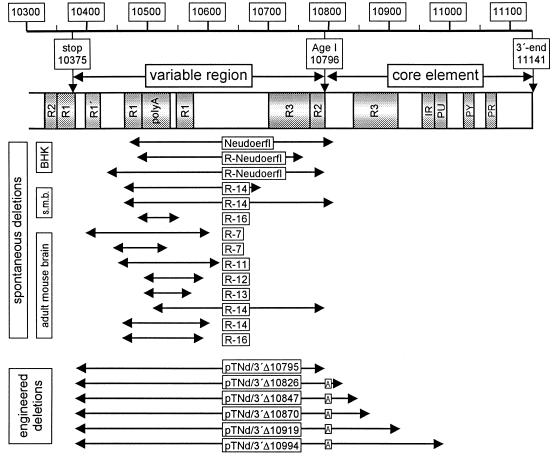
Schematic representation of the 3′ NCR of TBE virus Neudoerfl. Numbers correspond to the full-length genomic sequence (GenBank accession no. U27495). Shaded areas depict sequence elements that were described previously (15, 31): R1, R2, R3, direct repeats; polyA, internal poly(A) sequence, here 49 residues long, as it is in the infectious cDNA clone of TBE virus; IR, inverted repeat; PU, homopurine box; PY, homopyrimidine box; PR, pyrimidine-rich box. The positions of spontaneous 3′-NCR deletions observed during cell culture growth (BHK), in suckling-mouse brain stock solutions (s.m.b.), and in virus isolated from adult mouse brains are aligned below the schematic. For the exact positions of the deletion boundaries, refer to Tables 1 and 2. The bottom section shows the engineered deletions which extend from nucleotide 10378 to the positions as given in the corresponding plasmid name. A boxed A represents the 6-nucleotide AgeI recognition sequence which was retained in the construction procedure.
Virus recovery and stock virus preparations.
The generation of recombinant virus from the TBE infectious cDNA clone system and the preparation of virus stocks were performed as described elsewhere (17). Briefly, plasmid pTNd/5′, which contains the 5′-terminal one-third of the TBE virus genome, and one of the derivatives of plasmid pTNd/3′ described above were joined by in vitro ligation at the unique ClaI restriction site to give a full-length cDNA template, which was subsequently used to generate a genome-length RNA by in vitro T7-mediated transcription. Virus was harvested from the supernatant 3 to 5 days after electroporation of this RNA into BHK-21 cells. To achieve a high-titer stock suspension, the virus was then passaged twice in suckling-mouse brains. A 20% (wt/vol) suspension prepared from the second suckling-mouse brain passage was used as the stock virus for all further characterizations.
Sequence analysis.
Sequencing was performed with an automated DNA sequencing system that uses fluorescently labeled dideoxynucleotides (ABI-Perkin Elmer). Newly constructed plasmids were analyzed at least over the range of the new PCR-derived sequence elements and in the vicinity of restriction sites used in the particular cloning step. Genomic RNA of virus from cell culture supernatants, suckling-mouse brain suspensions, or adult mouse brains was analyzed by reverse transcription-PCR (RT-PCR) by standard methods and as described previously (31). PCR-derived fragments were sequenced directly. Sequence analysis was always performed on both strands.
RNA secondary-structure prediction.
Preliminary theoretical folding studies with the entire TBE virus genome sequence indicated that the 3′-terminal portion starting at position 10361 represents an independently folding domain (27a). All computations were therefore performed exclusively on a 3′-terminal domain of the TBE virus genome (and the various 3′-NCR deletion mutants) starting at nucleotide 10361. The calculations were performed with the public-domain Vienna RNA Package, which contains a variety of programs for the computation and comparison of RNA secondary structures (10).
Cell cultures.
BHK-21 cells, porcine kidney (PS) cells, and primary chicken embryo cells were grown under standard conditions as described previously (12, 17). Infectivity values of virus stocks were determined by plaque titer determinations on PS cells (12) and confirmed by end-point dilution infection experiments on BHK-21 and primary chicken embryo cells. Temperature sensitivity was tested by plaque assays performed on PS cells at the standard incubation temperature (37°C) and at 40°C. Growth curves were determined with primary chicken embryo cell monolayers as described in detail recently (17). Essentially, this method monitors the amount of infectious virus released from cells infected at a multiplicity of infection (MOI) of 1 within 1-h periods between 3 and 21 h postinfection.
Animal model.
Virulence and infectivity characteristics were analyzed in outbred Swiss-albino mice. Groups of 7 to 12 1-day-old suckling mice or groups of 10 5-week-old (body weight, approximately 20 g) mice were inoculated intracranially or subcutaneously, and survival was recorded for 28 days. Then mice were bled, and seroconversion was investigated by a TBE-antibody enzyme-linked immunosorbent assay (8). Adult mice were inoculated with a challenge dose of 100 50% lethal doses (LD50) of the highly virulent TBE virus strain Hypr (32). For the determination of the LD50 and the 50% infectious dose (ID50), mice were inoculated with sequential 10-fold dilutions of virus ranging from 10−3 to 101 PFU for suckling mice and from 10−1 to 105 PFU for adult mice. LD50s and ID50s were calculated by the method of Reed and Muench (28). For ID50 calculations, the number of infected mice was taken to be the total of mice killed plus surviving mice with detectable seroconversion. Surviving mice without detectable serum antibody were scored as uninfected.
RESULTS
Spontaneous 3′-NCR deletions.
The 3′ NCR of TBE virus Western prototypic strain Neudoerfl is 767 nucleotides long [assuming that the internal poly(A) tract is 49 adenosine residues, as used in the construction of the infectious cDNA clone (17)]. As shown schematically in Fig. 1 and described in detail elsewhere, it contains a number of sequence elements of interest, including direct and inverted repeats, purine and pyrimidine-rich conserved boxes, and the internal poly(A) tract (15, 31).
To test the genetic stability of this 3′ NCR, which is unusually long for a flavivirus, both parent virus and virus recovered from the infectious cDNA clone (recombinant virus, termed R-Neudoerfl) were repeatedly passaged in BHK cell cultures and the lengths of the 3′ NCRs were monitored by RT-PCR analysis. After approximately 15 and 20 passages for parent virus and R-Neudoerfl, respectively, smaller PCR fragments suggesting the emergence of viral genomes with smaller 3′ NCRs were detected. Nucleotide sequence analysis of two of these PCR fragments that predominated in the 23rd passage of R-Neudoerfl showed that deletions of 274 and 362 nucleotides had occurred. After several additional rounds of cell culture passages (using an m.o.i. of <0.01) for each of the viruses, only a single 3′-NCR species was detected by PCR. Sequence analysis revealed 3′ NCRs carrying deletions of 342 and 274 nucleotides, respectively. These data, including the exact boundaries of the deletions, are summarized in Table 1. Note that the finally evolved 3′ NCR of virus R-Neudoerfl as analyzed in the 32nd passage is identical to the larger of the two 3′-NCR species detected in the 23rd passage.
TABLE 1.
Spontaneous 3′-NCR deletions during passage in BHK cells
| Virus | BHK cell passage no. | Deletion size (boundariesa) |
|---|---|---|
| Neudoerfl | 22 | 342 (10469–10810) |
| R-Neudoerfl | 23 | 274 (10484–10757) |
| 362 (10432–10793) | ||
| 32 | 274 (10484–10757) |
The first and last nucleotides missing in the deleted sequence; numbering is according to the genomic sequence of TBE virus strain Neudoerfl.
These observations prompted us to investigate the phenomenon of spontaneous 3′-NCR deletions on a larger scale. We took advantage of a panel of stock suckling-mouse brain suspensions containing recombinant TBE virus mutants bearing mutations at locations other than the 3′ NCR, which will be described in detail elsewhere (17a), as well as viruses recovered from the brains of adult mice infected with these mutants. The results of the analyses of a total of 17 suckling-mouse brain suspensions and virus recovered from 24 adult mice are summarized in Table 2. Genomes with deleted 3′-NCR sequences were detected in 2 of 17 suckling-mouse brain suspensions. In one of these cases, two distinct deletion products were observed. The investigation of virus recovered from adult mouse brains indicated the presence of deleted 3′ NCRs in 8 of 24 samples. Deletions were detected more frequently in viruses that had undergone several passages in BHK cells before inoculation of the mice (Table 2).
TABLE 2.
Spontaneous 3′-NCR deletions detected in a panel of recombinant TBE virus mutants
| Virusa | No. of passagesb | No. of deletions in suckling micec | Adult miced
|
|
|---|---|---|---|---|
| No. testede | No. of deletionsf | |||
| R-1 | 1 | 0 | 2 | 0 |
| R-2 | 1 | 0 | 0 | 0 |
| R-3 | 1 | 0 | 2 | 0 |
| R-4 | 1 | 0 | 1 | 0 |
| R-5 | 1 | 0 | 0 | 0 |
| R-6 | 1 | 0 | 2 | 0 |
| R-7 | 1 | 0 | 8 | 2 (10394–10604, 10441–10537) |
| R-8 | 1 | 0 | 0 | 0 |
| R-9 | 1 | 0 | 0 | 0 |
| R-10 | 1 | 0 | 1 | 0 |
| R-11 | 5 | 0 | 1 | 1 (10450–10620) |
| R-12 | 5 | 0 | 1 | 1 (10494–10596) |
| R-13 | 5 | 0 | 2 | 1 (10495–10574) |
| R-14 | 5 | 2 (10458–10689, 10458–10811) | 2 | 2 (10504–10794, 10453–10603) |
| R-15 | 5 | 0 | 1 | 0 |
| R-16 | 5 | 1 (10485–10552) | 1 | 1 (10454–10592) |
| R-17 | 9 | 0 | 0 | 0 |
| Total | 3 | 24 | 8 | |
Recombinant TBE virus Neudoerfl mutants bearing specific mutations in regions of the genome other than the 3′ NCR, consecutively numbered.
Number of BHK cell culture passages (at low MOI) prior to the generation of suckling-mouse brain stock suspensions.
Number of deletion mutants found in suckling-mouse brain stock suspensions; boundaries of deletions (positions of the first and last deleted nucleotides) are in parentheses.
Virus isolated from the brains of adult mice after infection with the suckling-mouse brain stock suspensions.
Number of viral isolates tested.
Number of deletion mutants found; boundaries of deletions in parentheses.
The positions of all of the deletions described above are summarized in Fig. 1, where they are aligned with the schematic drawing of the original strain Neudoerfl 3′ NCR. This representation allows several conclusions to be drawn with respect to the pattern of these spontaneously occurring deletions: (i) all deletions affect the variable region of the 3′ NCR exclusively; (ii) the deletions involve sequence elements from almost the entire variable region (the most extreme 5′ boundary is nucleotide 10394, and the most extreme 3′ boundary is nucleotide 10811); (iii) the poly(A) tract is removed in each deletion event, although a few of the adenosine residues are maintained in some cases; (iv) the deletion boundaries are variable to a large extent but do not seem to be completely random, since deletions seem to cluster at certain positions (e.g., around nucleotides 10450, 10600, and 10800).
Engineered 3′-NCR deletion mutants.
The observations described above prompted us to construct mutants of strain Neudoerfl with even larger 3′-NCR deletions than those occurring spontaneously. We wanted to address the questions of (i) how, if at all, removal of the variable region would influence the biological properties of TBE virus; (ii) how far these deletions could be extended into the core element without loss of virus viability; and (iii) how the biology of TBE virus would be influenced by deletions extending into the core element.
The bottom portion of Fig. 1 illustrates the sizes and positions of the deletions introduced into the infectious cDNA clone of TBE virus. The detailed information on how these clones were constructed is contained in Materials and Methods. All of the deletions start with the nucleotide immediately following the stop codon that terminates the long open reading frame (i.e., nucleotide 10378). In the mutant clone termed pTNd/3′Δ10795, the deletion extends to nucleotide 10795, thus removing the entire variable region but leaving the core element intact. The deletion in clone pTNd/3′Δ10826 extends approximately 20 nucleotides into the core element but does not affect the core copy of the R3 direct repeat. In pTNd/3′Δ10847 and pTNd/3′Δ10870, the deletions are increased a further 21 and 23 nucleotides, respectively, thus removing parts of the R3 repeat. Deletion mutant pTNd/3′Δ10919 lacks the entire R3 repeat, whereas in mutant pTNd/3′Δ10994 the inverted repeat element and the purine box were also removed. It should be mentioned that due to the mutagenesis strategy used, the mutant plasmids pTNd/3′Δ10826, pTNd/3′Δ10847, pTNd/3′Δ10870, pTNd/3′Δ10919, and pTNd/3′Δ10994 contain an additional 6 nucleotides, forming an AgeI restriction cleavage site at the site of the deletion, as shown in Fig. 1.
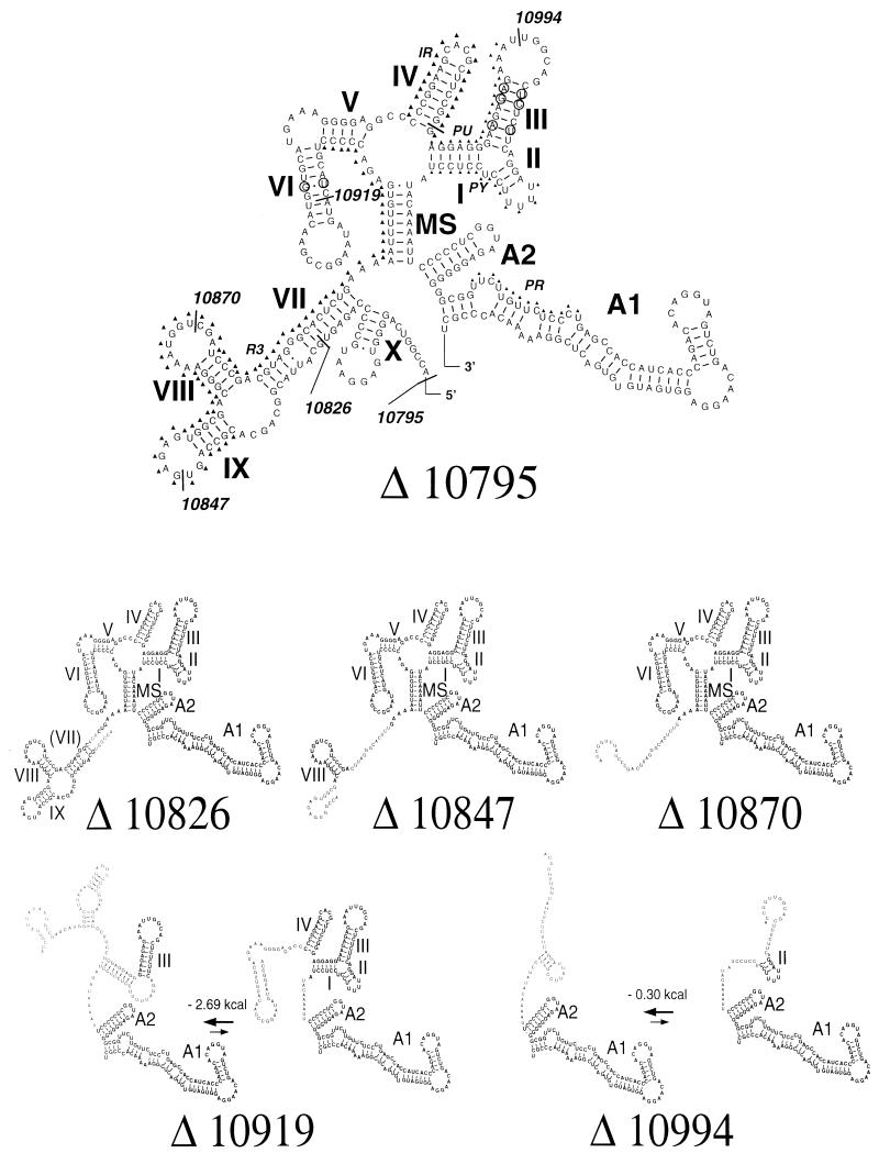
As reported recently, the entire core element of the TBE virus 3′ NCR is predicted to fold into a mostly well-defined and highly conserved secondary structure (27). This model, depicted in Fig. 2, contains a number of base-pairing elements and a multiloop-stem in addition to the previously described stem-loop structures at the very 3′ end. The deletions introduced in our mutants extend into the core element, as indicated in the figure. We wanted to assess by computer-based modeling how each individual deletion is predicted to influence the formation of the various secondary-structure elements. This was achieved by calculating minimum free energy structures of each deletion mutant sequence, starting in every case with nucleotide 10361, which represents the border of a 3′-terminal independently folding domain (27a). Removal of the entire variable region is not predicted to affect the folding of the core element, and an authentic and complete secondary structure can fold in the case of the pTNd/3′Δ10795 deletion mutation. The deletion mutation as present in pTNd/3′Δ10826 removes structure X and causes a truncation of stem VII, but all the other structures are still predicted to fold in the minimum free energy model. The deletions as present in pTNd/3′Δ10847 and pTNd/3′Δ10870 would cause the loss of structures X, VII, and IX, and in the latter case also VIII, but in all of these mutants the authentic complex of structures I through VI, which is stabilized by the multiloop-stem, is maintained. In contrast, the most stable conformation predicted for RNA transcribed from plasmid pTNd/3′Δ10919 includes only the 3′-terminal structures A1 and A2, as well as structure III. Structures I, II, and IV may also occur in this RNA, but their formation is thermodynamically unfavorable (ΔG = 2.69 kcal/mol), and therefore these structures would be expected to exist only in a minor percentage of molecules. For the pTNd/3′Δ10994 sequence, two almost equally stable structures are predicted, each containing A1 and A2, but an authentic element II is formed in only one of these alternative structures.
FIG. 2.
RNA secondary-structure predictions of the 3′ NCRs of the TBE virus deletion mutants. Sequences are shown starting with the first nucleotide following the stop codon, which is also the first nucleotide of an AgeI restriction site (Fig. 1). At the top is the structure of the 3′ NCR as present in mutant R-Nd/3′Δ10795, corresponding exactly to the previously published structure of the TBE 3′-NCR core element (27). The borders of the individual engineered deletions are indicated by the corresponding nucleotide numbers. Positions of the sequence elements R3, IR, PU, PY, and PR (compare Fig. 1) are indicated by triangles next to the sequence. Base-pairing elements are denoted by Roman numerals (I to X), MS depicts a multiloop-stem, and A1 and A2 are the known conserved flavivirus 3′-terminal structures. Below are structures as predicted for the truncated sequences present in the various deletion mutants. Wild-type structural elements are shown in boldface type; sequences in undefined or nonauthentic folding patterns are shown in normal type. A truncated form of stem VII is shown in parentheses. The two sequences carrying the largest deletions may adopt two alternative structures in which the energetically favored ones (energy differences are shown) lack some of the wild-type base-pairing elements.
Viability of mutants and growth properties in cell cultures.
The clones carrying the deletions described above were used for the in vitro transcription of full-length genomic RNAs. BHK cells were transfected with these RNAs, and infectious virus was recovered from the supernatants for all of the deletion mutants except pTNd/3′Δ10994 (Table 3). Suckling-mouse brain suspensions of all of the viable recombinant viruses, named R-Nd/3′Δ10795, R-Nd/3′Δ10826, R-Nd/3′Δ10847, R-Nd/3′Δ10870, and R-Nd/3′Δ10919, were prepared, and the cell culture infectivity titers of these stocks were determined. As shown in Table 3, these mutant virus stocks exhibited infectivity titers similar to that of wild-type TBE virus strain Neudoerfl (i.e., between 1 × 10⁸ and 3 × 10⁸ PFU/ml), except for mutant R-Nd/3′Δ10919, whose titer was only 3 × 10⁶ PFU/ml.
TABLE 3.
Stock virus preparations and plaque morphologies for TBE virus mutants with engineered 3′-NCR deletions
| Plasmid with 3′-NCR deletion | Mutant virus | Infectivity titer of stock solution (PFU/ml) | Plaque morphologya |
|---|---|---|---|
| pTNd/3′Δ10795 | R-Nd/3′Δ10795 | 3 × 10⁸ | WT |
| pTNd/3′Δ10826 | R-Nd/3′Δ10826 | 1 × 10⁸ | WT |
| pTNd/3′Δ10847 | R-Nd/3′Δ10847 | 3 × 10⁸ | WT |
| pTNd/3′Δ10870 | R-Nd/3′Δ10870 | 1 × 10⁸ | WT |
| pTNd/3′Δ10919 | R-Nd/3′Δ10919 | 3 × 10⁶ | Turbid |
| pTNd/3′Δ10994 | Not viable | ||
| Neudoerflb | 3 × 10⁸ | WT | |
| R-Neudoerflb | 3 × 10⁸ | WT |
Determined at 37 and 40°C; morphologies were identical at both temperatures. WT, wild-type (strain Neudoerfl) morphology.
Control viruses.
Since these stocks were to be used as starting materials for all of the following experiments, we first confirmed their 3′-NCR sequences by RT-PCR and sequence analysis. The antigenic structures of the E proteins of all mutants were compared to that of the wild-type virus E protein by using a previously described panel of monoclonal antibodies (6, 11), and the entire E protein-coding regions of these stock viruses were also sequenced to ensure that no spontaneous mutations had occurred there. No differences from the expected sequences or antigenic structure profiles were found in the stock virus preparations (data not shown).
We continued the characterizations of the 3′-NCR deletion mutants by performing plaque assays at 37 and 40°C. Neither wild-type TBE virus nor any of the mutants were found to be temperature sensitive under the conditions used (data not shown). There was a difference, however, with respect to plaque morphology in one mutant virus: R-Nd/3′Δ10919 produced turbid plaques at both temperatures tested (Table 3).
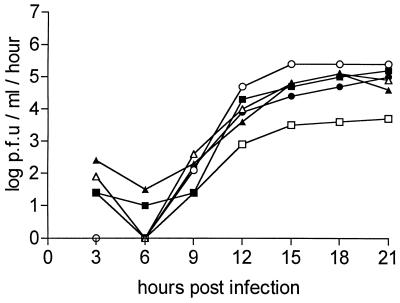
The growth properties of wild-type and mutant viruses were further compared by monitoring the release of newly synthesized infectious virus from primary chicken embryo fibroblasts (Fig. 3). The growth curves of the mutants were indistinguishable from that of the wild-type virus in this assay system, except for the curve of mutant R-Nd/3′Δ10919, which exhibited a growth capacity reduced by approximately 1.5 orders of magnitude.
FIG. 3.
Growth curves of wild-type and mutant TBE virus strains determined on primary chicken embryo cells infected at an m.o.i. of 1. The values plotted were derived from duplicate experiments. ▪, Neudoerfl; ▴, R-Nd/3′Δ10795; ▵, R-Nd/3′Δ10826; ○, R-Nd/3′Δ10847; •, R-Nd/3′Δ10870; □, R-Nd/3′Δ10919.
Virulence and induction of protective immunity.
Finally, the 3′-NCR mutants were tested in an animal model. Intracranial inoculation of suckling mice is the most sensitive assay system for TBE virus. Titer determinations for wild-type and mutant viruses and determinations of the LD50s as summarized in Table 4 indicated an approximately 100-fold-higher sensitivity of this system than that involving PFU determinations in cell culture. LD50s ranged from 10−1.7 to 10−2.5 PFU. The differences among these values are well within the expected range of inaccuracies and statistical deviations of the biological test systems used. Surviving mice were tested for seroconversion 4 weeks after inoculation. None of these mice had seroconverted. In other words, there was no occurrence of nonlethal infection in this system, and therefore lethality directly corresponds to infectivity. Thus, the use of this highly sensitive system allows us to conclude that all of the mutant viruses had retained a wild-type level of infectivity.
TABLE 4.
Inoculation of suckling mice and 5-week-old mice
| Virus | Suckling mice (intracranial inoculation)
|
5-week-old mice (subcutaneous inoculation)
|
||||
|---|---|---|---|---|---|---|
| LD50 (PFU) | Mean survival time (days) aftera:
|
LD50 (PFU) | ID50 (PFU) | LD50/ID50 | ||
| 1 PFUb | 0.1 PFUb | |||||
| Neudoerfl | 10−1.7 | 7.2 (±1.5) | 8.2 (±1.5) | 100.9 | 10−0.2 | 101.1 |
| R-Nd/3′Δ10795 | 10−2.5 | 7.7 (±1.4) | 8.5 (±0.9) | 101.0 | 10−0.4 | 101.4 |
| R-Nd/3′Δ10826 | 10−2.3 | 11.0 (±0.8) | 10.7 (±1.7) | 104.9 | 100.4 | 104.5 |
| R-Nd/3′Δ10847 | 10−1.9 | 11.1 (±0.9) | 11.4 (±0.9) | 105.0 | 101.0 | 104.0 |
| R-Nd/3′Δ10870 | 10−2.1 | 12.5 (±2.5) | 11.9 (±1.5) | >105.0 | 102.4 | >102.6 |
| R-Nd/3′Δ10919 | 10−2.4 | 12.6 (±0.6) | 11.5 (±0.5) | >105.0 | 100.5 | >104.5 |
 |
Inoculation dose/mouse. For each virus and dose, litters of 7 to 12 mice were inoculated. No mice survived at these inoculation doses.
However, differences in mean survival times were observed. Table 4 shows a comparison of these values determined at two different inoculation doses. Whereas the values determined for mutant R-Nd/3′Δ10795 are similar to those for the wild-type virus (around 8 days), survival times for all of the other mutants are increased by up to 5 days, implying that these mutants replicate more slowly in the brain cells of suckling mice.
It has been observed that a high percentage of adult Swiss albino mice inoculated peripherally with TBE virus develop lethal infections of the central nervous system that clinically resemble severe human TBE. This observation, together with data obtained with experimental tick-borne live-vaccine candidates tested in mice, primates, and humans, has established the mouse model as an appropriate animal model for studying human TBE (18, 19, 22). We used adult mice to determine the LD50 and infectious dose ID50 for the parent virus and each of the mutant viruses (ID50 calculations are based on the numbers of killed plus surviving mice exhibiting seroconversion versus surviving seronegative mice). As seen from the results summarized in Table 4, the infectivity values for adult mice were found to vary only moderately. They ranged from below 1 PFU for wild-type strain Neudoerfl and mutant R-Nd/3′Δ10795 to approximately 10-fold-higher values for all other mutants except for mutant R-Nd/3′Δ10870, which was found to be more than 100-fold less infectious than was the wild-type virus. In contrast, LD50s differed markedly among the viruses tested. As expected for virulent virus, the LD50s for wild-type virus and mutant virus R-Nd/3′Δ10795 were only about 10-fold higher than the ID50s. (This indicates that infection by these viruses is lethal in most cases, even at low inoculation doses.) For the other deletion mutants, however, the LD50s were 105 PFU or higher.
The most relevant parameter for judging the suitability of an attenuated mutant as a candidate for vaccine development is the LD50/ID50 ratio. Ideally, a vaccine strain infects the host and induces seroconversion at an inoculation dose far below its lethal dose, corresponding to a high LD50/ID50 ratio. As can be seen in Table 4, the LD50/ID50 ratios are around 10 for wild-type virus and mutant R-Nd/3′Δ10795 but are significantly higher for all other deletion mutants, ranging as high as 104.5. We conclude that removal of the entire variable region, as exemplified by mutant R-Nd/3′Δ10795, preserved the virulence properties of the parent virus but that all of the deletions extending into the core element caused significant attenuation.
To confirm that seroconversion corresponded to the induction of protective immunity, all surviving adult mice were challenged 4 weeks p.i. with a lethal dose (100 LD50) of the highly virulent TBE virus strain Hypr (32). Every animal that had seroconverted after the primary infection was found to be protected against disease. In fact, a few mice with antibody levels below the detection limit were also protected, whereas none of the mock-infected control mice survived infection (data not shown).
Finally, we addressed the question of the genetic stability of the 3′-NCR deletion mutants during the infection and invasion of the brains of adult mice. For each mutant, virus was isolated from the brains of two mice killed by the infection and the 3′ NCR and the E protein-coding regions were examined by RT-PCR and sequence analysis. Except for a single silent point mutation in the E protein-coding region of one R-Nd/3′Δ10919 isolate, there were no changes from the expected sequences (data not shown). Although it was not ruled out that mutations arising elsewhere in the genome could have also had an effect on the growth or virulence properties, none of the deletion mutants showed any genetic instability in the 3′ NCR itself.
DISCUSSION
Comparisons of the 3′-NCR structures of various TBE virus strains had previously led to a structural model that divided the 3′ NCR in two distinct regions: the 3′-terminal 340-nucleotide core element, which is highly conserved in its primary sequence and RNA-folding pattern, and the variable region, which is inserted between the core element and the coding region of the genome. In continuation of these studies, we now present evidence that this subdivision based on sequence comparisons also corresponds to functional differences between these two regions.
The variable region does not appear to have any relevant function for the growth of TBE virus in cell culture or in mice, as demonstrated by the occurrence of spontaneous deletions that affected almost all parts of this region. This notion is also supported by results with one of our engineered mutants, R-Nd/3′Δ10795, which lacks the entire variable region without exhibiting any significant change of its biology in cell culture or animal test systems. The observation of spontaneous deletions occurring in vitro raises the possibility that the shorter forms of 3′-NCR sequences detected in various TBE virus strains actually arose after the primary isolation of these virus strains during propagation in the laboratory. Size variability in the 5′ part of the 3′ NCR has also been observed in a few cases for other flaviviruses (24, 33). Our observations with TBE virus raise the question whether some of the 3′-NCR sequences determined for other flaviviruses, which are generally shorter than that of TBE virus prototype strain Neudoerfl, might also have originated from longer forms which underwent deletions during the isolation and laboratory propagation of these viruses. The purpose that the long variable region of TBE virus, which also includes an internal poly(A) tract, might serve under normal ecological conditions is unclear. It seems unlikely that an RNA virus would preserve this sequence if it were without any selective advantage in vivo, even though this advantage does not seem to be relevant under laboratory growth conditions. In this context, it should be noted, however, that our data suggest that the deletion boundaries are not totally random, and a mutant with a smaller 3′-NCR deletion was observed to prevail over another one with a larger deletion, suggesting subtle and as yet undefined differences in evolutionary fitness among various deletion mutants.
While the role of the variable region remains vague and its influence on the viral life cycle may be subtle, there is strong evidence for the functional relevance of the core element. The importance of this region had already been implied by its high degree of sequence conservation and its well-defined folding pattern (5, 25, 27). Results with our mutants now provide direct evidence for its functional role and define functional boundaries. Expanding the engineered deletion from position 10795 by only 31 nucleotides to position 10826 caused a pronounced reduction of virulence. The pTNd/3′Δ10826 deletion did not involve sequences of the R3 repeat, but on the secondary-structure level it caused the loss of one predicted short stem-loop element (X) and the truncation of another stem (VII). Stem-loop X, however, was also lost in two of the spontaneous deletion events (Neudoerfl in BHK passage, and R-14 in suckling-mouse brain) and is also missing in one of the TBE virus strains (RK 1424) that had been analyzed previously (31). We speculate, therefore, that the truncation of stem VII rather than the loss of structure X is the relevant cause of the observed attenuation. Further small increases in the deletion sizes tended to intensify the biological effects. The data summarized in Table 4 illustrate that there was a correlation between increased survival times of intracranially inoculated suckling mice and attenuation in adult mice. In all cases, survival times showed remarkably little dependence on dosage in this range. Interestingly, the marked attenuation in animals was not reflected in cell culture: three of the mutants with reduced virulence (R-Nd/3′Δ10826, R-Nd/3′Δ10847, and R-Nd/3′Δ10870) were indistinguishable from wild-type virus in our cell culture test systems. A significantly impaired growth behavior in these tests was observed only for mutant R-Nd/3′Δ10919, which lacks the entire R3 repeat. The 3′-NCR of this mutant was also predicted to preferentially fold in a nonauthentic manner, and this may also contribute to its growth restriction in cell culture. Surprisingly, even this mutant had retained the ability to establish infection in both suckling mice (with significantly prolonged survival times) and adult mice at almost wild-type levels. In contrast, mutant R-Nd/3′Δ10870 was more impaired than the others with respect to infectivity in adult mice (Table 4). Taken together, these results suggest that attenuation in mice, infectivity, and growth properties in cell culture are, at least to some extent, independent functional properties that may be governed by separable structural entities.
There is increasing evidence from work on other flaviviruses that the approximately 340 nucleotides at the extreme 3′ end of the genome form a functionally important entity. The analysis of several tick-borne flaviviruses by a different algorithm (5) yielded very similar folding patterns to those found by Rauscher et al. (27). Although conservation of certain sequence motifs is restricted to mosquito-borne flaviviruses and does not extend to the tick-borne group (16), there is significant conservation between these viruses at the secondary-structure level (25). Folding patterns involving the 3′-terminal 340 nucleotides were predicted for all major flavivirus subgroups, and the impact of different folding patterns on the attenuation of yellow fever virus was postulated recently (26). Direct evidence for the importance of this region and its suitability for achieving the attenuation of DEN-4 virus has been described by Men et al. (21), who reported the generation of growth-restricted mutants by introducing internal deletions in the 3′ NCR of this virus. They also demonstrated that the 5′-terminal part of the 3′ NCR could be removed without loss of viability and found the minimum requirement for viability to be the last 113 nucleotides at the 3′ end. In contrast, the minimum sequence required for TBE virus viability was found to begin between 147 (as in the nonviable deletion clone pTNd/3′Δ10994) and 222 (as in R-Nd/3′Δ10919) nucleotides from the 3′ terminus. Most of the DEN-4 virus deletion mutants exhibited restricted growth behavior in cell culture, and this seemed to correlate with a reduced immunogenicity in monkeys (21). In comparison, most of our TBE deletion mutants exhibited normal growth behavior in cell culture and satisfactory infectivity for mice, even though a high level of attenuation was achieved. One of the DEN-4 virus mutants (3′d 303–183), which retained an 80-nucleotide 5′-terminal section and the 183 3′-terminal nucleotides of the 3′ NCR, replicated well in cell culture and induced a good antibody response in monkeys (21). TBE mutant R-Nd/3′Δ10919, however, which contains the 222 3′-terminal nucleotides, was severely impaired with respect to cell culture growth but still was able to infect mice and induce seroconversion. Using Kunjin virus, Kromykh and Westaway (14) investigated the influence of 3′-NCR deletions on replication. In good agreement with our data, they found that a small (76-nucleotide) deletion that preserved the 3′-terminal 524 nucleotides had no measurable effect on replication whereas a larger deletion, extending to nucleotide 10774 (corresponding approximately to position 10893 of TBE virus strain Neudoerfl, i.e., between the 3′-terminal deletion boundaries of TBE virus mutants R-Nd/3′Δ10870 and R-Nd/3′Δ10919), caused significant impairment of RNA replication.
All of the available data taken together suggest that the use of 3′-NCR deletions may indeed be a suitable approach to the development of live flavivirus vaccines for several reasons. (i) Deletion mutants cannot revert to wild-type sequence, and so far we have not obtained any evidence for genetic instability in our mutants. (ii) Attenuation could be achieved without measurable loss of viral growth properties in cell culture and also while maintaining high in vivo infectivity. (iii) Seroconversion and protective immunity were induced at even very low inoculation doses. (iv) Our data suggest that the degree of attenuation can be modulated by small changes of the deletion size, thus making it feasible to specifically engineer a mutant exhibiting exactly the desired degree of attenuation. Future work will also aim toward the combination of 3′-NCR deletions with other genetic modifications to further explore the possibilities for specifically attenuating TBE virus and other flaviviruses.
ACKNOWLEDGMENTS
We gratefully acknowledge the excellent technical assistance of Melby Wilfinger and Jutta Ertl.
REFERENCES
- 1.Blackwell J L, Brinton M A. BHK cell proteins that bind to the 3′ stem-loop structure of the West Nile virus genome RNA. J Virol. 1995;69:5650–5658. doi: 10.1128/jvi.69.9.5650-5658.1995. [DOI] [PMC free article] [PubMed] [Google Scholar]
- 2.Cahour A, Pletnev A, Vazeille-Falcoz M, Rosen L, Lai C-J. Growth-restricted dengue virus mutants containing deletions in the 5′ noncoding region of the RNA genome. Virology. 1995;207:68–76. doi: 10.1006/viro.1995.1052. [DOI] [PubMed] [Google Scholar]
- 3.Chambers T J, Hahn C S, Galler R, Rice C M. Flavivirus genome organization, expression, and replication. Annu Rev Microbiol. 1990;44:649–688. doi: 10.1146/annurev.mi.44.100190.003245. [DOI] [PubMed] [Google Scholar]
- 4.Chen C-J, Kuo M-D, Chien L-J, Hsu S-L, Wang Y-M, Lin J-H. RNA-protein interactions: involvement of NS3, NS5, and 3′ noncoding regions of Japanese encephalitis virus genomic RNA. J Virol. 1997;71:3466–3473. doi: 10.1128/jvi.71.5.3466-3473.1997. [DOI] [PMC free article] [PubMed] [Google Scholar]
- 5.Gritsun T S, Venugopal K, de A. Zanotto P M, Mikhailov M V, Sall A A, Holmes E C, Polkinghorne I, Frolova T V, Pogodina V V, Lashkevich V A, Gould E A. Complete sequence of two tick-borne flaviviruses isolated from Siberia and the UK: analysis and significance of the 5′ and 3′-UTRs. Virus Res. 1997;49:27–39. doi: 10.1016/s0168-1702(97)01451-2. [DOI] [PubMed] [Google Scholar]
- 6.Guirakhoo F, Heinz F X, Kunz C. Epitope model of tick-borne encephalitis virus envelope glycoprotein E: analysis of structural properties, role of carbohydrate side chain, and conformational changes occurring at acidic pH. Virology. 1989;169:90–99. doi: 10.1016/0042-6822(89)90044-5. [DOI] [PubMed] [Google Scholar]
- 7.Hahn C S, Hahn Y S, Rice C M, Lee E, Dalgarno L, Strauss E G, Strauss J H. Conserved elements in the 3′ untranslated region of flavivirus RNAs and potential cyclization sequences. J Mol Biol. 1987;198:33–41. doi: 10.1016/0022-2836(87)90455-4. [DOI] [PubMed] [Google Scholar]
- 8.Heinz F X, Berger R, Tuma W, Kunz C. A topological and functional model of epitopes on the structural glycoprotein of tick-borne encephalitis virus defined by monoclonal antibodies. Virology. 1983;126:525–537. doi: 10.1016/s0042-6822(83)80010-5. [DOI] [PubMed] [Google Scholar]
- 9.Heinz F X, Mandl C W. The molecular biology of tick-borne encephalitis virus. Acta Pathol Microbiol Immunol Scand. 1993;101:735–745. doi: 10.1111/j.1699-0463.1993.tb00174.x. [DOI] [PubMed] [Google Scholar]
- 10.Hofacker I L, Fontana W, Stadler P F, Bonhoeffer S, Tacker M, Schuster P. Fast folding and comparison of RNA secondary structures. Monatsh Chem. 1994;125:167–188. [Google Scholar]
- 11.Holzmann H, Stiasny K, York H, Dorner F, Kunz C, Heinz F X. Tick-borne encephalitis virus envelope protein E-specific monoclonal antibodies for the study of low pH-induced conformational changes and immature virions. Arch Virol. 1995;140:213–222. doi: 10.1007/BF01309857. [DOI] [PubMed] [Google Scholar]
- 12.Holzmann H, Stiasny K, Ecker M, Kunz C, Heinz F X. Characterization of monoclonal antibody-escape mutants of tick-borne encephalitis virus with reduced neuroinvasiveness in mice. J Gen Virol. 1997;78:31–37. doi: 10.1099/0022-1317-78-1-31. [DOI] [PubMed] [Google Scholar]
- 13.Kolykhalov A A, Feinstone S M, Rice C M. Identification of a highly conserved sequence element at the 3′ terminus of hepatitis C virus genome RNA. J Virol. 1996;70:3363–3371. doi: 10.1128/jvi.70.6.3363-3371.1996. [DOI] [PMC free article] [PubMed] [Google Scholar]
- 14.Khromykh A A, Westaway E G. Subgenomic replicons of the flavivirus Kunjin: construction and applications. J Virol. 1997;71:1497–1505. doi: 10.1128/jvi.71.2.1497-1505.1997. [DOI] [PMC free article] [PubMed] [Google Scholar]
- 15.Mandl C W, Kunz C, Heinz F X. Presence of poly(A) in a flavivirus: significant differences between the 3′ noncoding regions of the genomic RNAs of tick-borne encephalitis virus strains. J Virol. 1991;65:4070–4077. doi: 10.1128/jvi.65.8.4070-4077.1991. [DOI] [PMC free article] [PubMed] [Google Scholar]
- 16.Mandl C W, Holzmann H, Kunz C, Heinz F X. Complete genomic sequence of Powassan virus: evaluation of genetic elements in tick-borne versus mosquito-borne flaviviruses. Virology. 1993;194:173–184. doi: 10.1006/viro.1993.1247. [DOI] [PubMed] [Google Scholar]
- 17.Mandl C W, Ecker M, Holzmann H, Kunz C, Heinz F X. Infectious cDNA clones of tick-borne encephalitis virus European subtype prototypic strain Neudoerfl and high virulence strain Hypr. J Gen Virol. 1997;78:1049–1057. doi: 10.1099/0022-1317-78-5-1049. [DOI] [PubMed] [Google Scholar]
- 17a.Mandl, C. W., et al. Unpublished results.
- 18.Mayer V. A live vaccine against tick-borne encephalitis: integrated studies. I. Basic properties and behaviour of the E5"14" clone (Langat virus) Acta Virol. 1974;19:209–218. [PubMed] [Google Scholar]
- 19.Mayer V, Pogády J, Orolin D, Stárek M, Hudecová M, Buran J, Hrbka G. Further virological and clinical investigations on the attenuated E5"14" virus from the tick-borne encephalitis complex. Acta Virol. 1975;19:143–149. [PubMed] [Google Scholar]
- 20.McMinn P C. The molecular basis of virulence of the encephalitogenic flaviviruses. J Gen Virol. 1997;78:2711–2722. doi: 10.1099/0022-1317-78-11-2711. [DOI] [PubMed] [Google Scholar]
- 21.Men R, Bray M, Clark D, Chanock R M, Lai C-J. Dengue type 4 virus mutants containing deletions in the 3′ noncoding region of the RNA genome: analysis of growth restriction in cell culture and altered viremia pattern and immunogenicity in Rhesus monkeys. J Virol. 1996;70:3930–3937. doi: 10.1128/jvi.70.6.3930-3937.1996. [DOI] [PMC free article] [PubMed] [Google Scholar]
- 22.Monath T P, Heinz F X. Flaviviruses. In: Fields B N, Knipe D M, Howley P M, et al., editors. Fields virology. 3rd ed. Philadelphia, Pa: Lippincott-Raven Publishers; 1996. pp. 961–1034. [Google Scholar]
- 23.Moormann R J, van Gennip H G, Miedema G K, Hulst M M, van Rijn P A. Infectious RNA transcribed from an engineered full-length cDNA template of the genome of a pestivirus. J Virol. 1996;70:763–770. doi: 10.1128/jvi.70.2.763-770.1996. [DOI] [PMC free article] [PubMed] [Google Scholar]
- 24.Poidinger M, Hall R A, Mackenzie J S. Molecular characterization of the Japanese encephalitis serocomplex of the flavivirus genus. Virology. 1996;218:417–421. doi: 10.1006/viro.1996.0213. [DOI] [PubMed] [Google Scholar]
- 25.Proutski V, Gould E A, Holmes E C. Secondary structure of the 3′ untranslated region of flaviviruses: similarities and differences. Nucleic Acids Res. 1997;25:1194–1202. doi: 10.1093/nar/25.6.1194. [DOI] [PMC free article] [PubMed] [Google Scholar]
- 26.Proutski V, Gaunt M W, Gould E A, Holmes E C. Secondary structure of the 3′-untranslated region of yellow fever virus: implications for virulence, attenuation and vaccine development. J Gen Virol. 1997;78:1543–1549. doi: 10.1099/0022-1317-78-7-1543. [DOI] [PubMed] [Google Scholar]
- 27.Rauscher S, Flamm C, Mandl C W, Heinz F X, Stadler P F. Secondary structure of the 3′-noncoding region of flavivirus genomes: comparative analysis of base pairing probabilities. RNA. 1997;3:779–791. [PMC free article] [PubMed] [Google Scholar]
- 27a.Rauscher, S., et al. Unpublished observation.
- 28.Reed J L, Muench H. A simple method for estimating fifty percent endpoints. Am J Hyg. 1938;27:493. [Google Scholar]
- 29.Rey F A, Heinz F X, Mandl C, Kunz C, Harrison S C. The envelope glycoprotein from tick-borne encephalitis virus at 2 A resolution. Nature. 1995;375:291–298. doi: 10.1038/375291a0. [DOI] [PubMed] [Google Scholar]
- 30.Tanaka T, Kato N, Cho M-J, Sugiyama K, Shimotohno K. Structure of the 3′ terminus of the hepatitis C virus genome. J Virol. 1996;70:3307–3312. doi: 10.1128/jvi.70.5.3307-3312.1996. [DOI] [PMC free article] [PubMed] [Google Scholar]
- 31.Wallner G, Mandl C W, Kunz C, Heinz F X. The flavivirus 3′-noncoding region: extensive size heterogeneity independent of evolutionary relationships among strains of tick-borne encephalitis virus. Virology. 1995;213:169–178. doi: 10.1006/viro.1995.1557. [DOI] [PubMed] [Google Scholar]
- 32.Wallner G, Mandl C W, Ecker M, Holzmann H, Stiasny S, Kunz C, Heinz F X. Characterization and complete genome sequences of high- and low-virulence variants of tick-borne encephalitis virus. J Gen Virol. 1996;77:1035–1042. doi: 10.1099/0022-1317-77-5-1035. [DOI] [PubMed] [Google Scholar]
- 33.Wang E, Weaver S C, Shope R E, Tesh R B, Watts D M, Barrett A D T. Genetic variation in yellow fever virus: duplication in the 3′ noncoding region of strains from Africa. Virology. 1996;225:274–281. doi: 10.1006/viro.1996.0601. [DOI] [PubMed] [Google Scholar]
- 34.Wengler G, Bradley D W, Collett M S, Heinz F X, Schlesinger R W, Strauss J H. Flaviviridae. In: Murphy F A, Fauquet C M, Bishop D H L, Ghabrial S A, Jarvis A W, Martelli G P, Mayo M A, Summers M D, editors. Virus Taxonomy. Sixth Report of the International Committee on Taxonomy of Viruses. Vienna, Austria: Springer-Verlag KG; 1995. pp. 415–427. [Google Scholar]
- 35.Yamada N, Tanihara K, Takada A, Yorihuzi T, Tsutsumi M, Shimomura H, Tsuji T, Date T. Genetic organization and diversity of the 3′ noncoding region of the hepatitis C virus genome. Virology. 1996;223:255–261. doi: 10.1006/viro.1996.0476. [DOI] [PubMed] [Google Scholar]


