Abstract
Four mutants of the virulent Mahoney strain of poliovirus were generated by introducing mutations in nucleotides (nt) 128 to 134 of the genome, a region that contains a part of the stem-loop II (SLII) structure located within the internal ribosomal entry site (IRES; nt 120 to 590) (K. Shiroki, T. Ishii, T. Aoki, Y. Ota, W.-X. Yang, T. Komatsu, Y. Ami, M. Arita, S. Abe, S. Hashizume, and A. Nomoto, J. Virol. 71:1–8, 1997). These mutants (SLII mutants) replicated well in human HeLa cells but not in mouse TgSVA cells that had been established from the kidney of a poliovirus-sensitive transgenic mouse. Their neurovirulence in mice was also greatly attenuated compared to that of the parental virus. The poor replication activity of the SLII mutants in TgSVA cells appeared to be attributable to reduced activity of the IRES. Two and three naturally occurring revertants that replicated well in TgSVA cells were isolated from mutants SLII-1 and SLII-5, respectively. The revertants recovered IRES activity in a cell-free translation system from TgSVA cells and returned to a neurovirulent phenotype like that of the Mahoney strain in mice. Two of the revertant sites that affected the phenotype were identified as being at nt 107 and within a region from nt 120 to 161. A mutation at nt 107, specifically a change from uridine to adenine, was observed in all the revertant genomes and exerted a significant effect on the revertant phenotype. Exhibition of the full revertant phenotype required mutations in both regions. These results suggested that nt 107 of poliovirus RNA is involved in structures required for the IRES activity in mouse cells.
The single-stranded genome of poliovirus has mRNA polarity, is approximately 7,500 nucleotides (nt) in length, is polyadenylylated (45), and is linked covalently at its 5′ end to a small protein called VPg (30, 41). The RNA itself is infectious; cells transfected with the RNA produce progeny virions that are infectious. Poliovirus RNA harbors a long 5′ noncoding region of approximately 740 nt that is important for viral RNA and protein syntheses. A possible cloverleaf-like structure formed by the 5′-proximal end of the RNA (approximately 90 nt) is a probable cis element that regulates the synthesis of the plus-strand RNA (1). nt 120 to 590 of the poliovirus RNA make up the internal ribosomal entry site (IRES) (32), which directs the viral translation initiation step in a 5′-end- and cap-independent manner (17, 25, 29, 44). The IRES is assumed to carry a number of secondary structures (10, 40), and multiple host cellular factors are required for its functions.
Translation of poliovirus does not occur in a cell-free wheat germ translation system, and it occurs only inefficiently and usually incorrectly in rabbit reticulocyte lysates (RRL) (9). The poor translation in RRL, however, is markedly improved by the addition of factors from HeLa cells (5, 9, 33). Other IRESs, such as the IRESs of encephalomyocarditis virus RNA (18) and hepatitis C virus RNA (43), are highly functional in the RRL system. These observations indicate that individual IRESs with different structures may require quantitatively and/or qualitatively different sets of host factors for their activities.
Determinants for strain-specific neurovirulence (replication ability) of poliovirus type 1 in the central nervous system (CNS) have been mapped in the IRES region, particularly at nt 480 of the genome, by using monkey neurovirulence tests on recombinant viruses between the virulent Mahoney and attenuated Sabin 1 strains (19, 24). Similar results were obtained when the recombinants were tested for their relative neurovirulence levels by using transgenic (Tg) mice carrying the human gene for the poliovirus receptor (15, 20, 34). Thus, the IRES seems to be an important regulatory element for strain-specific expression of poliovirus neurovirulence. These two animal models show no difference in the development of the disease, even though replication of the virus in vivo must involve a number of biological interactions between viral and host factors. These results suggest that host factors of monkeys and mice, including IRES-related factors, support the expression of poliovirus neurovirulence (replication) in much the same way. However, it is possible that species differences between the IRES-related host factors of monkeys and mice exist.
Several mutants with alterations in the stem-loop II (SLII) region were constructed from an infectious cDNA clone of the virulent Mahoney strain of poliovirus type 1 (39). The mutants replicated well in primate cells and in the CNS of monkeys but did poorly in mouse cells expressing human poliovirus receptor and in the CNS of the Tg mice carrying the human PVR gene (39). The replication of the mutant strains in mouse cells was blocked at the IRES-dependent translation initiation step, indicating that the function of the SLII as a part of the IRES is deficient in mouse cells but still active in primate cells. These differences in how the SLII mutants acted in the two animal models point to an interaction between the SLII and SLII-related host factors that could be a determinant for host-specific replication of poliovirus.
To gain a deeper insight into the molecular basis of the function of the SLII region within the IRES, revertants that acquired the ability to replicate in mouse cells were isolated from the SLII mutants. Genetic analysis of mutation sites in the revertant genomes revealed that nt 107 within the 5′ noncoding region of poliovirus RNA influenced the efficiency of the IRES-dependent translation initiation process and that the remaining mutation sites (nt 120 to 161), in addition to nt 107, were required for the expression of the full revertant phenotype.
MATERIALS AND METHODS
Cells and viruses.
Suspension-cultured HeLa S3 cells were grown in RPMI 1640 medium supplemented with 5% newborn calf serum and used for plaque formation and preparation of poliovirus type 1 Mahoney strain, PV1(M)OM (38, 39), which is named WT in this paper. African green monkey kidney (AGMK) cells cultured in Dulbecco modified Eagle medium supplemented with 5% newborn calf serum were used for plaque formation and transfection with infectious poliovirus cDNAs. TgSVA cells established from the kidney of a Tg mouse carrying the human PVR gene (37) were cultured in Dulbecco modified Eagle medium supplemented with 5% fetal calf serum.
SLII mutants (39) were prepared in AGMK cells at 37°C. Titers of poliovirus preparations used as inocula were measured by a plaque assay in AGMK cells.
Mouse neurovirulence test.
At 6 weeks of age, line IQI-PVRTg21 (heterozygote) (20, 39) Tg mice were inoculated intracerebrally with 30 μl of poliovirus suspensions at various titers. The mouse line had been maintained under specific-pathogen-free conditions before use. The animals were observed every 12 h up to 14 days after inoculation for paralysis or death.
Nucleotide sequence analysis.
The cDNAs corresponding to the 5′-proximal portion of the revertant genomes were prepared by reverse transcriptase PCR of intracellular viral RNA; total RNAs were isolated from cells infected with revertants, and oligonucleotides 5′-CTGAGAATTCGTAATACGATCACTATAGGTTAAAACAGCTCTGGGGTTG-3′ (nucleotide sequences of EcoRI site, T7 φ10 promoter, and nt 1 to 20 of poliovirus RNA) and 3′-CCACCACCTTCAACGGACTA-5′ (antisense sequence of nt 1182 to 1201 of poliovirus RNA) were used as sense and antisense primers, respectively. The reverse transcriptase PCR product was digested with EcoRI and SphI (nt 1131/1132), and inserted into the EcoRI and SphI sites of pUC118. These plasmids were termed pT7-1131-WT, pT7-1131-RSLII-1(I), pT7-1131-RSLII-1(S), pT7-1131-RSLII-5(H), pT7-1131-RSLII-5(I), and pT7-1131-RSLII-5(S) (Table 1). Nucleotide sequences of cDNAs corresponding to approximately the 5′-proximal 500 nt of the genomes were determined by a dideoxy sequencing method with a Pharmacia LKB ALFred DNA sequencer.
TABLE 1.
Nomenclature of poliovirus and DNA clones
| Virus | Infectious cDNA clone | pT7-1131 cDNA clone |
|---|---|---|
| WT | pWT | pT7-1131-WT |
| HR mutants | ||
| SLII-1 | pSLII-1 | |
| SLII-4 | pSLII-4 | |
| SLII-5 | pSLII-5 | |
| SLII-6 | pSLII-6 | |
| Revertants | ||
| RSLII-1(I) | pT7-1131-RSLII-1(I) | |
| RSLII-1(S) | pT7-1131-RSLII-1(S) | |
| RSLII-5(H) | pRSLII-5(H) | pT7-1131-RSLII-5(H) |
| RSLII-5(I) | pT7-1131-RSLII-5(I) | |
| RSLII-5(S) | pT7-1131-RSLII-5(S) | |
| Recombinants | ||
| RSLII-1/WT | pRSLII-1/WT | |
| RSLII-5(H)/WT | pRSLII-5(H)/WT | |
| RSLII-5(I)/WT | pRSLII-5(I)/WT | |
| RSLII-5(S)/WT | pRSLII-5(S)/WT | |
| WT/RSLII-5(H) | pWT/RSLII-5(H) | |
| SLII-5/RSLII-5(H) | pSLII-5/RSLII-5(H) | |
| 107A-WT | p107A-WT | |
| 107G-WT | p107G-WT | |
| 107C-WT | p107C-WT | |
| 107del-WT | p107del-WT | |
| 107A-SLII-1 | p107A-SLII-1 | |
| 107A-SLII-4 | p107A-SLII-4 | |
| 107A-SLII-5 | p107A-SLII-5 | |
| 107A-SLII-6 | p107A-SLII-6 | |
| M-1-WT | pM-1-WT | |
| M-5(H)-WT | pM-5(H)-WT | |
| M-5(I)-WT | pM-5(I)-WT | |
| M-5(S)-WT | pM-5(S)-WT |
Construction of infectious cDNA clones.
cDNAs to the genome of revertant RSLII-5(H) were prepared with a cDNA Synthesis System Plus (Amersham) (38). The cDNAs with EcoRI adapters at both ends were inserted into the EcoRI site of the plasmid vector pSVA14 (11). Plasmids that carried inserted cDNAs nearly equivalent in length to full-length poliovirus RNA were chosen. The cDNAs thus constructed lacked short segments corresponding to the 5′-proximal portion of the genomes.
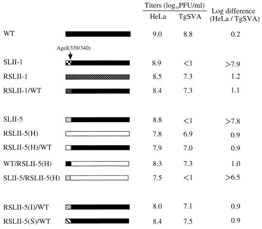
DNA segments complementary to the 5′-proximal region of nt 1 to 339 (AgeI site) were prepared from pRSLII-5(H)/WT (Table 1; see Fig. 3) (see below). To prepare infectious cDNA clones of RSLII-5(H), the DNA fragment was joined to the remaining cDNAs at the corresponding AgeI site at nt 339/340. Virus produced in cells transfected with the infectious cDNA clone pRSLII-5(H) phenotypically resembled the parental revertant RSLII-5(H).
FIG. 3.
Genome structures of the SLII mutants and revertants and
logarithmic differences between their virus titers in HeLa cells and
TgSVA cells. The genomes of poliovirus WT, RSLII-1, and RSLII-5(H) are
indicated by ▪,
, and □, respectively. Segments of nt 1 to 339
(AgeI fragment) for SLII-1, SLII-5, RSLII-5(I), and
RSLII-5(S) are shown by
, ░⃞,
 , and
, and
 ,
respectively. Virus titers in log10 PFU per milliliters in
HeLa cells and TgSVA cells and the logarithmic differences between the
titers are given on the right of the individual constructs.
Construction of recombinant cDNAs and viruses.
Recombinant infectious cDNA clones originating from poliovirus WT (pWT) and carrying revertant segments from nt 39 to 339 were constructed by replacing the PmlI (38/39) and AgeI (339/340) fragments of pWT with the corresponding revertant fragments; the resulting clones were designated pRSLII-1/WT, pRSLII-5(H)/WT, pRSLII-5(I)/WT, and pRSLII-5(S)/WT (Table 1). pWT/RSLII-5(H) and pSLII-5/RSLII-5(H) were constructed similarly.
Modifications of nt 107 in the cDNAs were carried out by PCR. The first PCR primers were a sense nucleotide sequence from nt 93 to 115 and an antisense sequence from nt 389 to 407, and the second primers were a sequence from the pSVA14 vector (nt 4639 to 4659) and an antisense sequence from nt 93 to 115. Plasmids, pWT, pSLII-1, pSLII-4, pSLII-5, and pSLII-6 were used as templates. The sense and antisense primers (nt 93 to 115) were designed to replace T at nt 107 with A, C, and G. A cDNA clone in which nt 107 is missing was also constructed by using a mutant primer.
Two overlapping fragments produced by the first and second PCRs were used in PCR along with primers of a sense sequence of pSVA14 vector (nt 4639 to 4659) and an antisense sequence from nt 389 to 407 to yield a longer fragment that spanned the full length of the overlapping fragments. The PvuI (nt 4704, vector sequence) -AgeI (nt 339) fragment of plasmid pWT was replaced by the corresponding fragments of the modified cDNAs. The infectious cDNAs thus constructed were designated p107A-WT, p107G-WT, p107C-WT, p107del-WT, p107A-SLII-1, p107A-SLII-4, p107A-SLII-5, and p107A-SLII-6. Plasmids pM-1-WT, pM-5(H)-WT, pM-5(I)-WT, and pM-5(S)-WT were constructed by the replacement of A at nt 107 with a T in pRSLII-1/WT, pRSLII-5(H)/WT, pRSLII-5(I)/WT, and pRSLII-5(S)/WT, respectively. As a way to confirm the presence of modified sequences, nucleotide sequences from nt 1 to 339 of the cDNAs were determined by dideoxy sequencing with a Pharmacia LKB ALFred DNA sequencer.
A DEAE-dextran method was used to transfect AGMK cells with cDNAs or with their RNA transcripts (11, 38). RNA transcripts were synthesized from cDNAs, which were linearized by digestion with PvuI, by using the MEGAscript T7 Kit (Ambion).
Cell-free translation.
Mouse TgSVA cell monolayers were collected and cultured in suspension for 4 to 6 h, and then a cytoplasmic extract (S10) was prepared as previously described (16, 39). Conditions for the translation reaction were those used by Iizuka et al. (16). After incubation at 32°C for 1 h, radioactive products in the translation mixtures were analyzed by sodium dodecyl sulfate-polyacrylamide gel electrophoresis.
RESULTS
Isolation of revertants from mutants SLII-1 and SLII-5.
Parental WT poliovirus grows well in primate and mouse systems (20, 21, 27, 39), and although SLII mutants grow well in primate cells and in the CNS of monkeys, they grow poorly in mouse TgSVA cells and in the CNS of the Tg mice carrying the human PVR gene (39). Apparently, SLII mutants are sensitive to species differences in the SLII-related host factors of primates and mice.
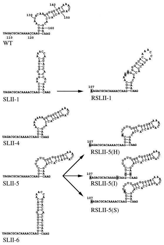
We isolated revertants of SLII-1 and SLII-5 mutants that were able to grow in TgSVA cells with the intention of learning about the molecular basis of SLII function. For possible secondary structures of the SLII region of WT and SLII mutants, see Fig. 2. TgSVA cells were infected with mutant SLII-1 or SLII-5 at a multiplicity of infection of approximately 1 and incubated at 37°C until most of the cells had detached. The virus was passaged three times, after which the virus preparations started to replicate efficiently in TgSVA cells, and the supernatants were checked for plaque formation on TgSVA cells. Single plaques formed in TgSVA cells were harvested and plaque purified again in TgSVA cells. Of these plaque-purified revertants, one revertant, RSLII-1(I), from SLII-1 and three revertants, RSLII-5(H), RSLII-5(I), and RSLII-5(S), from SLII-5 were chosen and used as revertants in this study (Table 1). Revertant RSLII-1(S) was obtained in a similar way from plaque-purified virus recovered from the spinal cord of a paralyzed Tg mouse intracerebrally inoculated with SLII-1 (39).
FIG. 2.
Secondary structures and sequences of SLII mutants and their revertants. Possible secondary structures and sequences of SLII regions of the genomes of WT virus, SLII mutants (SLII-1, SLII-4, SLII-5, and SLII-6), and their revertants [RSLII-1, RSLII-5(H), RSLII-5(I), and RSLII-5(H)] are shown. The mutation sites in the revertants are indicated by shadowed letters or an asterisk that represents a deleted nucleotide. Nucleotide positions are indicated on the WT genome.
The titers of these revertants were measured in HeLa cells and TgSVA cells, and the logarithmic difference between the titers was compared with that of WT to determine the extent to which the revertants overcame the limitations of host cell species differences on their capacity to replicate (Table 2). The log difference for each of the revertants was approximately 1, whereas that of WT was 0.2. The results suggested that all the revertants obtained in this study acquired at least a partial ability to replicate in mouse cells.
TABLE 2.
Plaque-forming ability of viruses
| Virus | Titer
(log10 PFU/ml) in:
|
Log differencea | |
|---|---|---|---|
| HeLa | TgSVA | ||
| WT | 9.0 | 8.8 | 0.2 |
| HR mutants | |||
| SLII-1 | 8.9 | <1b | >7.9 |
| SLII-4 | 8.7 | 4.5 | 4.2 |
| SLII-5 | 8.8 | <1 | >7.8 |
| SLII-6 | 7.8 | <1 | >6.8 |
| Revertants | |||
| RSLII-1 | 8.5 | 7.3 | 1.2 |
| RSLII-5(H) | 8.1 | 7.0 | 1.1 |
| RSLII-5(I) | 8.3 | 7.4 | 0.9 |
| RSLII-5(S) | 8.4 | 7.5 | 0.9 |
| Recombinant | |||
| 107A-WT | 7.8 | 7.0 | 0.8 |
| 107G-WT | 8.3 | 7.5 | 0.8 |
| 107C-WT | 8.3 | 7.3 | 1.0 |
| 107del-WT | 8.0 | 7.3 | 0.7 |
| 107A-SLII-1 | 7.3 | 4.0 | 3.3 |
| 107A-SLII-4 | 8.3 | 7.0 | 1.3 |
| 107A-SLII-5 | 7.8 | 3.5 | 4.3 |
| 107A-SLII-6 | 7.7 | <1 | >6.7 |
| M-1-WT | 8.0 | <1 | >7.0 |
| M-5(H)-WT | 7.8 | <1 | >6.8 |
| M-5(I)-WT | 8.0 | <1 | >7.0 |
| M-5(S)-WT | 8.3 | <1 | >7.3 |
Logarithmic difference in virus titers obtained in HeLa cells and TgSVA cells.
Plaques are not detected. Cytopathic effects were observed when the cells were infected with undiluted virus solution.
Mouse neurovirulence of revertants.
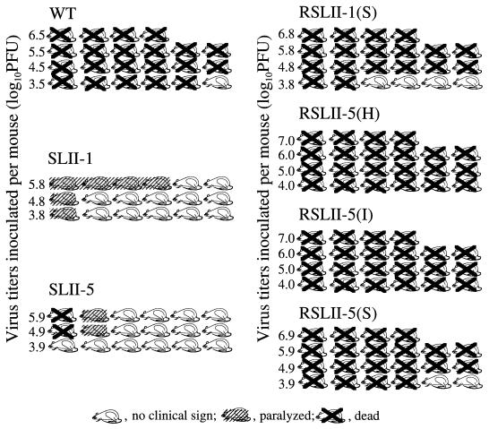
SLII mutants are known to be host range mutants; that is, they appeared to replicate well in the CNS of monkeys but not in the CNS of the Tg mice (39). Revertants obtained in this study grew well in TgSVA cells; therefore, they may have recovered the capacity to replicate in the CNS of the Tg mice. Accordingly, revertants were tested for their neurovirulence phenotypes in the Tg mice (Fig. 1). The Tg mice were inoculated intracerebrally with four revertants, RSLII-1(S), RSLII-5(H), RSLII-5(I), and RSLII-5(S). As shown in Fig. 1, these revertants exhibited a neurovirulence phenotype similar to that of WT whereas SLII-1 and SLII-5 showed much less of the neurovirulence phenotype than WT, as reported previously (39). These results suggested that the revertants overcame the host range restriction observed for SLII-1 and SLII-5 in the CNS of Tg mice.
FIG. 1.
Neurovirulence test on SLII mutants and their revertants in Tg mice after intracerebral inoculation. Neurovirulence tests were performed on SLII mutants and their revertants in Tg mice. Tg mice were inoculated intracerebrally with the WT, SLII-1, SLII-5, RSLII-1(S), RSLII-5(H), RSLII-5(I), or RSLII-5(S) and observed for paralysis and death for up to 14 days after the inoculation.
Genomic region influencing revertant phenotype.
We attempted to identify a mutation site(s) that influences the revertant phenotype, by preparing cDNA clones containing the segment located upstream of the SphI site (nt 1 to 1131) in all the revertants (Table 1) and then sequencing nt 1 to about 500. We found that all the mutations that occurred in this region of the revertant genomes resided between nt 107 and 161. The mutation sites are shown in Fig. 2. The mutations discovered in pT7-1131-RSLII-1(I) and pT7-1131-RSLII-1(S) were identical; therefore, revertants RSLII-1(I) and RSLII-1(S) are hereafter both simply referred to as RSLII-1 (Fig. 2 and 3; Tables 1 and 2). RSLII-5(H), RSLII-5(I), and RSLII-5(S) differed slightly from each other in this genome region (Fig. 2). Unexpectedly, all the revertants had A at nt 107, unlike WT and SLII, which had U at nt 107. Other mutation sites were observed in nt 120 to 161 (Fig. 2).
To find whether mutations seen in nt 107 to 161 conferred the revertant phenotype, a segment upstream of AgeI (nt 399) of pWT was replaced by the corresponding segment of pT7-1131-RSLII-1, pT7-1131-RSLII-5(H), pT7-1131-RSLII-5(I), or pT7-1131-RSLII-5(S); the resulting recombinant infectious cDNA clones were designated pRSLII-1/WT, pRSLII-5(H)/WT, pRSLII-5(I)/WT, and pRSLII-5(S)/WT, respectively. Recombinant cDNAs pWT/RSLII-5(H) and pSLII-5/RSLII-5(H) were constructed similarly. The recombinant viruses from these cDNA clones were designated RSLII-1/WT, RSLII-5(I)/WT, RSLII-5(S)/WT, WT/RSLII-5(H), and SLII-5/RSLII-5(H), respectively, and the log difference between their titers in HeLa cells and TgSVA cells was compared to that of WT (Fig. 3). The log difference between the titers of RSLII-1/WT in HeLa cells and TgSVA cells was similar to that of RSLII-1 (Fig. 3). An allele replacement experiment carried out for RSLII-5(H) also gave a very similar result (Fig. 3). Furthermore, WT/RSLII-5(H) showed a revertant phenotype, and SLII-5/RSLII-5(H) showed a phenotype like that of SLII-5. These data strongly suggested that the revertant phenotypes of RSLII-1 and RSLII-5(H) are due to mutations in nt 107 to 161.
Although all recombinant viruses that carried the 5′-proximal genome sequences of revertant or WT showed revertant phenotypes, the log differences in the viral titers of these viruses were slightly higher than that of WT. It should be noted that the reciprocal recombinants RSLII-5(H)/WT and WT/RSLII-5(H) had very similar log differences that were slightly higher than that of WT. Thus, all the revertants and the recombinants, except for SLII5/RSLII-5(H), constructed here appear to have a minor defect in their replication abilities in mouse cells. These observations may suggest that a mutation(s) other than in the 5′-proximal 339 nt existed in the revertant genomes and that such a mutation(s) influenced the viral replication efficiency to some extent in concert with a mutation(s) in nt 107 to 161.
Mutation sites influencing revertant phenotype.
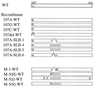
To narrow the number of possible sites that might be important for the revertant phenotype, we generated new recombinants and mutants for investigation (Fig. 4). First, U at nt 107 was changed to A in the genomes of SLII-1, SLII-4, SLII-5, and SLII-6, and these mutants were designated 107A-SLII-1, 107A-SLII-4, 107A-SLII-5, and 107A-SLII-6, respectively (Table 2; Fig. 4). The titers of these mutants were measured in HeLa and TgSVA cells, and the log differences were compared with those of the corresponding SLII mutants. The log differences of 107A-SLII-1, 107A-SLII-4, and 107A-SLII-5 were much lower than those of the parental SLII mutants (Table 2). Thus, mutation at nt 107 in the revertant genomes appeared to have a significant influence on the revertant phenotype, although mutation at nt 107 did not restore the replication capacity of SLII-6 in the TgSVA cells. We were unsuccessful in our attempts to isolate a revertant(s) from SLII-6 by the procedure described in Materials and Methods (data not shown). Only the mutation at nt 107 did not confer the full revertant phenotype on SLII-1 and SLII-5, suggesting that the remaining mutations observed in the region from nt 120 to 161 may affect the viral replication efficiency in TgSVA cells.
FIG. 4.
Genomic structures of nt 107 to 161 of recombinant viruses. The recombinant viruses shown were constructed as described in Materials and Methods. Genomic structures other than nt 107 to 161 were the same as that of the WT genome.
We tested the effect of mutations in the region from nt 120 to 161 by adding a U at nt 107 (as is found in WT and SLII mutants) into the background of RSLII-1/WT, RSLII-5(H)/WT, RSLII-5(I)/WT, and RSLII-5(S)/WT, thereby creating M-1-WT, M-5(H)-WT, M-5(I)-WT, and M-5(S)-WT, respectively. A schematic representation of nt 107 to 161 in these mutant genomes is shown in Fig. 4. As shown in Table 2, these mutants lacked any apparent plaque-forming activity in TgSVA cells, yet they manifested cytopathic effects that were obviously stronger than those of SLII-1 and SLII-5 when the cells were infected with the undiluted virus solutions (data not shown). These data suggested that the remaining mutations also contributed to better revertant replication in TgSVA cells. The mutation at nt 107 apparently affected the expression of the revertant phenotype more than the region spanning nt 120 to 161, although both sites seem to have been involved in the full revertant phenotype. Point mutations at nt 107 were introduced into the WT genome to yield a series of mutants designated 107A-WT, 107C-WT, 107G-WT, and 107del-WT (Fig. 4). The replication of these mutants was changed little from that of the replication phenotype of WT (Table 2). Thus, the role that A at nt 107 plays in poliovirus replication was detected by using host range mutants SLII-1, SLII-4, and SLII-5.
Cell-free translation.
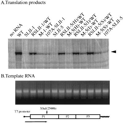
A possible change in the IRES activity of revertant RNAs was investigated by using a cell-free translation system. RNAs with free 5′ ends were synthesized from cDNA clones of pWT, pSLII-1, pRSLII-1/WT, pM-1-WT, p107A-SLII-1, pSLII-5, pRSLII-5(H)/WT, pM-5(H)-WT, pRSLII-5(I)/WT, pM-5(I)-WT, pRSLII-5(S)/WT, pM-5(S)-WT, and p107A-SLII-5, which had been linearized by digestion with XbaI (Fig. 5B), and were used as templates in S10 extracts prepared from TgSVA cells. The amounts of RNA templates used are shown in Fig. 5B. The 66-kDa protein was hardly detected in the products from RNAs of SLII mutants but was visible in the products of the WT RNA and revertant RNAs (Fig. 5A). The 66-kDa protein was detected weakly in the products directed by the recombinant RNAs. These observations indicated that the IRESs of the SLII mutant RNAs do not function in the mouse system but that the IRESs of the revertants recover much of their function. The intensities of the bands observed in Fig. 5A seemed to be compatible with growth phenotypes of individual viruses in TgSVA cells. Enhanced efficiency of translation initiation is therefore highly likely to be responsible for the regained growth ability of these revertants and recombinants.
FIG. 5.
Cell-free translation products of RNAs from WT, SLII mutants, and their recombinants. (B) Template RNAs were transcribed by T7 RNA polymerase from pWT, pSLII-1, pSLII-5, and recombinant cDNAs of WT and the revertants that had been cleaved with XbaI. The length of the template RNA is shown at the bottom. (A) The in vitro transcripts were incubated at 32°C for 1 h in S10 extract from TgSVA cells. The position of the 66-kDa product is indicated by an arrowhead on the right.
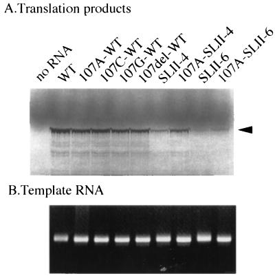
The translation directed by the RNAs from recombinants p107A-SLII-1 and p107A-SLII-5 was more efficient than that from pSLII-1 and pSLII-5. We examined the effect of mutation at nt 107 on the IRES activity by measuring the levels of mRNA translation from various RNAs carrying mutations at nt 107 (Fig. 6). Translational efficiency relative to WT RNA appeared not to be changed much by any of the mutations at nt 107. The translation from 107A-SLII-4 RNA was more efficient than that from SLII-4 RNA. In the case of 107A-SLII-6, the translation efficiency appeared not to be recovered. The translation efficiencies were compatible with the plaque-forming ability of the viruses. These experiments indicated that nt 107 is involved in the structure required for IRES-dependent translation initiation, a function that was detectable only by using SLII mutants.
FIG. 6.
Effect of nt 107 on IRES activity in cell-free translation. (B) Template RNAs were transcribed by T7 RNA polymerase from pWT, pSLII-4, pSLII-6, and their mutants concerning nt 107 that had been cleaved with XbaI, as in Fig. 5. (A) The in vitro transcripts were incubated at 32°C for 1 h in S10 extract from TgSVA cells. The position of the 66-kDa product is indicated by an arrowhead on the right.
DISCUSSION
Molecular genetic analysis of naturally occurring revertant viruses from two mutants (SLII-1 and SLII-5), which were able to grow in TgSVA cells, revealed that all the revertant genomes had a change at nt 107 from U to A. Mutants 107A-SLII-1 and 107A-SLII-5 showed more efficient plaque-forming ability in TgSVA cells than did SLII-1 and SLII-5, respectively. The in vitro translation directed by the RNA from p107A-SLII-1 and p107A-SLII-5 was also more efficient than that directed by RNAs from pSLII-1 and pSLII-5. These results suggested that nt 107 functions in IRES-dependent translation initiation in TgSVA cells. Trono et al. (42) reported that an insertion of 3, 11, or 15 bases between nt 108 and nt 109 of poliovirus RNA did not result in any alteration of the replication phenotype. Pelletier et al. (31) showed that the change of U to A at nt 119 in the poliovirus type 2 genome was a silent mutation. We also showed here that mutations at nt 107 of WT had no effect on the wild-type phenotype. These data suggested that the translational function of nt 107 can be detected only by using the host range mutants and their revertants.
In addition to nt 107, the remaining mutation sites (nt 120 to 161) were required for the expression of the full revertant phenotype. This observation suggested that nt 107 works with the SLII region to maintain the IRES activity in mouse cells. One or more host-specific factors may interact with nt 107 and the other mutation sites. At present, the structural relationship between nt 107 and SLII is not clear. Le and Zuker (22) reported a predicted consensus secondary structure that consists of nt 103 to 179 of poliovirus type 3 in which nt 107 and SLII are involved in a stem-loop structure. Cellular factors may recognize such a structure formed by a longer nucleotide sequence including nt 107 and the SLII (nt 124 to 162) region.
The initiation events directed by the IRES probably require many protein factors including most of the same set of initiation factors that are used by typical capped cellular mRNAs (36). Cap-binding activity of eIF-4F is not required for translation of the uncapped poliovirus RNAs, but the RNA helicase activity manifested by this protein complex may still play an important role in internal initiation (35). If eIF-4F is required for IRES-dependent initiation, it may function in a structurally modified form. Within the 5′ NCR of poliovirus RNA, eIF-2α associates with nt 97 to 182 and nt 510 to 629 (7); similarly, eukaryotic initiation factor 4B (eIF-4B) binds to the 5′ NCR of foot-and-mouse disease virus RNA (28). In addition to the standard initiation factors, other trans-acting protein factors probably mediate IRES-dependent translation initiation of poliovirus. La protein, polypyrimidine tract-binding protein, and poly(rC)-binding protein 2 are reported as being possible host factors for IRES-dependent translation initiation (2, 3, 6, 14, 26). Many unidentified host proteins are reported to bind to defined IRES regions of poliovirus RNA, as determined by UV cross-linking analysis or a gel shift assay (4, 7, 8, 12). Host cellular proteins that can be UV cross-linked to the SLII structure are now being investigated.
Although the revertant phenotypes observed for RSLII-1, RSLII-5(H), RSLII-5(I), and RSLII-5(S) were shown to be due to mutations in nt 107 to 161 of the revertant genomes, these revertants appeared to have a very minor defect in replication efficiency in TgSVA cells compared with that of WT as judged by logarithmic differences of virus titers in HeLa cells and TgSVA cells (Fig. 3). This phenomenon was reproducibly observed in repeated experiments. However, this discrepancy was very small and is almost negligible considering the range of error for plaque assays. Nevertheless, we determined the nucleotide sequence of the genome region encoding viral protein 2A of the revertant RSLII-5(H), because protein 2A is known to enhance the efficiency of poliovirus translation initiation (13, 23). The results demonstrated that the protein 2A coding sequence was not changed (data not shown). The reason for this phenomenon is not clear at present.
Expression of IRES function in mouse cells must be influenced by the efficiency of interactions with mouse host factors; therefore, the IRESs of SLII mutants may be recognized by such a putative mouse factor(s) while the revertant IRESs are recognized. Although the SLII mutants did not show efficient IRES activity in mouse cells and their S10 extracts (Fig. 4) (39), they did have IRES activities in HeLa cells and their S10 extracts (39). This difference in IRES activity may be due to species difference in host factors. If so, the two different systems may provide powerful tools for identifying a host factor(s) that restricts IRES activity in murine systems.
ACKNOWLEDGMENTS
We are grateful to S. Kuge and H. Toyoda for helpful suggestions and discussions. We thank Y. Sasaki and K. Iwasaki for expert technical assistance and E. Suzuki and M. Watanabe for help in preparation of the manuscript.
This work was supported in part by a grant-in-aid from the Ministry of Education, Science, Sports, and Culture of Japan and the Ministry of Health and Welfare of Japan and by funds from the Science and Technology of the Japanese Government.
REFERENCES
- 1.Andino R, Rieckhof G E, Baltimore D. A functional ribonucleoprotein complex forms around the 5′ end of poliovirus RNA. Cell. 1990;63:369–380. doi: 10.1016/0092-8674(90)90170-j. [DOI] [PubMed] [Google Scholar]
- 2.Belsham G J, Sonenberg N, Svitkin Y V. The role of the La autoantigen in internal initiation. Curr Top Microbiol Immunol. 1995;203:85–98. doi: 10.1007/978-3-642-79663-0_4. [DOI] [PubMed] [Google Scholar]
- 3.Blyn L B, Swiderek K M, Richards O, Stahl D C, Semler B L, Ehrenfeld E. Poly(rC) binding protein 2 binds to stem-loop IV of the poliovirus RNA 5′ noncoding region: identification by automated liquid chromatography-tandem mass spectrometry. Proc Natl Acad Sci USA. 1996;93:11115–11120. doi: 10.1073/pnas.93.20.11115. [DOI] [PMC free article] [PubMed] [Google Scholar]
- 4.Blyn L B, Chen R, Semler B L, Ehrenfeld E. Host cell proteins binding to domain IV of the 5′ noncoding region of poliovirus RNA. J Virol. 1995;69:4381–4389. doi: 10.1128/jvi.69.7.4381-4389.1995. [DOI] [PMC free article] [PubMed] [Google Scholar]
- 5.Brown B A, Ehrenfeld E. Translation of poliovirus RNA in vitro: changes in cleavage pattern and initiation sites by ribosomal salt wash. Virology. 1979;97:396–405. doi: 10.1016/0042-6822(79)90350-7. [DOI] [PubMed] [Google Scholar]
- 6.Craig A W B, Svitkin Y V, Lee H S, Belsham G J, Sonenberg N. The La autoantigen contains a dimerization domain that is essential for enhancing translation. Mol Cell Biol. 1997;17:163–169. doi: 10.1128/mcb.17.1.163. [DOI] [PMC free article] [PubMed] [Google Scholar]
- 7.del Angel R M, Papavassiliou A G, Fernandez-Tomas C, Silverstein S J, Racaniello V R. Cell proteins bind to multiple sites within the 5′ untranslated region of poliovirus RNA. Proc Natl Acad Sci USA. 1989;86:8299–8303. doi: 10.1073/pnas.86.21.8299. [DOI] [PMC free article] [PubMed] [Google Scholar]
- 8.Dildine S L, Semler B L. Conservation of RNA-protein interactions among picornaviruses. J Virol. 1992;66:4364–4376. doi: 10.1128/jvi.66.7.4364-4376.1992. [DOI] [PMC free article] [PubMed] [Google Scholar]
- 9.Dorner A J, Semler B L, Jackson R J, Hanecak R, Duprey E, Wimmer E. In vitro translation of poliovirus RNA: utilization of internal initiation sites in reticulocyte lysate. J Virol. 1984;50:507–514. doi: 10.1128/jvi.50.2.507-514.1984. [DOI] [PMC free article] [PubMed] [Google Scholar]
- 10.Ehrenfeld E, Semler B L. Anatomy of the poliovirus internal ribosome entry site. Curr Top Microbiol Immunol. 1995;203:65–83. doi: 10.1007/978-3-642-79663-0_3. [DOI] [PubMed] [Google Scholar]
- 11.Hagino-Yamagishi K, Nomoto A. In vitro construction of poliovirus defective interfering particles. J Virol. 1989;63:5386–5392. doi: 10.1128/jvi.63.12.5386-5392.1989. [DOI] [PMC free article] [PubMed] [Google Scholar]
- 12.Haller A A, Semler B L. Stem-loop structure synergy in binding cellular proteins to the 5′ noncoding region of poliovirus RNA. Virology. 1995;206:923–934. doi: 10.1006/viro.1995.1015. [DOI] [PubMed] [Google Scholar]
- 13.Hambidge S J, Sarnow P. Translational enhancement of the poliovirus 5′ noncoding region mediated by virus-encoded polypeptide 2A. Proc Natl Acad Sci USA. 1992;89:10272–10276. doi: 10.1073/pnas.89.21.10272. [DOI] [PMC free article] [PubMed] [Google Scholar]
- 14.Hellen C U T, Witherell G W, Schmid M, Shin S H, Pestova T V, Gil A, Wimmer E. A cytoplasmic 57-kDa protein that is required for translation of picornavirus RNA by internal ribosomal entry is identical to the nuclear pyrimidine tract-binding protein. Proc Natl Acad Sci USA. 1993;90:7642–7646. doi: 10.1073/pnas.90.16.7642. [DOI] [PMC free article] [PubMed] [Google Scholar]
- 15.Horie H, Koike S, Kurata T, Sato-Yoshida Y, Ise I, Ota Y, Abe S, Hioki K, Kato H, Taya C, Nomura T, Hashizume S, Yonekawa H, Nomoto A. Transgenic mice carrying the human poliovirus receptor: new animal models for study of poliovirus neurovirulence. J Virol. 1994;68:681–688. doi: 10.1128/jvi.68.2.681-688.1994. [DOI] [PMC free article] [PubMed] [Google Scholar]
- 16.Iizuka N, Yonekawa H, Nomoto A. Nucleotide sequences important for translation initiation of enterovirus RNA. J Virol. 1991;65:4867–4873. doi: 10.1128/jvi.65.9.4867-4873.1991. [DOI] [PMC free article] [PubMed] [Google Scholar]
- 17.Jackson R J, Howell M T, Kaminski A. The novel mechanism of initiation of picornavirus RNA translation. Trends Biochem Sci. 1990;15:477–483. doi: 10.1016/0968-0004(90)90302-r. [DOI] [PubMed] [Google Scholar]
- 18.Jang S K, Kräusslich H G, Nicklin M J H, Duke G M, Palmenberg A C, Wimmer E. A segment of the 5′ nontranslated region of encephalomyocarditis virus RNA directs internal entry of ribosomes during in vitro translation. J Virol. 1988;62:2636–2643. doi: 10.1128/jvi.62.8.2636-2643.1988. [DOI] [PMC free article] [PubMed] [Google Scholar]
- 19.Kawamura N, Kohara M, Abe S, Komatsu T, Tago K, Arita M, Nomoto A. Determinants in the 5′ noncoding region of poliovirus Sabin 1 RNA that influence the attenuation phenotype. J Virol. 1989;63:1302–1309. doi: 10.1128/jvi.63.3.1302-1309.1989. [DOI] [PMC free article] [PubMed] [Google Scholar]
- 20.Koike S, Taya C, Kurata T, Abe S, Ise I, Yonekawa H, Nomoto A. Transgenic mice susceptible to poliovirus. Proc Natl Acad Sci USA. 1991;88:951–955. doi: 10.1073/pnas.88.3.951. [DOI] [PMC free article] [PubMed] [Google Scholar]
- 21.Koike S, Horie H, Ise I, Okitsu A, Yoshida M, Iizuka N, Takeuchi K, Takegami T, Nomoto A. The poliovirus receptor protein is produced both as membrane-bound and secreted forms. EMBO J. 1990;9:3217–3224. doi: 10.1002/j.1460-2075.1990.tb07520.x. [DOI] [PMC free article] [PubMed] [Google Scholar]
- 22.Le S-Y, Zuker M. Common structures of the 5′ non-coding RNA in enteroviruses and rhinoviruses. Thermodynamical stability and statistical significance. J Mol Biol. 1990;216:729–741. doi: 10.1016/0022-2836(90)90395-3. [DOI] [PubMed] [Google Scholar]
- 23.Macadam A J, Ferguson G, Fleming T, Stone D M, Almond J W, Minor P D. Role for poliovirus protease 2A in cap independent translation. EMBO J. 1994;13:924–927. doi: 10.1002/j.1460-2075.1994.tb06336.x. [DOI] [PMC free article] [PubMed] [Google Scholar]
- 24.Martin A, Wychowski C, Couderc T, Crainic R, Hogle J, Girard M. Engineering a poliovirus type 2 antigenic site on a type 1 capsid results in a chimaeric virus which is neurovirulent for mice. EMBO J. 1988;7:2839–2847. doi: 10.1002/j.1460-2075.1988.tb03140.x. [DOI] [PMC free article] [PubMed] [Google Scholar]
- 25.Meerovitch K, Nicholson R, Sonenberg N. In vitro mutational analysis of cis-acting RNA translational elements within the poliovirus type 2 5′ untranslated region. J Virol. 1991;65:5895–5901. doi: 10.1128/jvi.65.11.5895-5901.1991. [DOI] [PMC free article] [PubMed] [Google Scholar]
- 26.Meerovitch K, Svitkin Y V, Lee H S, Lejbkowicz F, Kenan D J, Chan E K L, Agol V I, Keene J D, Sonenberg N. La autoantigen enhances and corrects aberrant translation of poliovirus RNA in reticulocyte lysate. J Virol. 1993;67:3798–3807. doi: 10.1128/jvi.67.7.3798-3807.1993. [DOI] [PMC free article] [PubMed] [Google Scholar]
- 27.Mendelsohn C L, Wimmer E, Racaniello V R. Cellular receptor for poliovirus: molecular cloning, nucleotide sequence, and expression of a new member of the immunoglobulin superfamily. Cell. 1989;56:855–865. doi: 10.1016/0092-8674(89)90690-9. [DOI] [PubMed] [Google Scholar]
- 28.Meyer K, Petersen A, Niepmann M, Beck E. Interaction of eukaryotic initiation factor eIF-4B with a picornavirus internal translation initiation site. J Virol. 1995;69:2819–2824. doi: 10.1128/jvi.69.5.2819-2824.1995. [DOI] [PMC free article] [PubMed] [Google Scholar]
- 29.Nicholson R, Pelletier J, Le S-Y, Sonenberg N. Structural and functional analysis of the ribosome landing pad of poliovirus type 2: in vivo translation studies. J Virol. 1991;65:5886–5894. doi: 10.1128/jvi.65.11.5886-5894.1991. [DOI] [PMC free article] [PubMed] [Google Scholar]
- 30.Nomoto A, Detjen B, Pozzatti R, Wimmer E. The location of the polio genome protein in viral RNAs and its implication for RNA synthesis. Nature (London) 1977;268:208–213. doi: 10.1038/268208a0. [DOI] [PubMed] [Google Scholar]
- 31.Pelletier J, Flynn M E, Kaplan G, Racaniello V, Sonenberg N. Mutational analysis of upstream AUG codons of poliovirus RNA. J Virol. 1988;62:4486–4492. doi: 10.1128/jvi.62.12.4486-4492.1988. [DOI] [PMC free article] [PubMed] [Google Scholar]
- 32.Pelletier J, Sonenberg N. Internal initiation of translation of eukaryotic mRNA directed by a sequence derived from poliovirus RNA. Nature (London) 1988;334:320–325. doi: 10.1038/334320a0. [DOI] [PubMed] [Google Scholar]
- 33.Phillips B A, Emmert A. Modulation of the expression of poliovirus proteins in reticulocyte lysates. Virology. 1986;148:255–267. doi: 10.1016/0042-6822(86)90323-5. [DOI] [PubMed] [Google Scholar]
- 34.Ren R B, Costantini F, Gorgacz E J, Lee J J, Racaniello V R. Transgenic mice expressing a human poliovirus receptor: a new model for poliomyelitis. Cell. 1990;63:353–362. doi: 10.1016/0092-8674(90)90168-e. [DOI] [PubMed] [Google Scholar]
- 35.Rozen F, Edery I, Meerovitch K, Dever T E, Merrick W C, Sonenberg N. Bidirectional RNA helicase activity of eucaryotic translation initiation factors 4A and 4F. Mol Cell Biol. 1990;10:1134–1144. doi: 10.1128/mcb.10.3.1134. [DOI] [PMC free article] [PubMed] [Google Scholar]
- 36.Scheper G C, Voorma H O, Thomas A A M. Eukaryotic initiation factors-4E and -4F stimulate 5′ cap-dependent as well as internal initiation of protein synthesis. J Biol Chem. 1992;267:7269–7274. [PubMed] [Google Scholar]
- 37.Shiroki K, Kato H, Koike S, Odaka T, Nomoto A. Temperature-sensitive mouse cell factors for strand-specific initiation of poliovirus RNA synthesis. J Virol. 1993;67:3989–3996. doi: 10.1128/jvi.67.7.3989-3996.1993. [DOI] [PMC free article] [PubMed] [Google Scholar]
- 38.Shiroki K, Ishii T, Aoki T, Kobashi M, Ohka S, Nomoto A. A new cis-acting element for RNA replication within the 5′ noncoding region of poliovirus type 1 RNA. J Virol. 1995;69:6825–6832. doi: 10.1128/jvi.69.11.6825-6832.1995. [DOI] [PMC free article] [PubMed] [Google Scholar]
- 39.Shiroki K, Ishii T, Aoki T, Ota Y, Yang W-X, Komatsu T, Ami Y, Arita M, Abe S, Hashizume S, Nomoto A. Host range phenotype induced by mutations in the internal ribosomal entry site of poliovirus RNA. J Virol. 1997;71:1–8. doi: 10.1128/jvi.71.1.1-8.1997. [DOI] [PMC free article] [PubMed] [Google Scholar]
- 40.Simoes E A F, Sarnow P. An RNA hairpin at the extreme 5′ end of the poliovirus RNA genome modulates viral translation in human cells. J Virol. 1991;65:913–921. doi: 10.1128/jvi.65.2.913-921.1991. [DOI] [PMC free article] [PubMed] [Google Scholar]
- 41.Tobin G J, Young D C, Flanegan J B. Self catalyzed linkage of poliovirus terminal protein VPg to poliovirus RNA. Cell. 1989;59:511–519. doi: 10.1016/0092-8674(89)90034-2. [DOI] [PubMed] [Google Scholar]
- 42.Trono D, Andino R, Baltimore D. An RNA sequence of hundreds of nucleotides at the 5′ end of poliovirus RNA is involved in allowing viral protein synthesis. J Virol. 1988;62:2291–2299. doi: 10.1128/jvi.62.7.2291-2299.1988. [DOI] [PMC free article] [PubMed] [Google Scholar]
- 43.Tsukiyama-Kohara K, Iizuka N, Kohara M, Nomoto A. Internal ribosome entry site within hepatitis C virus RNA. J Virol. 1992;66:1476–1483. doi: 10.1128/jvi.66.3.1476-1483.1992. [DOI] [PMC free article] [PubMed] [Google Scholar]
- 44.Wimmer E, Hellen C U T, Cao X. Genetics of poliovirus. Annu Rev Genet. 1993;27:353–436. doi: 10.1146/annurev.ge.27.120193.002033. [DOI] [PubMed] [Google Scholar]
- 45.Yogo Y, Wimmer E. Polyadenylic acid at the 3′-terminus of poliovirus RNA. Proc Natl Acad Sci USA. 1972;69:1877–1882. doi: 10.1073/pnas.69.7.1877. [DOI] [PMC free article] [PubMed] [Google Scholar]


