Abstract
Background
Existing panels for lower respiratory tract infections (LRTIs) are slow and lack quantification of important pathogens and antimicrobial resistance, which are not solely responsible for their complex etiology and antibiotic resistance. BioFire FilmArray Pneumonia (PN) panels may provide rapid information on their etiology.
Methods
The bronchoalveolar lavage fluid of 187 patients with LRTIs was simultaneously analyzed using a PN panel and cultivation, and the impact of the PN panel on clinical practice was assessed. The primary endpoint was to compare the consistency between the PN panel and conventional microbiology in terms of etiology and drug resistance, as well as to explore the clinical significance of the PN panel. The secondary endpoint was pathogen detection using the PN panel in patients with community-acquired pneumonia (CAP) or hospital-acquired pneumonia (HAP).
Results
Fifty-seven patients with HAP and 130 with CAP were included. The most common pathogens of HAP were Acinetobacter baumannii and Klebsiella pneumoniae, with the most prevalent antimicrobial resistance (AMR) genes being CTX-M and KPC. For CAP, the most common pathogens were Haemophilus influenzae and Staphylococcus aureus, with the most frequent AMR genes being CTX-M and VIM. Compared with routine bacterial culture, the PN panel demonstrated an 85% combined positive percent agreement (PPA) and 92% negative percent agreement (NPA) for the qualitative identification of 13 bacterial targets. PN detection of bacteria with higher levels of semi-quantitative bacteria was associated with more positive bacterial cultures. Positive concordance between phenotypic resistance and the presence of corresponding AMR determinants was 85%, with 90% positive agreement between CTX-M-type extended-spectrum beta-lactamase gene type and phenotype and 100% agreement for mecA/C and MREJ. The clinical benefit of the PN panel increased by 25.97% compared with traditional cultural tests.
Conclusion
The bacterial pathogens and AMR identified by the PN panel were in good agreement with conventional cultivation, and the clinical benefit of the PN panel increased by 25.97% compared with traditional detection. Therefore, the PN panel is recommended for patients with CAP or HAP who require prompt pathogen diagnosis and resistance identification.
Supplementary Information
The online version contains supplementary material available at 10.1007/s15010-023-02144-2.
Keywords: BioFire FilmArray Pneumonia panel, Pneumonia, Conventional culture, Diagnostic efficacy, Clinical practice
Introduction
According to the World Health Organization report, lower respiratory tract infections (LRTIs) are the world’s deadliest infectious diseases and the fourth leading cause of death, with 2.6 million deaths reported in 2019 [1]. The pathogenic spectrum of respiratory infectious diseases is complex, and mixed infections are common with considerable heterogeneity [2, 3]. Delaying the timely and accurate treatment of pneumonia by even an hour leads to increased mortality [4]. However, traditional etiological methods, such as sputum culture with lengthy culture cycles, fail to detect coinfections and atypical pathogens. Currently, rapid molecular detection methods mostly target respiratory viruses, with limited focus on bacteria [5, 6]. Few assays are available for LRTI diagnostics [7–10]. Moreover, these combined panels lack semi-quantification of bacterial targets, which could enable the differential analysis of infection from colonization.
Antibiotic resistance exacerbates clinical complexity and increases mortality [11]. An estimated 4.95 million people died from bacterial antimicrobial resistance (AMR) in 2019, with 1.27 million deaths attributed to bacterial AMR [12]. Pathogen resistance in community-acquired pneumonia is also on the rise [13]. Escherichia coli, Staphylococcus aureus, Klebsiella pneumoniae, Streptococcus pneumoniae, Acinetobacter baumannii, and Pseudomonas aeruginosa are among the primary pathogens responsible for bacterial AMR-related deaths. Production of extended-spectrum beta-lactamase (ESBL) and carbapenem by multidrug-resistant bacteria is the drug-resistant mechanism that needs to be emphasized on, as β-lactam antibiotics constitute the most common drugs and account for 65% of the total antibiotic market [14]. Delayed detection of drug resistance can lead to pathogen dissemination in hospitals and an increase in treatment costs. However, traditional microbial diagnosis may take 3–4 days to 1 week for bacterial culture, strain isolation, and antimicrobial susceptibility testing (AST). Consequently, physicians are often compelled to prescribe antibiotics based on empirical guidelines and local epidemiological data. Accordingly, the detection of key AMR genes holds potential for the rapid identification of resistance information.
The BioFire FilmArray Pneumonia (PN) panel can concurrently detect 15 bacteria, 3 atypical pathogens, 9 viruses, and 7 drug-resistant genes through multiple PCR detections and has received approval from the US Food and Drug Administration (Supplementary Table S1). It incorporates sample preparation steps that limit manual operation time to < 5 min and can be run for approximately 1 h. This study aimed to assess the diagnostic value and potential clinical significance of the PN panel by comparing the consistency of etiology and drug resistance between the PN panel and traditional detection methods for acute LRTIs.
Materials and methods
This prospective observational study was conducted at the Department of Pulmonary and Critical Care Medicine, Guangdong Second Provincial General Hospital, Guangzhou, China, between June 2021 and January 2023, and included patients diagnosed with acute LRTIs. Bronchoalveolar lavage fluid was collected using bronchoscopy and immediately underwent bedside surgery or was stored in a 4 °C refrigerator. This study received approval from the Ethics Committee of the Guangdong Second Provincial General Hospital (No. 2021-KY-167-02). All patients, or their guardians, provided informed consent. The inclusion criteria were (1) age older than 18 years old; (2) diagnosis of pneumonia according to the guidelines of the American Thoracic Society and the American Association for Infectious Diseases Society of America (ATS/IDSA) [2, 15]. Briefly, CAP occurs in the community, but HAP occurs in a patient who is not in the incubation period of a pathogenic infection but develops pneumonia within 48 h after admission [2]; (3) indications for bronchoalveolar lavage examination. The exclusion criteria were (1) fever and pneumonia caused by known non-infectious lung diseases, such as lung tumors, interstitial lung disease, pulmonary embolism, and other non-infectious pulmonary infiltrates; (2) contraindications to bronchial examination; (3) incomplete medical record information; and (4) lack of paired sputum culture.
Routine bacterial culture
After collection, the samples were promptly sent to the central laboratory for qualitative cultivation according to the Technical guide WS/T 499-2017 of China [16]. Pathogenic bacteria that tested positive in culture were automatically identified using the Vitek 2 system. Following identification, antimicrobial susceptibility testing was performed using the broth dilution method. The drug sensitivity results of the positive cultures were interpreted as sensitive (S), intermediate (I), or resistant (R) according to the criteria [17].
Metagenomic next-generation sequencing (mNGS)
mNGS primarily analyzes through the reversible terminator sequencing method. The process begins with nucleic acid extraction and concentration assessment of the biological sample’s DNA. Then, a DNA library is constructed, and a 50µL reaction system is established. This is followed by a series of PCR, after which the amplified products are sequenced using the NextSeq 550 platform. With the sequence data obtained, a database comparison is performed to ascertain the presence of any pathogens within the sample.
BioFire FilmArray PN panel
The PN panel was operated in accordance with the guidelines provided by the manufacturer [18]. The detection reagent was fully enclosed and capable of completing DNA/RNA extraction, DNA/RNA purification, RNA transcription, nested PCR amplification, and real-time detection simultaneously. It also facilitated three repeated tests for each target and ensured comprehensive process quality control. Positive or negative results were identified by analyzing the melting curve of the pathogen, and an automated detection report was generated.
We utilized a semi-quantitative report of 104–107 genomic copies/mL to estimate the relative abundance of nucleic acids in these common bacteria. Absence of measurable amplification or a calculated value below 103.5 copies/mL was deemed negative and reported as “undetected”. Viruses and atypical bacteria were qualitatively reported as “detected” or “undetected”. The presence of the AMR gene was also qualitatively reported as “detected” or “undetected”, provided that one or more relevant bacteria (i.e., potential carriers of the AMR gene) were detected in the sample. If no suitable bacteria were detected, the AMR gene result was reported as “N/A” (not applicable).
Genotype-to-phenotype prediction of antibiotic resistance
When the same pathogen was detected using both the culture method and the PN panel simultaneously, we compared the consistency between genotype and phenotype resistance. If the PN panel detected the CTX-M gene and the bacterial culture results indicated resistance (including intermediate or resistant) to penicillins and first-, second-, or third-generation cephalosporins [19, 20], it was defined as consistent genotypic and phenotypic resistance of CTX-M. Otherwise, the results were considered inconsistent. If the PN panel identified one or more carbapenem resistance genes and the culture test suggested resistance to either meropenem or imipenem, the two results were considered consistent. If the PN panel detected the presence of mecA/C and MREJ resistance genes and the bacterial culture identified methicillin-resistant S. aureus, the drug resistance findings from both methods were considered consistent.
Clinical benefits of the PN panel
After discharge, the microbiological etiology of each case and the clinical impact of PN panel were assessed by two senior physicians according to the clinical data. Based on the low sensitivity of culture to atypical pathogens and viruses, only bacterial PN panel are compared with cultural method. If the PN panel results for bacteria and culture were in positive or negative agreement, it indicated that the clinical detection efficacy of the two methods was comparable [21]. When the PN panel was in positive agreement with other PCR tests or mNGS but negative for culture, or even positive for culture but antimicrobial drugs were adjusted (such as initiation of targeted treatment, pathogen identification or treatment confirmation, treatment de-escalation) based on PN results, the clinical benefit of the PN panel was considered greater than that of the culture method. The PN panel was deemed to have no clinical benefit when clinical antibiotic adjustment was based on the culture method, regardless of the PN panel’s negative or positive results [22].
Statistical analysis
Continuous variables are presented as median (interquartile range [IQR]) or mean (SD), while categorical variables are presented as numbers or numbers (percentages). Fisher’s exact test was used to compare categorical data. For continuous data, Student’s t test and non-parametric tests, such as the Mann–Whitney and Kruskal–Wallis tests, were used, as appropriate.
All statistical analyses were conducted using SPSS statistical software (version 23.0; IBM Corp.). The detection of targeted bacteria and AMR of the PN panel were compared with those of bacterial culture methods. Positive and negative percent agreements (PPA and NPA) were calculated as follows: PPA, the number of concordant positive results divided by the total number of positive results by culture-based methods; NPA, the number of concordant negative results divided by the total number of negative results by culture-based methods. The 95% confidence intervals were calculated using R software (version 4.0.3). Two-sided P values < 0.05 were considered statistically significant.
Results
Patient characteristics
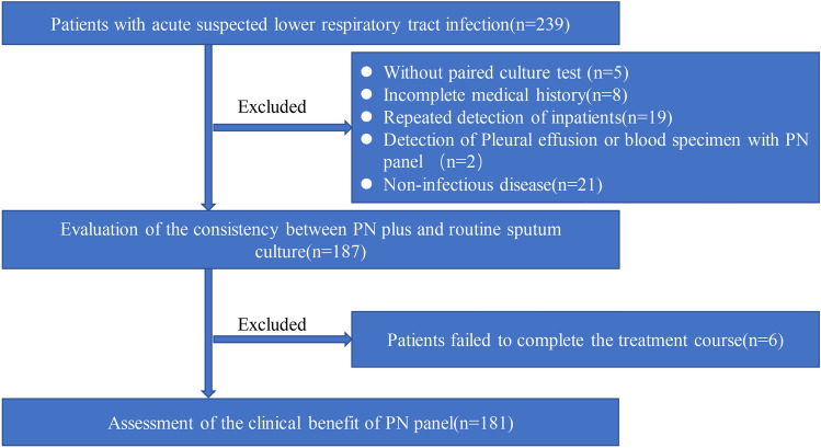
This study initially included a total of 239 patients suspected of having acute LRTIs. However, 52 patients without a paired culture test, incomplete medical history, repeated sample detection, or with non-infectious diseases were excluded from the analysis. Consequently, 187 patients were included for the comparison of etiology and drug resistance genes, and 181 patients were evaluated for the clinical efficacy of the PN panel (Fig. 1).
Fig. 1.
Flowchart of the study. A total of 181 patients completed the clinical efficacy evaluation using the PN panel test
Table 1 presents the demographic characteristics of the 132 male and 55 female participants included in the study. The average age was 64.00 ± 20.00 years, and 79.14% (148/187) of patients had underlying diseases, which included cardiovascular disease (19 cases), cerebrovascular disease (47 cases), Alzheimer’s disease (18 cases), diabetes (28 cases), hypertension (62 cases), chronic structural lung disease (49 cases), autoimmune diseases (9 cases), chronic kidney disease (10 cases), chronic liver disease (2 cases), malignant tumors (22 cases), indwelling endotracheal tubes (14 cases), and long-term bed rest (16 cases).
Table 1.
Demographic characteristics and basic diseases
| Subject | Cases (%) |
|---|---|
| Age (year, mean, SD) | 64.00 ± 20.00 |
| Male | 132 (70.60%) |
| Comorbidity | n (%) |
| Diabetes | 28 (14.98%) |
| Hypertension | 62 (33.16%) |
| Alzheimer disease | 18 (9.63%) |
| Structural lung diseases | 49 (26.20%) |
| Cardiac disease | 19 (10.16%) |
| Malignancy | 22 (11.76%) |
| Cerebrovascular diseases | 47 (25.13%) |
| Connective tissue disease | 9 (4.81%) |
| Tolerance of endotracheal tube | 14 (7.49%) |
| Long immobilization | 16 (8.56%) |
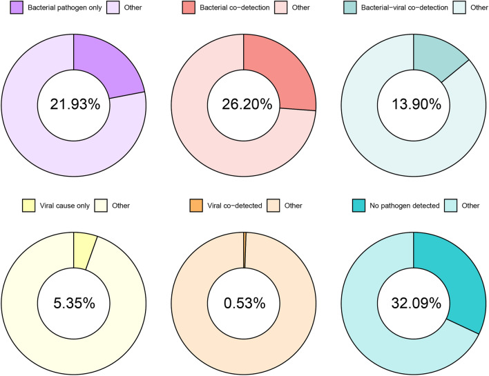
The PN panel revealed that bacteria (including atypical bacteria) were the predominant pathogens, identified in 116 patients (62.03%). Concurrent detection of bacterial and viral pathogens was observed in 26 patients (13.9%). However, no pathogens were detected in 60 patients (32.09%; see Fig. 2).
Fig. 2.
The ratio of pathogens detected in the PN panel bacteria were the predominant pathogens detected in 116 patients (62.03%). Bacterial and viral pathogens were detected simultaneously in 26 patients (13.9%)
Detection of pathogens in patients with CAP and HAP using the PN panel
The PN panel demonstrated a significantly higher detection rate in the HAP group (51/57 [89.47%]) than in the CAP group (76/130 [58.46%]; P = 0.000). Additionally, the detection abundance in the HAP group was 2 (range 1–4), which was significantly higher than that in the CAP group (P = 0.000).
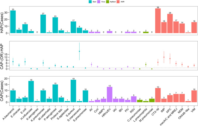
Among patients with CAP, H. influenzae (18/130 [13.85%]) and S. aureus (19/130 [14.62%]) were the most prevalent pathogens. In contrast, among patients with HAP, A. baumannii (33/57 [57.89%]) and K. pneumoniae (27/57 [47.37%]) were the most commonly identified pathogens. Figure 3 illustrates the discrepancies in pathogen detection between patients with CAP and those with HAP.
Fig. 3.
Differences in pathogens and AMR detected using the PN panel between patients with CAP and those with HAP. Blue bars represent bacteria, purple bars represent viruses, teal bars indicate atypical bacteria, and orange bars represent AMR genes
In the bronchoalveolar lavage fluid (BALF) samples from the HAP group, CTX-M and KPC were the most frequently detected AMR targets. In contrast, in the CAP group, CTX and VIM were the most commonly identified. Notably, the OXA48-like enzyme was the least detected in both groups.
Comparison of detection results against bacteria and AMR between the PN panel and cultural methods
The typical positive bacterial rate of the PN panel (110/187 [58.82%]) was significantly higher than that of the cultural method (65/187 [34.76%]; P = 0.000). Table 2 displays the consistency of the bacterial detection results between the PN panel and culture methods. An assessment of the overall performance of the PN panel for the detection of bacterial targets revealed an 85% (51/60) PPA with routine culture results. The PPA for H. influenzae, P. aeruginosa, S. marcescens, and S. aureus was 100%, whereas that for K. pneumoniae was 73%. The NPAs ranged from 84 to 99%, with an overall NPA for bacterial detection of 92%.
Table 2.
The concordance of bacteria between the Pneumonia panel and cultural methods
| Tag | CM + PN + | CM + PN- | CM − PN + | CM − PN − | PPA (95%CI) | NPA (95%CI) |
|---|---|---|---|---|---|---|
| A. baumannii complex | 19 | 4 | 24 | 140 | 0.83 (0.63–0.93) | 0.85 (0.79–0.9) |
| E. cloacae | 0 | 0 | 8 | 179 | NA | 0.96 (0.92–0.98) |
| E. coli | 4 | 1 | 13 | 169 | 0.8 (0.38–0.96) | 0.93 (0.88–0.96) |
| H. influenzae | 1 | 0 | 20 | 166 | 1 (0.21–1) | 0.89 (0.84–0.93) |
| K. aerogenes | 0 | 0 | 1 | 186 | NA | 0.99 (0.97–1) |
| K. pneumoniae | 11 | 4 | 26 | 146 | 0.73 (0.48–0.89) | 0.85 (0.79–0.89) |
| M. catarrhalis | 0 | 0 | 4 | 183 | NA | 0.98 (0.95–0.99) |
| Proteus spp. | 0 | 0 | 4 | 183 | NA | 0.98 (0.95–0.99) |
| P. aeruginosa | 9 | 0 | 29 | 149 | 1 (0.7–1) | 0.84 (0.78–0.88) |
| S. marcescens | 3 | 0 | 6 | 178 | 1 (0.44–1) | 0.97 (0.93–0.98) |
| S. aureus | 4 | 0 | 32 | 151 | 1 (0.51–1) | 0.83 (0.76–0.87) |
| S. agalactiae | 0 | 0 | 1 | 186 | NA | 0.99 (0.97–1) |
| S. pneumoniae | 0 | 0 | 14 | 173 | NA | 0.93 (0.88–0.95) |
| Total | 51 | 9 | 182 | 2189 | 0.85 (0.74–0.92) | 0.92 (0.91–0.93) |
Data are presented as n
CM cultural method; PN panel Pneumonia panel; PPA positive percent agreement; NPA negative percent agreement; CI confidence interval; NA the clinical features were not suitable for the control group
Table 3 presents the semi-quantitative results of the PN panel compared with the culture results. When the PN group exhibited an elevated semi-quantitative value of bacterial targets, the likelihood of consistently detecting the same positive bacteria using cultivation methods also increased. When the PN group detected bacterial targets of ≥ 107 copies/mL, the proportion of positive bacterial culture was 45.83% (33/72), with E. coli accounting for 100%, followed by K. pneumoniae (76.9%), and the lowest consistent bacteria being Proteus spp. (0%) and S. pneumoniae (0%). When the PN group detected bacterial target values of 107, 106, 105, and 104 copies/mL, the proportions of positive bacterial cultures were 45.83% (33/72), 21.74% (10/46), 11.76% (8/68), and 0% (0/59), respectively (P = 0.000).
Table 3.
The result of semiquantitative values of bacteria measured by the Pneumonia panel and the positive cultural method
| Bacteria | Methods | Methods | Methods | Methods | ||||
|---|---|---|---|---|---|---|---|---|
| PN (107) | CM | PN (106) | CM | PN (105) | CM | PN (104) | CM | |
| A. baumannii | 21 | 12 | 11 | 5 | 6 | 2 | 4 | 0 |
| Enterobacter cloacae complex | 0 | 0 | 0 | 0 | 4 | 0 | 3 | 0 |
| Escherichia coli | 3 | 3 | 6 | 0 | 3 | 1 | 5 | 0 |
| Haemophilus influenzae | 5 | 1 | 2 | 0 | 9 | 0 | 8 | 0 |
| Klebsiella aerogenes | 0 | 0 | 0 | 0 | 1 | 0 | 0 | 0 |
| Klebsiella pneumoniae group | 13 | 9 | 3 | 0 | 10 | 2 | 16 | 0 |
| Moraxella catarrhalis | 0 | 0 | 2 | 0 | 0 | 0 | 2 | 0 |
| Proteus spp. | 2 | 0 | 2 | 0 | 1 | 0 | 2 | 0 |
| Pseudomonas aeruginosa | 9 | 5 | 11 | 2 | 11 | 2 | 7 | 0 |
| Serratia marscens | 2 | 1 | 3 | 2 | 2 | 0 | 1 | 0 |
| Staphylococcus aureus | 11 | 2 | 5 | 1 | 13 | 1 | 10 | 0 |
| Streptococcus agalactiae | 0 | 0 | 0 | 0 | 1 | 0 | 0 | 0 |
| Streptococcus pneumoniae | 6 | 0 | 1 | 0 | 7 | 0 | 1 | 0 |
| Total overall | 72 | 33 | 46 | 10 | 68 | 8 | 59 | 0 |
Data are presented as n
CM cultural method; PN panel Pneumonia panel
AMR genotype–phenotype associations
The PN panel and culture methods resulted in 51 cases of consistent bacterial identification, with A. baumannii, K. pneumoniae, and P. aeruginosa being the most frequently identified species. Among these, 46 cases exhibited potential carbapenemase and ESBL AMR in various bacteria, four cases involved S. aureus, and one case involved H. influenzae. Table 4 illustrates the qualitative comparison of bacterial targets between the BioFire FilmArray PN panel and culture methods. Out of the 30 strains demonstrating carbapenem resistance in the culture method, 24 strains exhibited carbapenem resistance genes in the PN panel. Overall, 90% (27/30) of bacterial strains detected using the PN panel indicated consistency between genotype and phenotype resistance for CTX-M genes. The PN panel accurately detected mecA/C and MREJ resistance genes when methicillin-resistant S. aureus was isolated using the culture methods, indicating that the PPA was 100%. Additionally, the NPA of mecA/C and MREJ was 100%.
Table 4.
Consistency of genotype and phenotype resistance to antimicrobial resistance genes between the Pneumonia panel and the cultural methods
| Tag | CM + PN + | CM + PN− | CM − PN + | CM − PN− | PPA (95%CI) | NPA (95%CI) |
|---|---|---|---|---|---|---|
| CTX-Ma | 27 | 3 | 2 | 14 | 0.9 (0.74–0.97) | 0.88 (0.64–0.97) |
| Carbapenenase AMRb | 24 | 6 | 12 | 4 | 0.8 (0.63–0.9) | 0.25 (0.1–0.49) |
| mecA/C and MREJc | 1 | 0 | 0 | 3 | 1 (0.21–1) | 1 (0.44–1) |
| Overall | 52 | 9 | 14 | 21 | 0.85 (0.74–0.92) | 0.6 (0.44–0.74) |
Data are presented as n
CM cultural method; PN panel Pneumonia panel; AMR antimicrobial resistance; PPA positive percent agreement; NPA negative percent agreement; CI confdence interval
aresistance associated with CTX-M genes
bresistance associated with one or more carbapenenum resistance genes, including (KPC, NDM, Oxa48-like, VIM, IMP)
cresistance associated with mecA/C and MREJ genes
Clinical practice of the PN panel
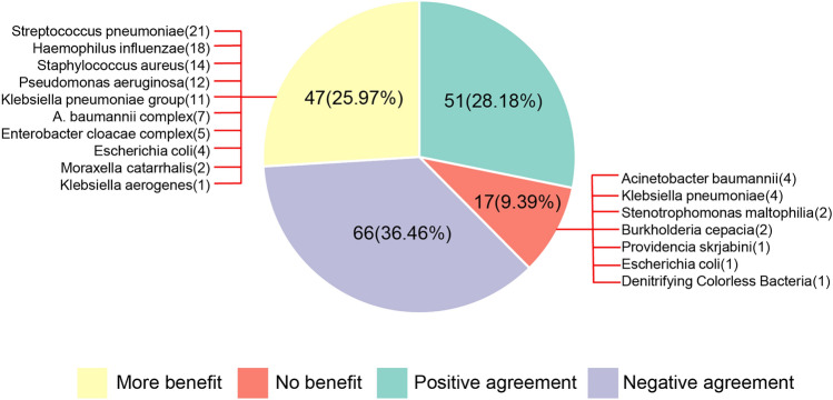
Among the 181 patients who completed treatment, the PN panel consistently maintained negative results in 36.46% (66/181) of cases and consistently positive results in 28.18% (51/181) of cases compared with the culture methods. Compared with the culture methods, 25.97% (47/181) of patients experienced additional clinical benefits from the PN panel, while 9.39% (17/181) of patients did not experience any clinical benefit (Fig. 4). Among the pathogens that can demonstrate clinical benefits when using PN reports, the most frequently missed pathogens when using culture methods were S. pneumoniae, H. influenzae, S. aureus, P. aeruginosa, and K. pneumoniae. The pathogens missed by the PN panel were isolated from BALF samples using culture methods, including A. baumannii, K. pneumoniae, S. maltophila, Burkholderia cepacia, Providencia skrjabini, and Achromobacter denitrificans.
Fig. 4.
The clinical practice and missed pathogen of PN panel and culture method. The light green part represents positive agreement, the purple part represents negative agreement, the yellow part represents additional clinical benefit, and the red part represents no clinical advantages
Among the 181 patients, there were 109 cases of bacterial detection (71 cases with AMR and 38 cases without AMR; Table 5). Hospitalization costs (P = 0.002) and antimicrobial drug use (P = 0.001) were higher in patients with drug resistance genes than in those without drug resistance genes. Compared with patients without AMR, those with AMR had a longer hospitalization time (14 days [interquartile range 10–19] vs 10 days [interquartile range 8–13]) and were more likely to develop complications (64.79% vs 21.05%). Adverse outcomes did not show significant differences between patients with and without AMR.
Table 5.
Comparision of clinical outcome between patients with and without AMR genes
| Outcomes | AMR (n = 71) | Non-AMR (n = 38) | P-value |
|---|---|---|---|
| Total hospitalization cost (RMB) | 74,288.53 ± 6127.93 | 41,631.86 ± 7633.84 | 0.002 |
| Antibiotic cost (RMB) | 14,453.69 ± 1803.09 | 5971.02 ± 1109.07 | 0.001 |
| Hospitalization duration (days) | 14 (10.19) | 10 (8.13) | 0.010 |
| Comorbidities (%) | 46 (64.79%) | 8 (21.05%) | 0.002 |
| Adverse outcomea (%) | 12 (16.90%) | 3 (7.89%) | 0.193 |
Data are presented as median (interquartile range [IQR]) or mean (SD) or n (percentage)
AMR,antimicrobial resistance
adeath or exacerbation of illness transferred to the ICU
Discussion
In this study, we designed a PN panel to evaluate the consistency between bacterial pathogens and AMR using routine culture methods. This is the first study to assess the significance of such a panel in clinical practice in China. Additionally, we analyzed the detection of the PN panel in cases of CAP and HAP. The bacterial panel and AMR both demonstrated a PPA of 85% compared with the conventional culture method. Apart from the advantages of point-of-care testing and rapid cultivation, the bacterial panel exhibited 25% more clinical benefits than the culture method. Also, early detection of AMR genes helps to adjust antibiotics in a timely manner, thereby avoiding complications and reducing the cost of antibiotics.
Bacteria were the most frequently detected pathogens in the PN panel, with A. baumannii and K. pneumoniae being the bacteria most strongly associated with HAP, in line with findings from previous studies [23, 24]. Unlike in a previous study, we found that S. aureus (14.62%) was more prevalent in patients with CAP than S. pneumoniae (7.69%), which might be related to the inclusion of nearly one-third of immunosuppressed patients in our study. As previously reported, S. aureus, P. aeruginosa, and K. pneumoniae are the most common bacterial infections in immunocompromised patients [25]. Hence, rapid bedside etiological testing is crucial as the etiology is linked to the patient’s underlying disease, disease severity, and even the site of onset. We observed that coinfection was more prevalent in patients with HAP than in those with CAP. Previous research has indicated that coinfection is significantly associated with disease severity and high mortality [26]. A study from Korea reported that 13.6% of patients with CAP had coinfection, while the proportion increased to 21.9% in patients with severe CAP [27]. The PN panel can simultaneously detect various bacteria, viruses, and even atypical pathogens, which may serve as an early warning indicator for the patient’s condition, whereas sputum bacterial cultures can only detect one pathogen at a time. In conclusion, the PN panel, as a multiplex detection reagent, may provide enhanced clinical practice guidance for physicians.
Consistent with the findings of a previous study [28], our study indicated that the PN panel detected more bacterial targets than the culture method, resulting in a 24.06% increase in patients reported as positive using the PN panel, with relatively high PPA (85%) and NPA (92%). Consequently, negative results may be employed for early antibiotic de-escalation, as the negative predictive value exceeded 90% in the PN panel. Similarly, prior research has reported superior performance of PN panels for bacterial detection, with PPAs ranging from 90.0 to 98.4% and NPAs ranging from 93.8 to 98.1% [21, 29–31]. While four K. pneumoniae strains were detected in the bacterial culture but not in the PN panel, resulting in a PPA of only 73%, Stenotrophomonas maltophilia, B. cepacia, and Providencia skrjabini were only detected using the culture method, whereas S. pneumoniae, H. influenzae, S. aureus, and P. aeruginosa were frequently missed.
Previous studies have demonstrated that quantitative PCR can distinguish symbiosis from pathogenicity by observing the charge [32], such as 103 CFU/mL used for the protected specimen brush or 104 CFU/mL of BAL used as an indicator to discontinue antibiotics against VAP [32]. The PN panel is semi-quantitative, with levels of 104, 105, 106, and 107 for bacterial targets, which is significant for guiding the initiation of antibiotic therapy in patients with HAP. In our study, an increasing number of semi-quantitative PN panels improved the likelihood of sputum cultures containing the same pathogen. The highest proportion of culture methods producing the same pathogen was observed when the PN panel detected bacterial targets of 107 copies/mL or greater. Conversely, the corresponding pathogen was not detected in the sputum culture when the bacterial target was 104 copies/mL, as detected using the PN panel. This suggests that it may be challenging to detect crucial organisms at extremely low concentrations, even though they are still associated with diseases. Further research is needed to explore whether the detection of unidentified, low-abundance, cultured microorganisms in the PN panel is of prognostic importance. Studies have also indicated that a high level of semi-quantitative signal intensity of positive microorganisms detected using multiple PCRs is closely related to positive bacterial cultures [29, 33], which may be useful for interpretation in the clinical applications of PN panels.
Molecular tests for genetic markers associated with antibiotic resistance, such as mecA, carbapenemases, and ESBLs, have been associated with positive outcomes, including reduced duration of optimal antibiotic therapy, shorter ICU stays, and decreased mortality rates [28, 34]. Our study demonstrated that hospitalization costs, antibiotic consumption, and the incidence of complications were higher in patients with drug-resistant genes than in those without drug-resistant genes. This emphasizes that early identification of drug resistance information and corresponding clinical interventions can help reduce economic costs and the occurrence of complications. Prior research has shown that the concordance rate for accessible resistance targets was 79% (14/18), consistent with phenotypic susceptibility testing [35], whereas in our study, the proportion of consistency in the phenotypic sensitivity test was 85% (52/61). Notably, mecA/C and MREJ of the PN panel exhibited extremely high predictive values for methicillin resistance, with 100% PPA and NPA in patients with positive S. aureus culture. Previous studies have indicated that the PPA for mecA/C and MREJ detection with PN panels was 100%, but NPA was < 90% [31]. However, further research is required to fully evaluate the PN panel, as our study included only four samples with positive S. aureus cultures.
We have previously described real-time PCR for the detection of NDM, KPC, VIM, IMP, and OXA-48, which are currently the most prevalent carbapenemase-producing genes [36]. In this study, the rate of phenotypic carbapenem resistance was relatively high, with 78.26% (36/46) of the specimens showing carbapenem resistance, the most common strains being A. baumannii and K. pneumoniae. Among the 30 carbapenem-resistant strains cultured, carbapenem-resistant genes were detected in 24 samples using the PN panels, while the remaining 6 were not detected. The six resistant strains were P. aeruginosa (three cases) and A. baumannii (three cases). This may be mediated by mechanisms other than carbapenem enzymes, such as the overexpression of efflux pumps or reduction of outer membrane pore proteins in Pseudomonas spp. [37]. Additionally, the overexpression of efflux pumps plays a significant role in the resistance of A. baumannii to tigecycline and imipenem [38]. Similarly, CTX-M testing demonstrated a positivity rate of 90%. However, 67% of these patients harbored concomitant carbapenemase genes. Considering that carbapenemase resistance often results in cephalosporin resistance [39, 40], the actual predictive efficacy of this measure may be diminished. These genetic tests facilitate the prompt addition of antibiotics and the implementation of appropriate isolation measures.
Our study has several limitations. First, we did not compare multiple specific etiological methods but rather culture results for bacteria with PN results. For example, bacterial culture is the primary method for the clinical diagnosis of S. pneumoniae; however, the detection rate of this method is relatively low and is influenced by various factors. Hence, urine antigen or other PCR tests should also be considered. Second, the methods of both analyses in this study were derived from the same alveolar lavage, but not the same specimen, which may have led to slight differences in the study results, although it is more in line with real-world research. Additionally, in this prospective study, we did not compare the clinical outcomes of the PN panel with those of standard methods. Our study revealed that patients in the HAP group had higher detection rates, a greater abundance of pathogens, and higher rates of resistance; however, the specific differentiation of clinical benefits was not achievable. Resistance genes influenced the clinical outcomes in our study, strongly supporting the necessity of detecting resistance genes in patients with LRTIs, though the cost of the panel will be higher than conventional culture. More prospective randomized studies are necessary to assess the impact of PN panels on the clinical outcomes of infected patients, including the types of pathogens and coinfections.
Conclusions
In summary, the PN panel, functioning as a rapid molecular diagnostic technology, demonstrated superior performance in detecting and quantifying HAP pathogens compared to the CAP panel. The PN panel exhibited a higher microbial detection rate and abundance compared to conventional bacterial culture, with an overall agreement of 85%. The AMR indicators provided by the PN panel proved to be reliable predictors of bacterial resistance phenotypes, particularly in the case of S. aureus. Notably, the presence of these indicators was correlated with increased hospitalization costs and a higher incidence of complications.
Supplementary Information
Below is the link to the electronic supplementary material.
Acknowledgements
We are grateful to all participants included in this study.
Abbreviations
- PN
Pneumonia
- HAP
Hospital-acquired pneumonia
- CAP
Community-acquired pneumonia
- PPA
Positive percent agreement
- NPA
Negative percent agreement
- AMR
Antimicrobial resistance
- AST
Antimicrobial susceptibility testing
- IQR
Interquartile range
- ATS/IDSA
American thoracic society and the American association for infectious diseases
- BALF
Bronchoalveolar lavage fluid
- ESBL
Extended-spectrum beta-lactamase
- PCR
Polymerase chain reaction
- Copies/mL
Copies per milliliter
Author contributions
JG and JY: design, analysis, operations, manuscript writing, and manuscript revision. LL and XC: figures and manuscript writing. GY: document translation. YH: blood samples collection and paper writing. RS: design and manuscript revision.
Funding
This study was supported by the Youth Research Project Fund of Guangdong Second Provincial General Hospital (No. YQ2019-012), the Science and Technology Projects in Guangzhou (No. 202102010078), and the Medical and Health Foundation Projects in Beijing (No. B20078DS).
Data availability
The datasets presented in this study are available upon reasonable request.
Declarations
Conflict of interest
The authors state that the study has no potential conflict of interest.
Consent for publication
Not applicable.
Ethical approval
The procedures of this study were in line with the Helsinki Declaration and approved by the Ethics Committee of the Second People’s Hospital of Guangdong Province (No. 2021-KY-167-02).
Footnotes
Publisher's Note
Springer Nature remains neutral with regard to jurisdictional claims in published maps and institutional affiliations.
Gong Jinru and Yang Jiasheng contributed equally.
Change history
2/15/2024
A Correction to this paper has been published: 10.1007/s15010-024-02207-y
References
- 1.World Health Organization. https://www.who.int/news-room/fact-sheets/detail/the-top-10-causes-of-death. Accessed 7 Aug 2023.
- 2.Metlay JP, Waterer GW, Long AC, Anzueto A, Brozek J, Crothers K, et al. Diagnosis and treatment of adults with community-acquired pneumonia. An official clinical practice guideline of the American thoracic society and infectious diseases society of America. Am J Respir Crit Care Med. 2019;200:e45–e67. doi: 10.1164/rccm.201908-1581ST. [DOI] [PMC free article] [PubMed] [Google Scholar]
- 3.Lieberman D, Schlaeffer F, Boldur I, Lieberman D, Horowitz S, Friedman MG, et al. Multiple pathogens in adult patients admitted with community-acquired pneumonia: a one year prospective study of 346 consecutive patients. Thorax. 1996;51:179–184. doi: 10.1136/thx.51.2.179. [DOI] [PMC free article] [PubMed] [Google Scholar]
- 4.Textoris J, Wiramus S, Martin C, Leone M. Antibiotic therapy in patients with septic shock. Eur J Anaesthesiol. 2011;28:318–324. doi: 10.1097/EJA.0b013e328346c0de. [DOI] [PubMed] [Google Scholar]
- 5.Osborne CM, Langelier C, Kamm J, Williamson K, Ambroggio L, Reeder RW, et al. Viral detection by reverse transcriptase polymerase chain reaction in upper respiratory tract and metagenomic RNA sequencing in lower respiratory tract in critically ill children with suspected lower respiratory tract infection. Pediatr Crit Care Med. 2023 doi: 10.1097/PCC.0000000000003336. [DOI] [PMC free article] [PubMed] [Google Scholar]
- 6.Zhang J, Yang T, Zou M, Wang L, Sai L. The epidemiological features of respiratory tract infection using the multiplex panels detection during COVID-19 pandemic in Shandong province, China. Sci Rep. 2023;13:6319. doi: 10.1038/s41598-023-33627-9. [DOI] [PMC free article] [PubMed] [Google Scholar]
- 7.Gadsby NJ, McHugh MP, Forbes C, MacKenzie L, Hamilton SKD, Griffith DM, et al. Comparison of Unyvero P55 pneumonia cartridge, in-house PCR and culture for the identification of respiratory pathogens and antibiotic resistance in bronchoalveolar lavage fluids in the critical care setting. Eur J Clin Microbiol Infect Dis. 2019;38:1171–1178. doi: 10.1007/s10096-019-03526-x. [DOI] [PubMed] [Google Scholar]
- 8.Miners L, Huntington S, Lee N, Turner KME, Adams E. An economic evaluation of two PCR-based respiratory panel assays for patients admitted to hospital with community-acquired pneumonia (CAP) in the UK, France and Spain. BMC Pulm Med. 2023;23:220. doi: 10.1186/s12890-023-02516-2. [DOI] [PMC free article] [PubMed] [Google Scholar]
- 9.Roh EJ, Lee MH, Lee JY, Kim HB, Ahn YM, Kim JK, et al. Analysis of national surveillance of respiratory pathogens for community-acquired pneumonia in children and adolescents. BMC Infect Dis. 2022;22:330. doi: 10.1186/s12879-022-07263-z. [DOI] [PMC free article] [PubMed] [Google Scholar]
- 10.Serigstad S, Knoop ST, Markussen DL, Ulvestad E, Bjørneklett RO, Ebbesen MH, et al. Diagnostic utility of oropharyngeal swabs as an alternative to lower respiratory tract samples for PCR-based syndromic testing in patients with community-acquired pneumonia. J Clin Microbiol. 2023;61:e0050523. doi: 10.1128/jcm.00505-23. [DOI] [PMC free article] [PubMed] [Google Scholar]
- 11.Hu FP, Guo Y, Zhu DM, Wang F, Jiang XF, Xu YC, et al. Resistance trends among clinical isolates in China reported from CHINET surveillance of bacterial resistance, 2005–2014. Clin Microbiol Infect. 2016;22:S9–14. doi: 10.1016/j.cmi.2016.01.001. [DOI] [PubMed] [Google Scholar]
- 12.Collaborators AR. Global burden of bacterial antimicrobial resistance in 2019: a systematic analysis. Lancet. 2022;399:629–655. doi: 10.1016/S0140-6736(21)02724-0. [DOI] [PMC free article] [PubMed] [Google Scholar]
- 13.Ho J, Ip M. Antibiotic-resistant community-acquired bacterial pneumonia. Infect Dis Clin North Am. 2019;33:1087–1103. doi: 10.1016/j.idc.2019.07.002. [DOI] [PubMed] [Google Scholar]
- 14.Thakuria B, Lahon K. The beta lactam antibiotics as an empirical therapy in a developing country: an update on their current status and recommendations to counter the resistance against them. J Clin Diagn Res. 2013;7:1207–1214. doi: 10.7860/JCDR/2013/5239.3052. [DOI] [PMC free article] [PubMed] [Google Scholar]
- 15.Kalil AC, Metersky ML, Klompas M, Muscedere J, Sweeney DA, Palmer LB, et al. Management of adults with hospital-acquired and ventilator-associated pneumonia: 2016 clinical practice guidelines by the infectious diseases society of America and the American thoracic society. Clin Infect Dis. 2016;63:e61–e111. doi: 10.1093/cid/ciw353. [DOI] [PMC free article] [PubMed] [Google Scholar]
- 16.National Health and Family Planning Commission of the People's Republic of China. Performance guideline for bacterial culture of lower respiratory tract infections:WS/T 499-2017[S]. China Quality Inspection Publishing House. 2017.
- 17.Clinical and Laboratory Standards Institute (CLSI). performance standards for antimicrobial 8 susceptibility testing; Thirty-second informational supplement. CLSI document M100. 2022.
- 18.BioFire. https://www.biofiredx.com/products/the-filmarray-panels/filmarray-pneumonia/. Accessed 7 Aug 2023.
- 19.Ruppé E, Cherkaoui A, Lazarevic V, Emonet S, Schrenzel J. Establishing genotype-to-phenotype relationships in bacteria causing hospital-acquired pneumonia: a prelude to the application of clinical metagenomics. Antibiotics (Basel) 2017;6:30. doi: 10.3390/antibiotics6040030. [DOI] [PMC free article] [PubMed] [Google Scholar]
- 20.Bauernfeind A, Stemplinger I, Jungwirth R, Ernst S, Casellas JM. Sequences of beta-lactamase genes encoding CTX-M-1 (MEN-1) and CTX-M-2 and relationship of their amino acid sequences with those of other beta-lactamases. Antimicrob Agents Chemother. 1996;40:509–513. doi: 10.1128/AAC.40.2.509. [DOI] [PMC free article] [PubMed] [Google Scholar]
- 21.Buchan BW, Windham S, Balada-Llasat JM, Leber A, Harrington A, Relich R, et al. Practical comparison of the BioFire FilmArray Pneumonia panel to routine diagnostic methods and potential impact on antimicrobial stewardship in adult hospitalized patients with lower respiratory tract infections. J Clin Microbiol. 2020 doi: 10.1128/JCM.00135-20. [DOI] [PMC free article] [PubMed] [Google Scholar]
- 22.Zhan Y, Xu T, He F, Guan WJ, Li Z, Li S, et al. Clinical evaluation of a metagenomics-based assay for pneumonia management. Front Microbiol. 2021;12:751073. doi: 10.3389/fmicb.2021.751073. [DOI] [PMC free article] [PubMed] [Google Scholar]
- 23.Kyriazopoulou E, Karageorgos A, Liaskou-Antoniou L, Koufargyris P, Safarika A, Damoraki G, et al. BioFire(®) FilmArray(®) Pneumonia panel for severe lower respiratory tract infections: subgroup analysis of a randomized clinical trial. Infect Dis Ther. 2021;10:1437–1449. doi: 10.1007/s40121-021-00459-x. [DOI] [PMC free article] [PubMed] [Google Scholar]
- 24.Yang T, Mei Q, Fang X, Zhu S, Wang Y, Li W, et al. Clinical value of metagenomics next-generation sequencing in Bronchoalveolar lavage fluid for patients with severe hospital-acquired pneumonia: a nested case-control study. Infect Drug Resist. 2022;15:1505–1514. doi: 10.2147/IDR.S356662. [DOI] [PMC free article] [PubMed] [Google Scholar]
- 25.Sun T, Wu X, Cai Y, Zhai T, Huang L, Zhang Y, et al. Metagenomic next-generation sequencing for pathogenic diagnosis and antibiotic management of severe community-acquired pneumonia in immunocompromised adults. Front Cell Infect Microbiol. 2021;11:661589. doi: 10.3389/fcimb.2021.661589. [DOI] [PMC free article] [PubMed] [Google Scholar]
- 26.Carbonell R, Urgelés S, Salgado M, Rodríguez A, Reyes LF, Fuentes YV, et al. Negative predictive value of procalcitonin to rule out bacterial respiratory co-infection in critical covid-19 patients. J Infect. 2022;85:374–381. doi: 10.1016/j.jinf.2022.06.024. [DOI] [PMC free article] [PubMed] [Google Scholar]
- 27.Lim YK, Kweon OJ, Kim HR, Kim TH, Lee MK. Impact of bacterial and viral coinfection in community-acquired pneumonia in adults. Diagn Microbiol Infect Dis. 2019;94:50–54. doi: 10.1016/j.diagmicrobio.2018.11.014. [DOI] [PMC free article] [PubMed] [Google Scholar]
- 28.Bauer KA, West JE, Balada-Llasat JM, Pancholi P, Stevenson KB, Goff DA. An antimicrobial stewardship program's impact with rapid polymerase chain reaction methicillin-resistant Staphylococcus aureus/S. aureus blood culture test in patients with S. aureus bacteremia. Clin Infect Dis. 2010;51(9):1074–1080. doi: 10.1086/656623. [DOI] [PubMed] [Google Scholar]
- 29.Kosai K, Akamatsu N, Ota K, Mitsumoto-Kaseida F, Sakamoto K, Hasegawa H, et al. BioFire FilmArray Pneumonia panel enhances detection of pathogens and antimicrobial resistance in lower respiratory tract specimens. Ann Clin Microbiol Antimicrob. 2022;21:24. doi: 10.1186/s12941-022-00512-8. [DOI] [PMC free article] [PubMed] [Google Scholar]
- 30.Lee SH, Ruan SY, Pan SC, Lee TF, Chien JY, Hsueh PR. Performance of a multiplex PCR pneumonia panel for the identification of respiratory pathogens and the main determinants of resistance from the lower respiratory tract specimens of adult patients in intensive care units. J Microbiol Immunol Infect. 2019;52:920–928. doi: 10.1016/j.jmii.2019.10.009. [DOI] [PMC free article] [PubMed] [Google Scholar]
- 31.Webber DM, Wallace MA, Burnham CA, Anderson NW. Evaluation of the BioFire FilmArray Pneumonia panel for detection of viral and bacterial pathogens in lower respiratory tract specimens in the setting of a tertiary care academic medical center. J Clin Microbiol. 2020 doi: 10.1128/JCM.00343-20. [DOI] [PMC free article] [PubMed] [Google Scholar]
- 32.Johansson N, Kalin M, Giske CG, Hedlund J. Quantitative detection of Streptococcus pneumoniae from sputum samples with real-time quantitative polymerase chain reaction for etiologic diagnosis of community-acquired pneumonia. Diagn Microbiol Infect Dis. 2008;60:255–261. doi: 10.1016/j.diagmicrobio.2007.10.011. [DOI] [PubMed] [Google Scholar]
- 33.Sun L, Li L, Du S, Liu Y, Cao B. An evaluation of the Unyvero pneumonia system for rapid detection of microorganisms and resistance markers of lower respiratory infections-a multicenter prospective study on ICU patients. Eur J Clin Microbiol Infect Dis. 2021;40:2113–2121. doi: 10.1007/s10096-021-04259-6. [DOI] [PubMed] [Google Scholar]
- 34.Walker T, Dumadag S, Lee CJ, Lee SH, Bender JM, Cupo Abbott J, et al. Clinical impact of laboratory implementation of verigene BC-GN microarray-based assay for detection of gram-negative bacteria in positive blood cultures. J Clin Microbiol. 2016;54:1789–1796. doi: 10.1128/JCM.00376-16. [DOI] [PMC free article] [PubMed] [Google Scholar]
- 35.Collins ME, Popowitch EB, Miller MB. Evaluation of a novel multiplex PCR panel compared to quantitative bacterial culture for diagnosis of lower respiratory tract infections. J Clin Microbiol. 2020 doi: 10.1128/JCM.02013-19. [DOI] [PMC free article] [PubMed] [Google Scholar]
- 36.Cohen Stuart J, Leverstein-Van Hall MA. Guideline for phenotypic screening and confirmation of carbapenemases in Enterobacteriaceae. Int J Antimicrob Agents. 2010;36:205–210. doi: 10.1016/j.ijantimicag.2010.05.014. [DOI] [PubMed] [Google Scholar]
- 37.Köhler T, Michea-Hamzehpour M, Epp SF, Pechere JC. Carbapenem activities against Pseudomonas aeruginosa: respective contributions of OprD and efflux systems. Antimicrob Agents Chemother. 1999;43:424–427. doi: 10.1128/AAC.43.2.424. [DOI] [PMC free article] [PubMed] [Google Scholar]
- 38.Nikaido H. Preventing drug access to targets: cell surface permeability barriers and active efflux in bacteria. Semin Cell Dev Biol. 2001;12:215–223. doi: 10.1006/scdb.2000.0247. [DOI] [PubMed] [Google Scholar]
- 39.Giddins MJ, Macesic N, Annavajhala MK, Stump S, Khan S, McConville TH, et al. Successive emergence of ceftazidime–avibactam resistance through distinct genomic adaptations in bla(KPC-2)-Harboring Klebsiella pneumoniae sequence type 307 isolates. Antimicrob Agents Chemother. 2018 doi: 10.1128/AAC.02101-17. [DOI] [PMC free article] [PubMed] [Google Scholar]
- 40.García-Betancur JC, Appel TM, Esparza G, Gales AC, Levy-Hara G, Cornistein W, et al. Update on the epidemiology of carbapenemases in Latin America and the Caribbean. Expert Rev Anti Infect Ther. 2021;19:197–213. doi: 10.1080/14787210.2020.1813023. [DOI] [PubMed] [Google Scholar]
Associated Data
This section collects any data citations, data availability statements, or supplementary materials included in this article.
Supplementary Materials
Data Availability Statement
The datasets presented in this study are available upon reasonable request.


