ABSTRACT
Respiratory syncytial virus (RSV) is a highly contagious virus that affects the lungs and respiratory passages of many vulnerable people. It is a leading cause of lower respiratory tract infections and clinical complications, particularly among infants and elderly. It can develop into serious complications such as pneumonia and bronchiolitis. The development of RSV vaccine or immunoprophylaxis remains highly active and a global health priority. Currently, GSK’s Arexvy™ vaccine is approved for the prevention of lower respiratory tract disease in older adults (>60 years). Palivizumab and currently nirsevimab are the approved monoclonal antibodies (mAbs) for RSV prevention in high-risk patients. Many studies are ongoing to develop additional therapeutic antibodies for preventing RSV infections among newborns and other susceptible groups. Recently, additional antibodies have been discovered and shown greater potential for development as therapeutic alternatives to palivizumab and nirsevimab. Plant expression platforms have proven successful in producing recombinant proteins, including antibodies, offering a potential cost-effective alternative to mammalian expression platforms. Hence in this study, an attempt was made to use a plant expression platform to produce two anti-RSV fusion (F) mAbs 5C4 and CR9501. The heavy-chain and light-chain sequences of both these antibodies were transiently expressed in Nicotiana benthamiana plants using a geminiviral vector and then purified using single-step protein A affinity column chromatography. Both these plant-produced mAbs showed specific binding to the RSV fusion protein and demonstrate effective viral neutralization activity in vitro. These preliminary findings suggest that plant-produced anti-RSV mAbs are able to neutralize RSV in vitro.
KEYWORDS: Respiratory syncytial virus; RSV-fusion protein; monoclonal antibody; recombinant expression; immunoprophylaxis, viral infection; germinivirus; Nicotiana benthamiana
Introduction
Respiratory syncytial virus (RSV) is a contagious virus that can spread from person to person through aerosols and causes lower respiratory tract infections (LTRI) in infants, children, and elders. In some cases, infection leads to pneumonia and bronchiolitis that requires hospitalization. In the United States, RSV leads to the hospitalization of approximately 57,000 children under 5 years of age,1,2 and 100–500 deaths are reported to be due to RSV infection per year.3 Approximately 177,000 persons above the age of 65 require hospitalization and approximately 14,000 per year die due to RSV.4 Though there is no official report documenting the number of deaths from RSV in Thailand, hospitalizations for RSV numbered about 20,000 cases from 2015 to 20205 and 70% of those cases developed pneumonia.1
RSV belongs to Pneumoviridae family, Orthopneumovirus genus.6 Its genome is negative-sense single-stranded RNA. RSV is classified into two major antigenic subgroups, A and B based on antigenic and genomic differences.7 The two major glycoproteins of RSV are the fusion glycoprotein (F protein) and attachment glycoprotein (G protein) which are displayed on the surface of the virion. The primary function of G and F proteins is mediating the attachment between the virus and the host cell and fusion of the virion envelope with the cell membrane to initiate infection. The F protein is more highly conserved than the G protein between two subtypes.8
The trimeric F protein contains six antigenic sites which are classified as sites I, II, III, IV, Ø, and V. Antigenic sites I, II, III, and IV are present on both the pre- and post-fusion F protein conformations, whereas antigenic sites Ø and V are only present on the pre-fusion conformation.9–12 A study conducted by Hause et al.13 found that antigenic site IV is highly conserved in over 1000 clinical isolates.13 Additionally, Mas et al.14 found that antigenic sites III and IV are the most conserved regions of the F protein.14
The U.S. Food and Drug Administration (FDA) recent authorization of Arexvy™, the first RSV vaccine, marks an important step forward in the fight against RSV-induced LTRI. This vaccine was approved for those aged 60 and above, and it has a remarkable success rate of up to 82.6% in preventing RSV-related LTRI. Arexvy™ is effective against both RSV A and B subtypes. In instances of RSV-related LTRI, it has effectiveness rates of 84.6% and 80.9% against RSV A and B subtypes, respectively. Furthermore, it has effectiveness rates for RSV-related acute respiratory illness of 71.9% and 70.6% against the subtypes A and B. These findings highlight the vaccine’s ability to successfully tackle both RSV subtypes, reducing the burden of RSV-related diseases.15
While Arexvy™ is a useful preventative measure for those aged 60 and above, passive vaccination can also be an option for infants and children aged 8–19 months infected with RSV. Palivizumab, an anti-RSV monoclonal antibody (mAb), was approved by the U.S. FDA for use prophylactically to reduce morbidity in a high-risk population of infants and children. Palivizumab binds to RSV-F at antigenic site II. Clinical trials demonstrated reductions in hospitalization of 45–55% compared to placebo.16 Nirsevimab is a recently approved monoclonal antibody used to prevent RSV infection in infants. Like palivizumab, nirsevimab provides passive immunity against RSV by targeting the F protein and neutralizing the ability of RSV to infect cells. Nirsevimab binds to RSV-F at antigenic site Ø and it is 50-fold more potent than palivizumab in vitro and ninefold more potent in vivo at equivalent concentrations. This increased potency is not only due to its binding to a prefusion-specific epitope but also to the engineering of three-point mutations on the heavy chain. These mutations enhance stability and circulation, resulting in a threefold increase in its half life.17,18 Nirsevimab is administered as an injection once per RSV season and demonstrated 74.5% effectiveness against LTRI caused by RSV compared to placebo.17,19 Many research groups have developed the monoclonal antibodies that have greater potency than palivizumab. Monoclonal antibodies such as 5C4 binds to antigenic site Ø20 and CR9501 binds to antigenic site V.21 As these antibodies specifically target the prefusion form of the F protein, they exhibit greater potency than palivizumab22 and have been developed for clinical use.
Generally, mammalian cell expression platforms are used to produce therapeutic proteins including monoclonal antibodies. However, protein production in mammalian cells requires specialized equipment and facilities that contribute to the cost of the antibody product. Since 2014, plant expression systems have been considered as a cost-effective alternative for the production of therapeutic proteins. Plants have several advantages over traditional protein production systems. Whole plants can be grown in large quantities quickly and economically, resulting in a scalable manufacturing system. Plants can also perform post-translational modifications of proteins, such as glycosylation, which are important for protein function and stability. Additional benefits of plant transient expression platforms include faster production times, and lower downstream processing costs.23–26
Plant expression platforms have demonstrated remarkable success in the production of a diverse range of monoclonal antibodies, including those targeting significant diseases such as dengue fever,27 Ebola virus,28 and SARS-CoV-229,30 by the adoption of transient expression. Transient expression is faster and simpler than generating transgenic plants and avoids the need for integrating foreign DNA into the host genome. This approach overcomes the limitations associated with stable expression such as low expression, gene silencing, position effects, and tissue specificity.31,32 In the recent decade, plants have been successfully used to efficiently produce therapeutic antibodies, significantly advancing the field of biopharmaceuticals.33,34
Since 2020, there has been a growing interest in using plants as a platform for producing recombinant proteins for various purposes, including biopharmaceuticals, vaccines, and industrial enzymes. Recombinant proteins such as immunoglobulin, growth factors, cytokines, enzymes, diagnostic reagents, and vaccines have been produced by plants and are proven to be safe and effective in functional studies.35–39 In this study, two anti-RSV monoclonal antibodies (5C4 and CR9501), which specifically bind to the pre-fusion conformation (sites Ø and V respectively) of the F protein and exhibit the highest neutralizing potency compared to the other sites,40 were selected for the production in Nicotiana benthamiana and functional studies of their product mAbs.
Materials and methods
Vector construction
Protein sequences encoding the heavy chain (HC) and light chain (LC) of 5C4 and CR9501 were obtained from protein data bank (PDB) accession numbers 5W24 and 6OE4, respectively. The murine 5C4 chimeric mAb and human CR9501 mAb heavy and light variable sequences were codon optimized. The sequences encoding variable heavy and variable light chain regions were fused to the constant regions of the human heavy and human light chains, respectively. For the HC, the human IgG1 (gamma heavy chain) constant region (GenBank accession number: AAX09634.1) was linked to the C-terminus of the variable region. For the light chain (LC), the human IgG1 (kappa light chain) constant region (GenBank accession number: AAD29610.1) was linked to the C-terminus of the variable region. The barley alpha-amylase signal peptide (SP2) was added to the N-terminus, and SEKDEL was added to the C-terminus of both HC and LC genes and cloned into a geminiviral plant expression vector (pBYR2eK2Md; pBYR2e) as shown in Figure 1. Restriction sites XbaI, BmtI for HC and XbaI, Aflll for LC were added in the variable sequence for cloning, BmtI, SacI for HC and AflII, SacI for LC were added in the constant sequence (Figure 1). The recombinant vectors were further confirmed by DNA sequencing before transforming into Agrobacterium tumefaciens GV3101 via electroporation and expressed in Nicotiana benthamiana.
Figure 1.
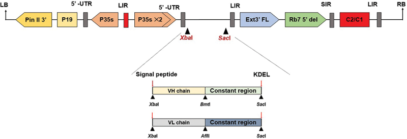
Schematic representation of the T-DNA region of the pBYR2e RSV-F Fc fusion plant expression vector. The T-DNA region plays a crucial role in facilitating the transfer of the gene of interest into plant cells. It includes the left border (LB) and right border (RB) which serve as the boundaries for gene transfer. The Pin II 3’ sequence derived from potato proteinase inhibitor II acts as a border element helping to facilitate the insertion of the desired genes into the plant genome. The vector also incorporates several important components, such as the Tomato Bushy Stunt Virus (TBSV) RNA silencing suppressor, P19; the Cauliflower Mosaic Virus (CaMV) 35s promoter, P35s; the CaMV enhancer, P35s×2; the tobacco extension gene region, Ext3’ FL, 3‘; the tobacco RB7 promoter, Rb7 5’ del; the Bean Yellow Dwarf Virus (BeYDV) short intergenic region, SIR; the BeYDV long intergenic region, LIR; and the BeYDV replication initiation proteins, Rep and RepA, along with C2/C1.41.
Transient expression of mAbs in N. benthamiana
Agrobacterium tumefaciens GV3101 harboring either HC or LC expression vector were cultured in Luria – Bertani (LB) medium containing 50 µg/ml rifampicin, gentamicin, and kanamycin at 28°C with continuous shaking at 200 rpm, overnight. The Agrobacterium culture was diluted in infiltration buffer [10 mM MES and 10 mM MgSO4, pH 5.5] to yield a final optical density (OD600) of 0.2. The diluted Agrobacterium suspension containing HC and LC was mixed equally before infiltrating into the 6- to 8-week-old wild-type plants (N. benthamiana) by vacuum infiltration. Infiltrated leaves were harvested on day 4 after infiltration.
mAbs extraction and purification
Infiltrated leaves were homogenized with phosphate-buffered saline, pH 7.4 (PBS), and centrifuged at 18,000 g and 4°C for 30 min. The supernatant was then filtered through a 0.45 µm membrane filter (Merck, Rahway, New Jersey, USA) before loading into a column that was previously packed with MabSelectSURE® protein A beads (Cytiva, Buckinghamshire, UK). After 10 column volumes of washing with PBS, the mAb was eluted with 0.1 M glycine, pH 2.7, and neutralized with 1.5 M Tris pH 8.0. The neutralized mAbs were further dialyzed against PBS using Slide-A-Lyzer G2 Dialysis Cassettes (30K MWCO) (ThermoFisher Scientific, MA, USA).
Protein quantification by ultra-high-performance liquid chromatography (UHPLC)
For antibody quantification, the MAbPac® Protein A column (ThermoFisher Scientific, MA, USA) was connected to the Waters Acquity Arc UHLPC system. The mobile phase A was PBS, pH 7.4. and mobile phase B was PBS +30 mM HCl, pH 2.5. The flow rate was 2 mL/min, the gradient elution profile was 0% B for 0.2 min, 100% B for 0.60 min, 0%B for 1.20 min and the column temperature was 25°C. The UV absorbance was recorded at 280 nm. The 280 nm antibody peak (after elution) was integrated and compared with a standard curve generated by injecting known quantities of Human IgG (Sigma, MA, USA) in the range of 0.025 to 2 mg/mL.
SDS-PAGE and Western blot analysis
Two micrograms of purified antibodies were separated on sodium dodecyl sulfate polyacrylamide gel electrophoresis (SDS – PAGE) under non-reducing conditions with NuPage® 4–12% gel (ThermoFisher Scientific, MA, USA). For reducing conditions, the antibodies were reduced with a buffer containing 10 mM DTT. The protein bands were then visualized by InstantBlue™ (Abcam, Cambridge, UK) staining. For Western blot analysis, the proteins on SDS-PAGE gel were transferred into nitrocellulose membrane (Bio-Rad, Hercules, USA) and blocked with 5% BSA in PBS before probing with 1:5000 HRP-conjugated goat anti-human IgG (Southern Biotech, USA) or otherwise as stated in the figure. The membranes were then developed and visualized by chemiluminescence using West Pico ECL reagent (ThermoFisher Scientific, MA, USA) and visualized by the ImageQuantTM LAS500 imaging system (GE Health care Uppsala, Sweden).
Purity analysis
Size exclusion chromatography (SEC-UHPLC) was performed on Waters Acquity Arc UHLPC system coupled with XBridge Protein BEH SEC Column, 200Å, 2.5 µm 4.6 mm × 300 mm (Waters, MA, USA). The mobile phase was PBS, pH 7.4. The flow rate was 0.3 mL/min, the run time was 20 min, and the column temperature was maintained at 25°C. The UV absorbance signals were recorded at 280 nm. Peaks area have been automatically integrated and calculated as percentage (%) relative area under the curves of each peak (High molecular weight, Dimer, and Monomer) using Empower3 software (Waters, MA, USA).
N-linked glycan analysis of anti-RSV mAbs
Glycosylation of RSV antibodies was analyzed using liquid chromatography-mass spectrometry (LC/MS). The purified anti-RSV antibodies were reduced, alkylated, and then digested with trypsin (Promega, USA) following the manufacturer’s instructions. The reaction was stopped by adding 5% formic acid and drying under a vacuum for 4 h. The resulting dried tryptic peptides were reconstituted with water and injected into a Vanquish™ Neo UHPLC system (ThermoFisher Scientific, USA) coupled with an Orbitrap Exploris™ 480 mass spectrometer (ThermoFisher Scientific, USA). Positive peptides with m/z = 350–3200 were recorded. The extracted ion chromatogram (XIC) of the glycopeptide (EEQYNSTYR, 1189.51 Da) was manually detected, deconvoluted, and analyzed using the FreeStyle 1.8 program (Thermo Scientific, USA).
In vitro binding of plant-produced mAbs to RSV-F protein
96-well ELISA plates (Corning, NY, USA) were coated with 2 µg/ml of RSV-F his-tagged monomer protein (Sino biological, Beijing, China) in phosphate buffer (20 mM, pH 7.4), 25 µl per well, overnight at 4°C. After washing three times with PBS + 0.05% Tween 20 (PBS-T), the plate was blocked with 200 µL of 5% nonfat dried milk in PBS at 37°C for 1 h. Then, the plate was washed three times with PBS-T and incubated with a serially diluted plant-produced mAbs sample and incubated at 37°C for another 2 h. After sample incubation, the solution was removed and washed with PBS-T. After washing, the plate was incubated with 1:2,500 HRP-conjugated goat anti-human IgG (Southern Biotech, Birmingham, AL, USA), and incubation was continued at 37°C for another hour. After secondary antibody incubation, the wells were emptied and washed. A TMB one substrate solution (Promega, Madison, WI, USA) was added and incubated for 10 min, the reaction was stopped by adding 1 M sulfuric acid, and the absorbance was measured using a multimode reader (Perkin Elmer, MA, USA) at 450 nm. All experiments were performed in triplicate. The half maximal effective concentration (EC50) value was calculated using nonlinear regression in GraphPad. Prism software v9.3.
Cells and virus propagation
RSV strain A2 (VR-1540) was obtained from the American Type Culture Collection (ATCC, Rockville, MD) and propagated in HEp-2 cells (ATCC CCL-23) in Dulbecco’s Modified Eagle Medium (DMEM) supplemented with 2% FBS, 100 U/mL of penicillin, and 100 µg/mL of streptomycin. Vero cells (ATCC CCL-81) were used to perform the microneutralization assay. Vero cells were cultured in Minimum Essential Medium (MEM) supplemented with 10% FBS, 2 mM L-glutamine, non-essential amino acids, and 100 U/mL of penicillin/streptomycin. All culture media reagents and supplements were obtained from Gibco® (ThermoFisher Scientific, Detroit, MI, USA).
Microneutralization assay (MNA)
The MNA protocol was modified from an earlier study.42 In brief, the mAbs were serially diluted fourfold in triplicate, starting at 1 µg/mL. The D-MEM supplemented with 2% FBS, 100 U/mL of penicillin, and 100 µg/mL of streptomycin was used as the dilution buffer throughout the assay. The RSV strain A2 was diluted in dilution buffer to achieve an infectious dose of 100 TCID50 (50% tissue culture infectious dose) in the final volume of the assay. Equal volumes of 100 TCID50 RSV strain A2 were added to the serial dilution samples in 96-well U-bottom plates, which were then incubated at 37°C with 5% CO2 for 1 h. After 1 h of incubation, 100 µl of the sample-virus mixture were transferred to a 96-well flat bottom plate, then mixed with 50 µL of freshly trypsinized Vero cells in dilution buffer (20,000 cells/50 µL). The sample-virus-cell mixture was incubated for 3 days at 37°C with 5% CO2. The virus control, cell control, and virus back-titration were all present on each plate.
After 3 days of incubation, the medium was discarded and washed once with D-PBS. The cells were fixed with ice-cold 80% acetone/20% D-PBS for 20 min at 4°C. The plates were washed three times with 1×PBS-T before being blocked for 1 h with a blocking buffer (4% BSA and 0.01% Tween-20 in 1×PBS). Human RSV fusion glycoprotein was detected with rabbit anti-RSV-F mAb (Cat:11049-R302-H, Sino Biological, Beijing, China) diluted 1:3000 in PBS containing 0.5% BSA and 0.01% Tween-20 added to each well and incubated for 1 h at 37°C. The detection antibody was removed by washing the plate three times, then 1:2000 HRP-conjugated goat anti-rabbit polyclonal antibody (Dako, Glostrup, Denmark A/S) was added, and the plate was incubated at 37°C for 1 h. Plates were washed three times, and then TMB substrate was added (KPL, MA, USA) for 10 min. The reaction was stopped with 1N HCl. Absorbance was measured at 450 and 620 nm (reference wavelength) with an ELISA plate reader (Tecan Sunrise®, Männedorf, Switzerland). The average OD450/620 values of cell control and virus control were used to normalize 0% and 100% of infection, respectively. The inhibitory concentration (IC50) was calculated using a normalized response with a variable slope in GraphPad Prism software version 9.3.
Results
mAb expression and purification
To produce anti-RSV mAbs, A. tumefaciens harboring the heavy chain (HC) and the light chain (LC) plant expression vectors (Figure 1) were diluted to the OD600 0.2 and mix with equal quantity then co-delivered into N. benthamiana leaves through vacuum infiltration. Co-expression of the HC and LC resulted in the assembly of the full structure of the antibody in the infiltrated leaves. Within 4 days post-infiltration, both 5C4 and CR9501 had accumulated in N. benthamiana leaves, which were extracted and purified using protein A affinity chromatography. The final yields after purification resulted up to 33.5 mg/kg of fresh leaf weight for 5C4 and 12.0 mg/kg of fresh leaf weight for CR9501.
The purified antibodies were analyzed using SDS-PAGE and western blot to confirm their molecular weight and identity. Under non-reducing conditions, both mAbs were observed in SDS-PAGE and western blot with the expected molecular weight of approximately 150 kDa (Figure 2a,b). Both anti-human gamma and anti-human kappa antibodies were used in the western blot to assess the antibody assembly in the plants. Under reducing conditions, the protein bands were detected at a molecular weight of approximately 50 kDa and 25 kDa, respectively (Figure 2c,d). These results showed that the co-infiltration of anti-RSV gene constructs encoding HC and LC can produce intact IgG structures that can be purified by protein A chromatography. Furthermore, we also assessed the purity of the mAb size variants using SEC-UHPLC. The size variant of the anti-RSV mAbs contains both high molecular weight (HMW) and monomeric forms as dominant peaks (Figure 3). The observed HMW structures were mostly dimers and oligomers that are present in small amounts (approx. 3%) suggest that these mAbs have a high level of purity (up to 95%) after single-step protein A chromatography as summarized in Table 1.
Figure 2.
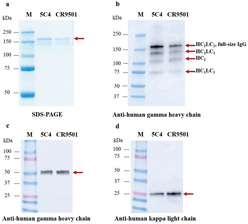
SDS–PAGE (a) and Western blot (b) for purified plant-produced anti-RSV monoclonal antibodies (mAbs) under nonreducing conditions. The Western blot (b) was probed using HRP-conjugated anti-human gamma chain antibody. For reducing conditions, the membrane was probed with either HRP-conjugated anti-human gamma chain antibody (c) or anti-human kappa chain antibody (d). Lane M, protein ladder; lane 1, purified plant-produced 5C4; lane 2, purified plant-produced CR9501.
Figure 3.
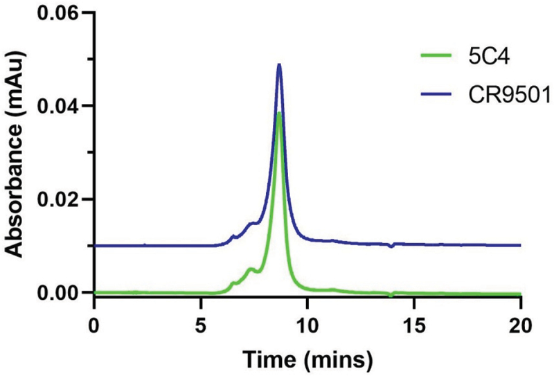
Size exclusion analysis of purified plant-produced anti-RSV mAbs. Purified mAbs were separated in the XBridge protein BEH 200 column. A total of 5 µg of purified mAb was loaded on the column and run at 0.3 mL/min for 20 minutes, UV detection at 280 nm, and the column temperature was maintained at 25°C.
Table 1.
Purity of plant-produced anti-RSV antibodies using SEC-UHPLC.
| Antibody | Monomer (%) | Dimer (%) | Higher molecular weight (%) |
|---|---|---|---|
| 5C4 | 96.61 | 2.91 | 0.48 |
| CR9501 | 98.25 | 1.25 | 0.50 |
N-linked glycans analysis of anti-RSV mAbs
We utilized liquid chromatography-mass spectrometry (LC/MS) to examine the N-glycan profile of anti-RSV mAbs produced in Nicotiana benthamiana. The high presence of oligo-mannose glycan residues in these antibodies suggests the processing of glycoprotein in the endoplasmic reticulum (ER) (Figure 4). The deliberate inclusion of ER retention signals in the production process resulted in the distinctive mannose-rich glycosylation pattern observed in these antibodies.
Figure 4.
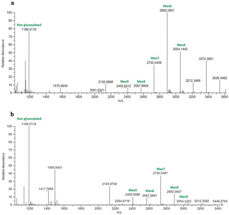
Liquid chromatography mass spectrometry (LC/MS) analysis illustrating the N-glycosylation profiles of the heavy chain peptide EEQYNSTYR (mass: 1189.51 Da) from plant-produced anti-RSV monoclonal antibodies. The peaks specifically assigned to mannose N-glycans (Man5-Man9) are illustrated, providing insights into the glycan composition associated with the analysed glycopeptide. Figure 4a corresponds to the 5C4 antibody, while Figure 4b represents the CR9501 antibody.
In vitro binding of anti-RSV mAbs
The kinetic binding of plant-produced RSV mAbs was determined by ELISA. Two plant-produced mAbs were fivefold serially diluted starting from 100 µg/ml and incubated on plates that were overnight-coated with RSV-F his-tagged monomer protein. The plant-produced Nivolumab (anti PD-1) was used as a negative control. Both plant-produced anti-RSV mAbs showed positive signals for specific binding to RSV-F his-tagged protein (Figure 5) and plant-produced Nivolumab (anti PD-1) does not show reactivity with RSV-F his-tagged monomer protein as expected. This result suggested that the plant-produced RSV mAbs can recognize the RSV-F protein even at low concentrations. The potency of the RSV antibodies was quantified by EC50 which was calculated using nonlinear regression. The EC50 value of 5C4 was 8.212 µg/ml, whereas the EC50 value of CR9501 was 6.182 µg/ml. These results suggest that CR9501 binds with higher affinity than 5C4.
Figure 5.
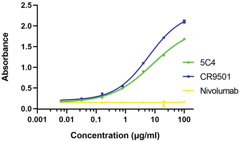
Binding characteristics of plant-produced anti-RSV mAbs. ELISA was used to quantify the specific binding to the RSV-F his-tagged monomer protein. The purified plant produced anti-RSV mAbs, and the plant produced nivolumab (anti PD-1, as a negative control). The binding activity of the antibody was detected with HRP-conjugated anti-human IgG antibody. The data are shown as the mean of triplicates and error bars represent standard deviation. The EC50 was calculated by using GraphPad Prism software 9.3.
Microneutralization
Plant-produced anti-RSV mAbs were tested for their ability to neutralize RSV infection using a microneutralization assay. Two plant-produced mAbs were fourfold serially diluted starting from 1 µg/ml and pre-incubated with RSV strain A2 before inoculating Vero cells. Both mAbs show the ability to neutralize RSV in a dose-dependent manner (Figure 6). The inhibitory concentration (IC50) was calculated using nonlinear regression. In this study, the plant-produced nivolumab (anti-PD-1) and H4 (anti-SARS-CoV-2) were used as negative controls (data in supplementary file). The neutralizing activity of plant-produced CR9501 was found to be superior to 5C4 with an IC50 value of 0.007 and 0.032 µg/ml, respectively. These findings demonstrate that the anti-RSV mAbs produced in N. benthamiana are functional and can prevent RSV infection in a cell culture model.
Figure 6.
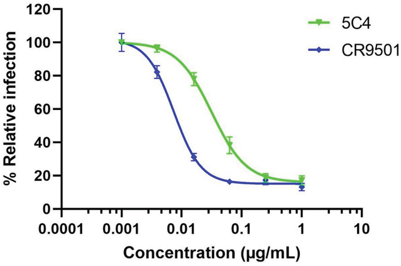
The neutralizing activity of plant-produced anti-RSV mAbs. Both plant-produced mAbs show a dose-dependent viral inhibitory effect. Error bars denote standard deviation of triplicate samples. The inhibitory concentration (IC50) was calculated using a normalized response with a variable slope in GraphPad Prism software version 9.3.
Discussion
Monoclonal antibodies have been used for decades, not only as immunodetection reagents but also in therapeutics. Palivizumab and nirsevimab are the only FDA-approved antibodies for the prevention of RSV disease in infants. Due to the high cost of these drugs,43 access to treatment are limited especially in developing countries. Traditionally, therapeutic antibodies are produced using mammalian platforms, which is expensive. In this study, we aimed to use plants as an alternative platform to produce monoclonal antibodies for RSV prevention and treatment. The major advantage of the plant expression platform, compared to the mammalian platform, is that plant cannot act as host for human or animal pathogens, so the risk of contamination with human or animal pathogens in the product is far lower.23,44,45 Plants also have the ability to produce large quantities of proteins and they do not require complex media for cultivation. This reduces the cost of production, making them a suitable choice for industrial-scale production in a short period of time.
The utilization of plant expression platforms for monoclonal antibody production holds immense potential for significant cost reduction. Techno-economic analyses from previous studies46–49 have addressed the economic advantages of plant expression systems. However, we did not perform such analysis in this proof-of-concept experimental phase. Nandi et al.46 projected that leveraging plant expression could lead to cost reductions of up to 50% compared to other platforms operating at similar scales.46 Furthermore, Ridgley et al.50 found that the production costs of plant-produced anti-immune checkpoint antibodies were 15–75 times lower than those derived from mammalian cells.50 Notably, other research supports these cost advantages, stating that when compared to Chinese Hamster Ovary (CHO) cell-based production, where operational costs were estimated at $65 M/year or $260/g of purified antibody, plant-based production was estimated at $33 M/year or $131/g of purified antibody, representing a potential 50% reduction,51 which similarity to Nandi et al. findings.46 Moreover, conventional mammalian cell culture incurred downstream processing costs of approximately $232/g of purified antibody, whereas the plant-based system’s cost was approximately $99/g, demonstrating a > 50% reduction in manufacturing costs.52 These findings further highlight the significant cost-effectiveness of plant-based expression systems for antibody production.
Plant expression platforms have been used to successfully produce a wide variety of mAbs, including those targeting RSV. Palivizumab53,54 has been produced in plants and showed promising results in preclinical studies. In vivo protection in RSV challenge studies conducted in the cotton rat model demonstrated similar protection of plant-produced compared to mammalian-produced palivizumab.53 These findings highlight the potential of plant expression platforms for the efficient and effective production of therapeutic antibodies. In fact, studies have shown that plant-produced palivizumab has a serum pharmacokinetic profile similar to mammalian-derived palivizumab, suggesting that different expression systems have minimal effect on the pharmacokinetics.53 Furthermore, to reduce plant-specific complex N-glycan modifications, we utilized an ER retention signal, which effectively reduces plant-specific N-glycans and promotes the formation of high-mannose type N-glycans (Figure 4). Notably, other studies also support the effectiveness of this approach. Loos et al.55 found that a KDEL-tagged anti-HIV antibody (2G12) was deposited in protein storage vacuoles, bypassing the Golgi apparatus and carrying mainly high-mannose N-glycans.55 Similarly, Triguero et al.56 demonstrated that a plant-derived mouse IgG mAb fused to the KDEL ER-retention signal displayed homogeneous N-glycosylation throughout the plant, primarily comprising high-mannose-type N-glycans.56 As a result, these modifications in plant-specific glycosylation are minimized, reducing the likelihood of inducing allergic reactions.
In this study, we successfully demonstrated the production and functional characterization of two potential anti-RSV mAbs, 5C4 and CR9501, using N. benthamiana as the expression platform. Our findings revealed that plant-produced mAbs, 5C4 and CR9501, exhibited binding activity to the RSV-F protein and possess neutralizing activity against RSV strain A2. The neutralizing ability of these mAbs demonstrated their effectiveness in vitro in neutralizing the virus.
Previously, 5C4 and CR9501 were produced using a mammalian expression platform and exhibit significant neutralizing activity. The concentration required to achieve 50% neutralization (IC50) of RSV strain A2 of 5C4 and CR9501 were 0.015 µg/mL and 0.004 µg/mL, respectively.20,21 The neutralizing activity of these antibodies was tested and found to be effective in our plant expression platform as well with IC50 of 0.032 µg/mL and 0.007 µg/mL for 5C4 and CR9501, respectively. These results suggest that the antibodies produced from the mammalian expression platform can also be effectively produced using the plant expression platform while maintaining similar neutralizing activity. Taken together, these results suggest that the plant-produced anti-RSV mAbs were functional, exhibiting both antigen recognition and neutralizing activity. Further studies are warranted to test the efficacy of these candidates in suitable animal models.
In the context of therapeutic development, CR9501 shows promising neutralizing activity against RSV and should be a lead candidate for further investigation. However, it is important to note that the efficacy of mAbs can be affected by various factors, including the formulation of the drug product. Therefore, to achieve maximum efficacy and safety profiles, improvements, optimization of these mAbs may be required. Furthermore, to ensure the safety and efficacy of plant-produced mAbs for clinical use, further studies are needed to evaluate their characteristics, pharmacokinetic profiles and in vivo protection in animal models. It is crucial to perform rigorous preclinical studies to demonstrate the safety and efficacy of these plant-produced mAbs before clinical trials.
Conclusion
In conclusion, we have successfully produced functional anti-RSV mAbs in N. benthamiana by transient expression. The plant-produced mAbs showed specific binding to the RSV-F protein and demonstrated effective viral neutralization activity in vitro. Further studies in animal models to test the safety, pharmacokinetics, and efficacy need to be determined before a plant-produced anti-RSV antibody could be used clinically.
Supplementary Material
Acknowledgments
We appreciate the technical assistance provided by the technicians and staff during the experimental study. The author would like to thank the 72nd anniversary of His Majesty King Bhumibol Adulyadej for the graduate school doctoral fellowship and Overseas Research Experience Scholarship (ORES) for Graduate Students from both the Graduate School and the Faculty of Pharmaceutical Science, Chulalongkorn University. We also thank Rudolf Figl for conducting MS experiments. The MS equipment was kindly provided by the EQ-BOKU VIBT GmbH and the BOKU Core Facility Mass Spectrometry.
Funding Statement
This research is funded by Thailand Science research and Innovation Fund Chulalongkorn University and the NSRF via the Program Management Unit for Human Resources & Institutional Development, Research, and Innovation [grant number B13F660137].
Disclosure statement
No potential conflict of interest was reported by the author(s).
Data availability statement
All the data are available from the corresponding author upon request.
Supplementary material
Supplemental data for this article can be accessed on the publisher’s website at https://doi.org/10.1080/21645515.2024.2327142
References
- 1.Rha B, Curns AT, Lively JY, Campbell AP, Englund JA, Boom JA, Azimi PH, Weinberg GA, Staat MA, Selvarangan R.. Respiratory Syncytial Virus-Associated Hospitalizations Among Young Children: 2015-2016. Pediatrics. 2020;146(1):e20193611. doi: 10.1542/peds.2019-3611. [DOI] [PubMed] [Google Scholar]
- 2.Hall CB, Weinberg GA, Iwane MK, Blumkin AK, Edwards KM, Staat MA, Auinger P, Griffin MR, Poehling KA, Erdman D, et al. The burden of respiratory syncytial virus infection in young children. N Engl J Med. 2009;360(6):588–10. doi: 10.1056/NEJMoa0804877. [DOI] [PMC free article] [PubMed] [Google Scholar]
- 3.Centers for Disease Control and Prevention . Increased interseasonal respiratory syncytial virus (RSV) activity in parts of the Southern United States. CDC Health Alert Network; 2021. [Google Scholar]
- 4.Falsey AR, Hennessey PA, Formica MA, Cox C, Walsh EE. Respiratory syncytial virus infection in elderly and high-risk adults. N Engl J Med. 2005;352(17):1749–59. doi: 10.1056/NEJMoa043951. [DOI] [PubMed] [Google Scholar]
- 5.Shi T, McAllister DA, O’Brien KL, Simoes EAF, Madhi SA, Gessner BD, Polack FP, Balsells E, Acacio S, Aguayo C, et al. Global, regional, and national disease burden estimates of acute lower respiratory infections due to respiratory syncytial virus in young children in 2015: a systematic review and modelling study. Lancet. 2017;390(10098):946–58. doi: 10.1016/S0140-6736(17)30938-8. [DOI] [PMC free article] [PubMed] [Google Scholar]
- 6.Battles MB, McLellan JS. Respiratory syncytial virus entry and how to block it. Nat Rev Microbiol. 2019;17(4):233–45. doi: 10.1038/s41579-019-0149-x. [DOI] [PMC free article] [PubMed] [Google Scholar]
- 7.Pandya MC, Callahan SM, Savchenko KG, Stobart CC. A contemporary view of respiratory syncytial virus (RSV) biology and strain-specific differences. Pathogens. 2019;8(2):67. doi: 10.3390/pathogens8020067. [DOI] [PMC free article] [PubMed] [Google Scholar]
- 8.Patel N, Tian JH, Flores R, Jacobson K, Walker M, Portnoff A, Gueber-Xabier M, Massare MJ, Glenn G, Ellingsworth L et al. Flexible RSV prefusogenic fusion glycoprotein exposes multiple neutralizing epitopes that may collectively contribute to protective immunity. Vaccines (Basel). 2020;8:607. [DOI] [PMC free article] [PubMed] [Google Scholar]
- 9.Tang A, Chen Z, Cox KS, Su H-P, Callahan C, Fridman A, Zhang L, Patel SB, Cejas PJ, Swoyer R, et al. A potent broadly neutralizing human RSV antibody targets conserved site IV of the fusion glycoprotein. Nat Commun. 2019;10(1):4153. doi: 10.1038/s41467-019-12137-1. [DOI] [PMC free article] [PubMed] [Google Scholar]
- 10.McLellan JS, Chen M, Joyce MG, Sastry M, Stewart-Jones GBE, Yang Y, Zhang B, Chen L, Srivatsan S, Zheng A, et al. Structure-based design of a fusion glycoprotein vaccine for respiratory syncytial virus. Science. 2013;342(6158):592–8. doi: 10.1126/science.1243283. [DOI] [PMC free article] [PubMed] [Google Scholar]
- 11.McLellan JS, Yang Y, Graham BS, Kwong PD. Structure of respiratory syncytial virus fusion glycoprotein in the postfusion conformation reveals preservation of neutralizing epitopes. J Virol. 2011;85(15):7788–96. doi: 10.1128/JVI.00555-11. [DOI] [PMC free article] [PubMed] [Google Scholar]
- 12.Mousa JJ, Kose N, Matta P, Gilchuk P, Crowe JE. A novel pre-fusion conformation-specific neutralizing epitope on the respiratory syncytial virus fusion protein. Nat Microbiol. 2017;2(4):16271. doi: 10.1038/nmicrobiol.2016.271. [DOI] [PMC free article] [PubMed] [Google Scholar]
- 13.Hause AM, Henke DM, Avadhanula V, Shaw CA, Tapia LI, Piedra PA, Tregoning JS. Sequence variability of the respiratory syncytial virus (RSV) fusion gene among contemporary and historical genotypes of RSV/A and RSV/B. PLoS One. 2017;12(4):e0175792. doi: 10.1371/journal.pone.0175792. [DOI] [PMC free article] [PubMed] [Google Scholar]
- 14.Mas V, Nair H, Campbell H, Melero JA, Williams TC. Antigenic and sequence variability of the human respiratory syncytial virus F glycoprotein compared to related viruses in a comprehensive dataset. Vaccine. 2018;36(45):6660–73. doi: 10.1016/j.vaccine.2018.09.056. [DOI] [PMC free article] [PubMed] [Google Scholar]
- 15.Papi A, Ison MG, Langley JM, Lee D-G, Leroux-Roels I, Martinon-Torres F, Schwarz TF, van Zyl-Smit RN, Campora L, Dezutter N, et al. Respiratory syncytial virus prefusion F protein vaccine in older adults. N Engl J Med. 2023;388(7):595–608. doi: 10.1056/NEJMoa2209604. [DOI] [PubMed] [Google Scholar]
- 16.Viguria N, Navascués A, Juanbeltz R, Echeverría A, Ezpeleta C, Castilla J. Effectiveness of palivizumab in preventing respiratory syncytial virus infection in high-risk children. Hum Vaccines Immunother. 2021;17(6):1867–72. doi: 10.1080/21645515.2020.1843336. [DOI] [PMC free article] [PubMed] [Google Scholar]
- 17.Keam SJ. Nirsevimab: first approval. Drugs. 2023;83(2):181–7. doi: 10.1007/s40265-022-01829-6. [DOI] [PubMed] [Google Scholar]
- 18.Brady T, Cayatte C, Roe TL, Speer SD, Ji H, Machiesky L, Zhang T, Wilkins D, Tuffy KM, Kelly EJ, et al. Fc-mediated functions of nirsevimab complement direct respiratory syncytial virus neutralization but are not required for optimal prophylactic protection. Front Immunol. 2023;14:1283120. doi: 10.3389/fimmu.2023.1283120. [DOI] [PMC free article] [PubMed] [Google Scholar]
- 19.Hammitt LL, Dagan R, Yuan Y, Baca Cots M, Bosheva M, Madhi SA, Muller WJ, Zar HJ, Brooks D, Grenham A, et al. Nirsevimab for prevention of RSV in healthy late-preterm and term infants. N Engl J Med. 2022;386(9):837–46. doi: 10.1056/NEJMoa2110275. [DOI] [PubMed] [Google Scholar]
- 20.Tian D, Battles MB, Moin SM, Chen M, Modjarrad K, Kumar A, Kanekiyo M, Graepel KW, Taher NM, Hotard AL, et al. Structural basis of respiratory syncytial virus subtype-dependent neutralization by an antibody targeting the fusion glycoprotein. Nat Commun. 2017;8(1):1877. doi: 10.1038/s41467-017-01858-w. [DOI] [PMC free article] [PubMed] [Google Scholar]
- 21.Gilman M, Furmanova-Hollenstein P, Pascual G, Wout A, Langedijk J, McLellan J. Transient opening of trimeric prefusion RSV F proteins. Nat Commun. 2019;10(1):2105. doi: 10.1038/s41467-019-09807-5. [DOI] [PMC free article] [PubMed] [Google Scholar]
- 22.McLellan J, Chen M, Leung S, Graepel K, Du X, Yang Y, Zhou T, Baxa U, Yasuda E, Beaumont T, et al. Structure of RSV fusion glycoprotein trimer bound to a prefusion-specific neutralizing antibody. Science. 2013;340(6136):1113–17. doi: 10.1126/science.1234914. [DOI] [PMC free article] [PubMed] [Google Scholar]
- 23.Chen Q, Davis K. The potential of plants as a system for the development and production of human biologics. F1000 Resear. 2016;5. doi: 10.12688/f1000research.8010.1. [DOI] [PMC free article] [PubMed] [Google Scholar]
- 24.Gerasimova SV, Smirnova OG, Kochetov AV, Shumnyi VK. Production of recombinant proteins in plant cells. Russ J Plant Physiol. 2016;63(1):26–37. doi: 10.1134/S1021443716010076. [DOI] [Google Scholar]
- 25.Twyman RM, Schillberg S, Fischer R. Transgenic plants in the biopharmaceutical market. Expert Opin Emerg Drugs. 2005;10(1):185–218. doi: 10.1517/14728214.10.1.185. [DOI] [PubMed] [Google Scholar]
- 26.Sack M, Hofbauer A, Fischer R, Stoger E. The increasing value of plant-made proteins. Curr Opin Biotechnol. 2015;32:163–70. doi: 10.1016/j.copbio.2014.12.008. [DOI] [PMC free article] [PubMed] [Google Scholar]
- 27.Dent M, Hurtado J, Paul AM, Sun H, Lai H, Yang M, Esqueda A, Bai F, Steinkellner H, Chen Q, et al. Plant-produced anti-dengue virus monoclonal antibodies exhibit reduced antibody-dependent enhancement of infection activity. J Gen Virol. 2016;97(12):3280–90. doi: 10.1099/jgv.0.000635. [DOI] [PMC free article] [PubMed] [Google Scholar]
- 28.Zhang Y, Li D, Jin X, Huang Z. Fighting ebola with ZMapp: spotlight on plant-made antibody. Sci China Life Sci. 2014;57(10):987–8. doi: 10.1007/s11427-014-4746-7. [DOI] [PubMed] [Google Scholar]
- 29.Shanmugaraj B, Rattanapisit K, Manopwisedjaroen S, Thitithanyanont A, Phoolcharoen W. Monoclonal antibodies B38 and H4 produced in Nicotiana benthamiana neutralize SARS-CoV-2 in vitro. Front Plant Sci. 2020;11:11. doi: 10.3389/fpls.2020.589995. [DOI] [PMC free article] [PubMed] [Google Scholar]
- 30.Rattanapisit K, Shanmugaraj B, Manopwisedjaroen S, Purwono PB, Siriwattananon K, Khorattanakulchai N, Hanittinan O, Boonyayothin W, Thitithanyanont A, Smith DR, et al. Rapid production of SARS-CoV-2 receptor binding domain (RBD) and spike specific monoclonal antibody CR3022 in Nicotiana benthamiana. Sci Rep. 2020;10(1):17698. doi: 10.1038/s41598-020-74904-1. [DOI] [PMC free article] [PubMed] [Google Scholar]
- 31.Nosaki S, Hoshikawa K, Ezura H, Miura K. Transient protein expression systems in plants and their applications. Plant Biotechnol (Tokyo). 2021;38(3):297–304. doi: 10.5511/plantbiotechnology.21.0610a. [DOI] [PMC free article] [PubMed] [Google Scholar]
- 32.Tyurin AA, Suhorukova AV, Kabardaeva KV, Goldenkova-Pavlova IV, Goldenkova-Pavlova IV. Transient gene expression is an effective experimental tool for the research into the fine mechanisms of plant gene function: advantages, limitations, and solutions. Plants. 2020;9(9):1187. doi: 10.3390/plants9091187. [DOI] [PMC free article] [PubMed] [Google Scholar]
- 33.SBaR S. Plant expression platform for the production of recombinant pharmaceutical proteins. Austin J Biotechnol Bioeng. 2014;1:1–4. [Google Scholar]
- 34.Iyappan G, Shanmugaraj BM, Inchakalody V, Ma JKC, Ramalingam S. Potential of plant biologics to tackle the epidemic like situations – case studies involving viral and bacterial candidates. Int J Infect Dis. 2018;73:363. doi: 10.1016/j.ijid.2018.04.4236. [DOI] [Google Scholar]
- 35.He W, Baysal C, Lobato Gómez M, Huang X, Alvarez D, Zhu C, Armario‐Najera V, Blanco Perera A, Cerda Bennaser P, Saba‐Mayoral A, et al. Contributions of the international plant science community to the fight against infectious diseases in humans—part 2: affordable drugs in edible plants for endemic and re-emerging diseases. Plant Biotechnol J. 2021;19(10):1921–36. doi: 10.1111/pbi.13658. [DOI] [PMC free article] [PubMed] [Google Scholar]
- 36.Shanmugaraj B, Jirarojwattana P, Phoolcharoen W. Molecular farming strategy for the rapid production of protein-based reagents for use in infectious disease diagnostics. Planta Med. 2023;89(10):1010–1020. doi: 10.1055/a-2076-2034. [DOI] [PubMed] [Google Scholar]
- 37.Shanmugaraj B, Bulaon CJ, Phoolcharoen W. Plant molecular farming: a viable platform for recombinant biopharmaceutical production. Plants (Basel). 2020;9(7):842. doi: 10.3390/plants9070842. [DOI] [PMC free article] [PubMed] [Google Scholar]
- 38.Lobato Gómez M, Huang X, Alvarez D, He W, Baysal C, Zhu C, Armario‐Najera V, Blanco Perera A, Cerda Bennasser P, Saba‐Mayoral A, et al. Contributions of the international plant science community to the fight against human infectious diseases – part 1: epidemic and pandemic diseases. Plant Biotechnol J. 2021;19(10):1901–20. doi: 10.1111/pbi.13657. [DOI] [PMC free article] [PubMed] [Google Scholar]
- 39.Phoolcharoen W, Shanmugaraj B, Khorattanakulchai N, Sunyakumthorn P, Pichyangkul S, Taepavarapruk P, Praserthsee W, Malaivijitnond S, Manopwisedjaroen S, Thitithanyanont A, et al. Preclinical evaluation of immunogenicity, efficacy and safety of a recombinant plant-based SARS-CoV-2 RBD vaccine formulated with 3M-052-Alum adjuvant. Vaccine. 2023;41(17):2781–92. doi: 10.1016/j.vaccine.2023.03.027. [DOI] [PMC free article] [PubMed] [Google Scholar]
- 40.Graham BS. Vaccine development for respiratory syncytial virus. Curr Opin Virol. 2017;23:107–12. doi: 10.1016/j.coviro.2017.03.012. [DOI] [PMC free article] [PubMed] [Google Scholar]
- 41.Chen Q, He J, Phoolcharoen W, Mason HS. Geminiviral vectors based on bean yellow dwarf virus for production of vaccine antigens and monoclonal antibodies in plants. Hum Vaccin. 2011;7(3):331–8. doi: 10.4161/hv.7.3.14262. [DOI] [PMC free article] [PubMed] [Google Scholar]
- 42.Boukhvalova MS, Mbaye A, Kovtun S, Yim KC, Konstantinova T, Getachew T, Khurana S, Falsey AR, Blanco JCG. Improving ability of RSV microneutralization assay to detect G-specific and cross-reactive neutralizing antibodies through immortalized cell line selection. Vaccine. 2018;36(31):4657–62. doi: 10.1016/j.vaccine.2018.06.045. [DOI] [PubMed] [Google Scholar]
- 43.Shahabi A, Peneva D, Incerti D, McLaurin K, Stevens W. Assessing variation in the cost of Palivizumab for respiratory syncytial virus prevention in preterm infants. PharmacoEconomics Open. 2018;2(1):53–61. doi: 10.1007/s41669-017-0042-3. [DOI] [PMC free article] [PubMed] [Google Scholar]
- 44.Pogue GP, Vojdani F, Palmer KE, Hiatt E, Hume S, Phelps J, Long L, Bohorova N, Kim D, Pauly M, et al. Production of pharmaceutical-grade recombinant aprotinin and a monoclonal antibody product using plant-based transient expression systems. Plant Biotechnol J. 2010;8(5):638–54. doi: 10.1111/j.1467-7652.2009.00495.x. [DOI] [PubMed] [Google Scholar]
- 45.Xu J, Ge X, Dolan MC. Towards high-yield production of pharmaceutical proteins with plant cell suspension cultures. Biotechnol Adv. 2011;29(3):278–99. doi: 10.1016/j.biotechadv.2011.01.002. [DOI] [PubMed] [Google Scholar]
- 46.Nandi S, Kwong AT, Holtz BR, Erwin RL, Marcel S, McDonald KA. Techno-economic analysis of a transient plant-based platform for monoclonal antibody production. Mabs-austin. 2016;8(8):1456–66. doi: 10.1080/19420862.2016.1227901. [DOI] [PMC free article] [PubMed] [Google Scholar]
- 47.Tusé D, Tu T, McDonald KA. Manufacturing economics of plant-made biologics: case studies in therapeutic and industrial enzymes. Biomed Res Int. 2014;2014:256135. doi: 10.1155/2014/256135. [DOI] [PMC free article] [PubMed] [Google Scholar]
- 48.Walwyn DR, Huddy SM, Rybicki EP. Techno-economic analysis of horseradish peroxidase production using a transient expression system in Nicotiana benthamiana. Appl Biochem Biotechnol. 2015;175(2):841–54. doi: 10.1007/s12010-014-1320-5. [DOI] [PubMed] [Google Scholar]
- 49.Wilken LR, Nikolov ZL. Recovery and purification of plant-made recombinant proteins. Biotechnol Adv. 2012;30(2):419–33. doi: 10.1016/j.biotechadv.2011.07.020. [DOI] [PubMed] [Google Scholar]
- 50.Ridgley LA, Falci Finardi N, Gengenbach BB, Opdensteinen P, Croxford Z, Ma JK, Bodman‐Smith M, Buyel JF, Teh AYH. Killer to cure: expression and production costs calculation of tobacco plant-made cancer-immune checkpoint inhibitors. Plant Biotechnol J. 2023;21(6):1254–69. doi: 10.1111/pbi.14034. [DOI] [PMC free article] [PubMed] [Google Scholar]
- 51.Werner RG. Economic aspects of commercial manufacture of biopharmaceuticals. J Biotechnol. 2004;113(1–3):171–82. doi: 10.1016/j.jbiotec.2004.04.036. [DOI] [PubMed] [Google Scholar]
- 52.Xenopoulos A. A new, integrated, continuous purification process template for monoclonal antibodies: process modeling and cost of goods studies. J Biotechnol. 2015;213:42–53. doi: 10.1016/j.jbiotec.2015.04.020. [DOI] [PubMed] [Google Scholar]
- 53.Zeitlin L, Bohorov O, Bohorova N, Hiatt A, Kim DH, Pauly MH, Velasco J, Whaley K, Barnard D, Bates J, et al. Prophylactic and therapeutic testing of Nicotiana-derived RSV-neutralizing human monoclonal antibodies in the cotton rat model. MAbs. 2013;5(2):263–9. doi: 10.4161/mabs.23281. [DOI] [PMC free article] [PubMed] [Google Scholar]
- 54.Hiatt A, Bohorova N, Bohorov O, Goodman C, Kim D, Pauly MH, Velasco J, Whaley KJ, Piedra PA, Gilbert BE, et al. Glycan variants of a respiratory syncytial virus antibody with enhanced effector function and in vivo efficacy. Proc Natl Acad Sci USA. 2014;111(16):5992–7. doi: 10.1073/pnas.1402458111. [DOI] [PMC free article] [PubMed] [Google Scholar]
- 55.Loos A, Van Droogenbroeck B, Hillmer S, Grass J, Pabst M, Castilho A, Kunert R, Liang M, Arcalis E, Robinson DG, et al. Expression of antibody fragments with a controlled N-Glycosylation pattern and induction of endoplasmic reticulum-derived vesicles in seeds of arabidopsis. Plant Physiol. 2011;155(4):2036–48. doi: 10.1104/pp.110.171330. [DOI] [PMC free article] [PubMed] [Google Scholar]
- 56.Triguero A, Cabrera G, Cremata JA, Yuen C-T, Wheeler J, Ramírez NI. Plant-derived mouse IgG monoclonal antibody fused to KDEL endoplasmic reticulum-retention signal is N-glycosylated homogeneously throughout the plant with mostly high-mannose-type N-glycans. Plant Biotechnol J. 2005;3(4):449–57. doi: 10.1111/j.1467-7652.2005.00137.x. [DOI] [PubMed] [Google Scholar]
Associated Data
This section collects any data citations, data availability statements, or supplementary materials included in this article.
Supplementary Materials
Data Availability Statement
All the data are available from the corresponding author upon request.


