PLOS ONE logoLink to PLOS ONE
. 2024 Mar 21;19(3):e0300255. doi: 10.1371/journal.pone.0300255

The CHD family chromatin remodeling enzyme, Kismet, promotes both clathrin-mediated and activity-dependent bulk endocytosis

Emily L Hendricks 1, Faith L W Liebl 1,*
Editor: Alexander G Obukhov2
PMCID: PMC10956772  PMID: 38512854

Abstract

Chromodomain helicase DNA binding domain (CHD) proteins, including CHD7 and CHD8, remodel chromatin to enable transcriptional programs. Both proteins are important for proper neural development as heterozygous mutations in Chd7 and Chd8 are causative for CHARGE syndrome and correlated with autism spectrum disorders, respectively. Their roles in mature neurons are poorly understood despite influencing the expression of genes required for cell adhesion, neurotransmission, and synaptic plasticity. The Drosophila homolog of CHD7 and CHD8, Kismet (Kis), promotes neurotransmission, endocytosis, and larval locomotion. Endocytosis is essential in neurons for replenishing synaptic vesicles, maintaining protein localization, and preserving the size and composition of the presynaptic membrane. Several forms of endocytosis have been identified including clathrin-mediated endocytosis, which is coupled with neural activity and is the most prevalent form of synaptic endocytosis, and activity-dependent bulk endocytosis, which occurs during periods of intense stimulation. Kis modulates the expression of gene products involved in endocytosis including promoting shaggy/GSK3β expression while restricting PI3K92E. kis mutants electrophysiologically phenocopy a liquid facets mutant in response to paradigms that induce clathrin-mediated endocytosis and activity-dependent bulk endocytosis. Further, kis mutants do not show further reductions in endocytosis when activity-dependent bulk endocytosis or clathrin-mediated endocytosis are pharmacologically inhibited. We find that Kis is important in postsynaptic muscle for proper endocytosis but the ATPase domain of Kis is dispensable for endocytosis. Collectively, our data indicate that Kis promotes both clathrin-mediated endocytosis and activity-dependent bulk endocytosis possibly by promoting transcription of several endocytic genes and maintaining the size of the synaptic vesicle pool.

Introduction

Neural communication relies on the coordinated release of neurotransmitter, which diffuses across the synaptic cleft and binds to postsynaptic receptors [1, 2]. Synaptic proteins facilitate processes required for proper neurotransmission and synaptic plasticity by inducing cellular changes in response to an action potential [3]. The mobilization and trafficking of synaptic vesicles constitute the presynaptic response to action potential-induced Ca2+ influx [4]. Thus, Ca2+-dependent release of neurotransmitter-containing vesicles, and the proteins that comprise vesicle release machinery and postsynaptic receptors, are important for mediating neural communication. Presynaptic neurons maintain distinct pools of neurotransmitter-filled vesicles that can be quickly mobilized upon stimulation. The localization of vesicles into these pools influences their temporal availability for release [5, 6]. Vesicles in the readily releasable pool are docked at presynaptic release sites and are the first to be released upon stimulation [7]. The reserve pool, however, may be more distal to sites of release and is mobilized upon high frequency stimulation to replenish the readily releasable pool [8, 9]. Both the readily releasable and reserve pools are replenished by recycling vesicles through endocytosis [10] and disruptions in these endocytic pathways compromise neurotransmitter release.

Endocytosis is a process that involves the internalization and scission of the plasma membrane and functions to recycle membrane-associated proteins and phospholipids. Several mechanisms of endocytosis have been characterized including clathrin-mediated endocytosis (CME), activity-dependent bulk endocytosis (ADBE), kiss-and-run, fast compensatory endocytosis, and ultrafast endocytosis [11, 12]. CME involves the recruitment of clathrin scaffold proteins that surround vesicles upon endocytic uptake and is the primary mode of synaptic vesicle recycling during basal neurotransmission [12, 13]. Conversely, ADBE initially occurs independently of clathrin and is used during high frequency neuronal stimulation to replenish reserve pool vesicles [12, 14]. Both processes facilitate synaptic vesicle recycling and are important for overall neuronal function. Mutations in endocytic machinery-encoding genes are associated with neurodevelopmental disorders [15] and neurodegenerative diseases [16].

The expression of synaptic gene products, including those required for endocytosis, is dynamically regulated by chromatin states. Chromatin regulatory proteins establish and maintain the epigenome, which consists of the cellular chemical modifications of DNA and histones [17]. Epigenetic changes in gene expression are required for synaptic remodeling during learning [18] and occur in both neurodevelopmental disorders [19] and neurodegenerative diseases [20]. The ATP-dependent chromodomain helicase DNA-binding (CHD) family proteins regulate neurodevelopmental processes [21] but are also expressed in mature neurons [22]. Given the partial overlap of misregulated genes shared between autism spectrum disorder and Alzheimer’s disease [23], a better understanding of how chromatin remodeling influences synaptic processes during both neurodevelopment and neurodegeneration is warranted. The link between neurodevelopmental and neurodegenerative diseases and the molecular pathways underlying their shared pathology are becoming increasingly clear. For example, mutations in the catalytic γ-secretase subunit, presenilin, impair postsynaptic development [24] and are also linked to aberrant proteolytic processing in familial Alzheimer’s disease [25]. How dysfunctional CHD proteins specifically impact synaptic function throughout neurodevelopmental and neurodegenerative disease pathways, however, is still poorly characterized.

Kismet (Kis) is the Drosophila homolog of group III CHD proteins, including CHD7 and CHD8 [21, 26]. The full length Kis isoform possesses two chromodomains that bind methylated histone residues and an ATPase domain that catalyzes nucleosome remodeling [26, 27]. These nucleosome modifications may activate or repress transcription depending on the binding sites exposed [28]. At the Drosophila neuromuscular junction (NMJ), Kis restricts axonal branching and bouton formation [29] and promotes glutamate receptor localization [30] and synaptic vesicle endocytosis [31]. Here, we find that Kis regulates ADBE and CME at the Drosophila NMJ possibly by regulating the expression of endocytosis-related gene products and maintaining vesicle pool sizes. We also show that Kis is important in postsynaptic cells for presynaptic vesicle endocytosis and Kis’s modulation of endocytosis is independent of ATPase domain activity.

Materials and methods

Drosophila stocks and husbandry

Fly stocks were maintained at 25°C with a 12 h light:dark cycle and fed Jazz Mix food (Fisher Scientific AS153). Nutri-Fly Instant Food (Genesee Scientific 66–118) was used for experiments where flies were raised on food containing compounds. Male and female larvae and adults were used for all experiments except for sggEP1576. The sggEP1576 mutation is on the first chromosome necessitating the use of only females of this and the control genotypes. Most fly stocks were obtained from the Bloomington Drosophila stock center including w1118 (RRID:BDSC_3605), dap160EP2543 (RRID:BDSC_3605), kisk13416 (RRID:BDSC_10442), lqfKG03016 (RRID:BDSC_13766), sggEP1576 (RRID:BDSC_11008), Actin5c-Gal4 (RRID:BDSC_30558), elavC155-Gal4 (RRID:BDSC_458), elav-Gal4 (RRID:BDSC_8760), D42-Gal4 (RRID:BDSC_8816), 24B-Gal4 (RRID:BDSC_1767), and repo-Gal4 (RRID:BDSC_7415). kisLM27 and UAS-kis-L were gifts from Dan Marenda [32]. UAS-CHD7 and UAS-kisK2060R were generated for this work.

Generation and expression of UAS-CHD7 and UAS-kisK2060R

Human CHD7 (NM_017780) was used to determine the Drosophila optimized CHD7 cDNA sequence. The kis-L (NM_001258889) cDNA sequence was used for kisK2060R but was modified by substituting CGA (encoding R) for AAA (encoding K) at codon 2059. cDNAs were synthesized by Thermo Fisher and included a NotI restriction site on the 5’ end and a XbaI site on the 3’ end. A Drosophila optimized V5 cDNA sequence (GGT AAG CCC ATC CCG AAC CCC CTG CTG GGT TTG GAC TCC ACT) was included upstream of the stop codon and XbaI restriction site. cDNAs were inserted into pUAST using the NotI and XbaI restriction sites and insertion was confirmed by DNA sequencing. Both constructs were injected into w1118 embryos using standard germline transformation methods (BestGene, Inc., Chino Hills, CA).

Reverse transcription PCR and qPCR

Central nervous systems (CNSs) were dissected from male and female third instar larvae in Roger’s Ringer solution (135 mM NaCl, 5 mM KCl, 4 mM MgCl2*6H2O, 1.8 mM CaCl2*2H2O, 5 mM TES, 72 mM Sucrose, 2 mM glutamate, pH 7.15), placed in Invitrogen RNAlater Stabilization Solution (Fisher Scientific AM7020), and stored at -20°C. 30 CNSs or 8–12 larvae per genotype were used for each technical replicate. RNA was isolated using the Invitrogen Purelink RNA Mini Kit (Fisher Scientific 12-183-025). RNA concentrations were obtained using an Implen Nanophotometer N50. 100 ng of RNA was used for each reaction. Primers were designed using PerlPrimer (v. 1.1.21). qRT-PCR was performed using the iTaq Universal SYBR Green One Step Kit (Bio-Rad 1725151) and a CFX Connect Real-Time PCR Detection System (Bio-Rad). At least four biological replicates each including three technical replicates were used for data analysis. 2-ΔΔC(t) values [33] were calculated by first subtracting the C(t) value of the target transcript reaction from the C(t) value for GAPDH to obtain ΔC(t) for each transcript. Next, the difference between control and kis mutant ΔC(t)s was calculated to obtain the 2-ΔΔC(t). 2-ΔΔC(t) were calculated using RNA samples isolated the same day with RT-qPCR reactions executed simultaneously. Student’s t-tests were used to determine if there was a statistical difference between control and kis mutant ΔC(t)s.

Immunocytochemistry and FM labeling

Third instar larvae were fillet dissected in Roger’s Ringer solution (135 mM NaCl, 5 mM KCl, 4 mM MgCl2*6H2O, 1.8 mM CaCl2*2H2O, 5 mM TES, 72 mM Sucrose, 2 mM glutamate, pH 7.15) on Sylgard (World Precision Instruments)-coated 60 mm dishes. Larvae were fixed for 30 min with 4% paraformaldehyde (Fisher Scientific F79500) in 1 x phosphate buffered saline (PBS, Midwest Scientific QS1200). Fixed larvae were placed in 1.5 ml centrifuge tubes containing PTX (1 x PBS + 0.1% Triton X-100, Fisher Scientific AAA16046AP) and washed three times for 10 min in PTX. After two 30 min washes in PBTX (1 x PBS + 0.1% Triton X-100 + 1% Bovine Serum Albumin, Fisher Scientific BP1600-100), rabbit α-V5 (1:1000, Sigma AB3792, RRID: AB_91591) was diluted in PBTX and applied overnight at 4°C. After primary antibodies were removed, larvae were washed three times for 10 min and two times for 30 min in PBTX. α-rabbit FITC (Jackson ImmunoResearch; RRID: AB_2337972) was diluted 1:400 in PBTX and applied for 2 h at room temperature with Cy3 HRP (1:125, Jackson ImmunoResearch, RRID: AB_2338959) and DAPI (1:500, ThermoFisher D1306, RRID: AB_2629482).

Endocytosis was measured as described by Verstreken et al. [34]. Briefly, third instar larvae were fillet dissected in Ca2+-free HL-3 (100 mM NaCl, 5 mM KCl, 10 mM NaHCO3, 5 mM HEPES, 30 mM sucrose, 5 mM trehelose, 10 mM MgCl2, pH 7.2). After one wash with Ca2+-free HL-3 and cutting the motor neurons, 4 μM FM 1-43FX (Fisher Scientific F35355) in HL-3 containing 90 mM KCl and 1.0 mM CaCl2 was applied for one minute. For endocytosis inhibition experiments, larvae were exposed to DMSO, 100 μM BAPTA-AM for 10 min, 25 μM EGTA-AM for 10 min, 200 μM Chlorpromazine for 30 min, 100 μM Dynasore for 10 min, or 200 μM Roscovitine for 30 min prior to one min stimulation with 90 mM KCl. Concentrations and treatment times were determined using previously published protocols (see results). Next, larvae were washed five times over 5–10 min in Ca2+-free HL-3. Larvae were fixed in 3.7% paraformaldehyde (Fisher Scientific) in Ca2+-free HL-3 for 5 min and then placed in 1.5 ml centrifuge tubes. Larvae were washed with Ca2+-free HL-3 containing 2.5% goat serum several times and with Ca2+-free HL-3 10 times over 10–15 min. A647 HRP (1:125, Jackson ImmunoResearch) was applied for 30 min in Ca2+-free HL-3 containing 5% goat serum. Next, larvae were washed with Ca2+-free HL-3 10 times over 10–15 min and mounted on slides with Vectashield (Vector Laboratories H1000).

An Olympus FV1000 confocal microscope was used to acquire images of 6/7 NMJs within segments 3 or 4 and ventral nerve cords. The former were acquired using a 60x oil immersion objective while the latter were acquired using a 40x oil immersion objective. Genotypes were labeled using the same reagents for each experimental replicate. The mean of all control acquisition settings was used for each experimental animal and approximately equal numbers of controls and experimental animals were imaged each day. Image z-stacks were constructed using Fiji [35]. Measurement of FM 1-43FX signal intensities was accomplished by calculating the relative fluorescence intensity, which was the difference between the synaptic fluorescence and background fluorescence, from each max projected z-stack slice. The mean relative fluorescence value of all slices was used as the NMJ fluorescence intensity for each NMJ and this is represented as a point on bar graphs.

Electrophysiology

Third instar larvae were fillet dissected in ice cold HL-3 containing 0.25 mM Ca2+ and glued to Sylgard (World Precision Instruments)-coated round 18 mm coverslips. After the nervous system was removed, larvae were rinsed once with room temperature HL-3 containing 1.0 mM Ca2+, which was used for recordings. Muscle 6 of body wall segments 3 or 4 was used for two electrode voltage clamp recordings. Electrodes were filled with 3 M KCl and used for recordings provided they demonstrated resistances of 10–30 MΩ. Recordings were acquired in pClamp (Molecular Devices, v. 11.1) and obtained from muscles clamped at -60 mV using an Axoclamp 900A amplifier (Molecular Devices) with input resistances <5 MΩ. Segmental nerves were stimulated with suction electrodes filled with HL-3 containing 1.0 mM Ca2+. Suprathreshold stimuli were administered from a Grass S88 stimulator with a SIU5 isolation unit (Grass Technologies). High frequency stimulation included stimuli administered at 0.2 Hz for 50 s, 20 Hz for 60 s, and 0.2 Hz for 50 s. Low frequency stimulation included 5 Hz stimulation for five minutes. Stock concentrations of each compound were diluted to 100 μM BAPTA-AM, 25 μM EGTA-AM, and 100 μM Dynasore in HL-3 containing 1.0 mM Ca2+ the day of recording and kept on ice. Working concentrations were allowed to warm to room temperature immediately before use. Vesicle pool sizes were assessed by incubating dissected larvae at room temperature for 20 min in freshly prepared 2 μM Bafilomycin in HL-3 containing 1 mM Ca2+. Concentrations and treatment times for all compounds were determined using previously published protocols (see results). mEJCs were recorded for 3 min followed by stimulation of the segmental nerve at 3 Hz for 10 min or at 10 Hz for 5 min. Recordings were digitized with a Digidata 1443 digitizer (Molecular Devices).

Recordings from approximately the same number of controls and experimental animals were obtained each day. All experiments included at least three biological replicates with sample sizes of 9–11 larvae as indicated in figure legends. Data were analyzed in Clampfit (v 11.1, Molecular Devices) and GraphPad Prism (v. 9.3.0). When trains of stimulation were delivered, eEJC amplitudes were normalized to the first eEJC. Multiple unpaired t-tests were used to determine if eEJC amplitudes differed over time.

Behavior

Third instar larvae were obtained from vials with Jazz Mix food and briefly wiped with a damp paintbrush to remove any debris. Larvae were then transferred to a 1.6% agar plate and allowed to freely wander for one minute to acclimate to crawling surface. After the acclimation period, larvae were moved to the behavior arena (a backlit 1.6% agar slab with overhead camera mount) and video recorded for 30 seconds with a Cannon EOS M50 camera at 29.97 frames per second. Recording started after larvae engaged in forward-directed motion. 899 frames were analyzed in Fiji (NIH ImageJ) with the wrMTrck plugin by Jesper S. Pedersen [36] to obtain values for average distances travelled, average and maximum velocities, and body lengths per second. XY coordinates of larval crawling paths were extracted from wrMTrck and graphed in Excel (Microsoft) to generate representative path tracings.

Experimental design and statistical analyses

All experiments included at least two biological replicates with approximately equal numbers of controls and experimental animals. Data analyses were performed with GraphPad Prism (v. 9.3.0 and 10.0.0). When data sets included one control group, for example comparison of mutant genotypes to w1118 controls, means were compared using unpaired t-tests. When data sets included two control groups, a one-way ANOVA was used followed by Tukey’s post hoc tests. Bartlett’s Test for homogeneity of variance was used to assess the variances between data sets. For experiments that included more than one outcrossed Gal4 control groups, the control groups were compared to determine if they statistically differed. If the Gal4 control groups did not statistically differ, they were combined and treated as a single control group. Bar graphs in figures represent the means and show sample sizes as individual points. Sample sizes indicate individual larvae except for the RT-qPCR experiments where the points represent one technical replicate. Statistical significance is represented on bar graphs as follows: * = <0.05, ** = <0.01, *** = <0.001 with error bars representing standard error of the mean (SEM). S1 Table shows the statistically significant p-values corresponding to each figure.

Results

Kismet influences transcription of gene products encoding CME- and ADBE-associated proteins

Presynaptic endocytosis replenishes synaptic vesicles [37], influences protein localization, and preserves the size and composition of the presynaptic membrane [38, 39]. We previously showed that that Kis promotes endocytosis by regulating transcription of endocytic genes and synaptic Dynamin (Dyn) localization. Specifically, mutations in kis increased AP2α but decreased dap160 and endophilin B (endoB) transcripts [31]. Each of these gene products is required for CME. Dap160/Intersectin is a scaffolding protein that recruits the clathrin adapter AP2 to the membrane [40] while endophilins recruit the GTPase, Dyn [41], and help induce curvature of the plasma membrane [42]. In this study, we sought to better understand the role of Kis in the distinct regulation of CME, ADBE, or both using the Drosophila NMJ. The Drosophila NMJ provides a well-established system to model mammalian neurotransmission as these synapses share common structural features and mechanisms of neurotransmitter release, vesicle endocytosis, and postsynaptic signaling [43].

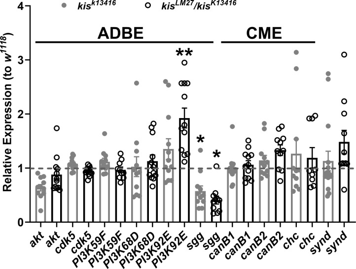
To further investigate Kis’ regulation of endocytosis, we extended our analysis to gene products involved in CME, ADBE, or both. We used two kis mutant alleles for our analysis including the hypomorphic allele, kisk13416, and kisLM27, a null allele [29]. Because the latter is embryonic lethal, we used animals heterozygous for kisk13416 and kisLM27 to examine endocytic transcript levels via RT-qPCR. Of the 10 transcripts examined, PI3K92E, which encodes a catalytic subunit of the phosphatidylinositol 3-kinase (PI3K) enzyme, was increased in kisLM27/kisk13416 but not kisk13416 mutants compared with controls. In contrast, shaggy (sgg), which encodes Glycogen Synthase Kinase 3 (GSK3) was decreased in both kis mutants relative to w1118 controls (Fig 1). PI3K indirectly inhibits GSK3 activity thereby activating ADBE [44]. These data, taken together with our previous observations [31], suggest that Kis may regulate the expression of several CME and ADBE transcripts.

Fig 1. Kismet affects the expression of gene products involved in both CME and ADBE.

Fig 1

PI3K92E and shaggy/GSK3β (sgg) transcripts are differentially expressed in kis mutants. Relative expression of CNS transcripts was assessed via RT-qPCR. 2-ΔΔC(t) values are indicated. Data includes at least four biological replicates each including three technical replicates. Technical replicates are represented by circles for the representative genotypes. Bars indicate means with the error bars showing the SEM.

Kismet promotes CME and ADBE

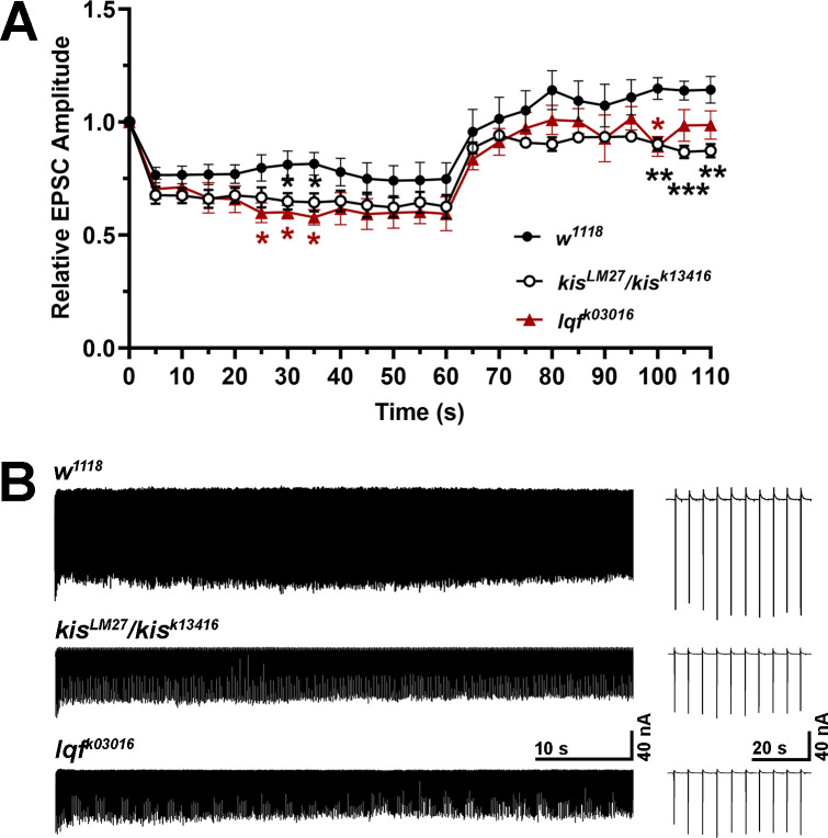
To determine whether Kis is functionally involved in CME and ADBE, we assessed endocytosis via electrophysiology and uptake of the lipophilic dye FM 1-43FX. We first used a 20 Hz high frequency stimulation (HFS) protocol in 1.0 mM Ca2+ for one minute to promote ADBE [45, 46]. This was followed by 0.2 Hz stimulation to assess recovery, which requires the readily releasable pool of vesicles to be replenished by mobilization of the reserve pool [47]. We also assessed endocytosis in liquid facets (lqf) mutants because of its roles in CME and ADBE. Lqf, which is the Drosophila homolog of Epsin 1 [48], remodels the membrane and recycles synaptic vesicles during CME [37] and is dephosphorylated along with several other proteins to trigger ADBE [49]. Both kis and lqf mutants showed decreased evoked endplate junctional current (eEJC) amplitudes during HFS compared to controls (Fig 2A and 2B). Recovery from HFS was impaired in kis mutants from 40–50 seconds after HFS and at 40 seconds after HFS in lqf mutants (100–110 seconds in Fig 2A). These data indicate that kis and lqf mutants show impaired ADBE and recycling of vesicles after HFS.

Fig 2. Kismet and Liquid Facets promote ADBE.

Fig 2

eEJCs were measured during 60 sec of 20 Hz HFS to induce ADBE and during a 50 sec recovery period with 0.2 Hz stimulation. (A) Mean eEJC amplitudes are shown relative to the amplitude measured after the first stimuli for w1118 controls (n = 10) and kis (n = 9) and lqf (n = 12) mutants. Error bars represent the SEM. (B) Representative eEJC recordings from the genotypes listed.

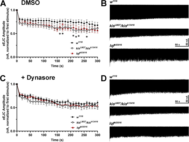
Next, we used a low frequency stimulation (LFS) protocol consisting of five minutes of 5 Hz stimulation in 1.0 mM Ca2+ to promote CME [50] in kis and lqf mutants. eEJC amplitudes in controls were approximately 80% of the initial stimulus amplitudes 10–50 seconds after the onset of stimulation. Amplitudes continued to decline until they were approximately 67% of the initial stimulus at the end of stimulation. Although lqfk03016 mutant eEJC amplitudes were reduced to approximately 76% of the initial stimulus at the beginning and 57% at the end of stimulation, there was only a significant reduction in eEJC amplitudes at 170 seconds after onset of stimulation (Fig 3A and 3B). kisLM27/kisk13416 mutants exhibited a similar reduction as lqfk03016 mutants in initial eEJC amplitudes, which were approximately 72% of the first stimulus. There was a significant reduction in eEJC amplitudes in kis mutants at several time points from 160–260 seconds after the onset of stimulation when eEJC amplitudes were approximately 60–62% of the initial stimulus (Fig 3A and 3B). Reduced eEJC amplitudes in kis mutants when CME was induced suggests that Kis promotes CME.

Fig 3. Inhibition of CME using Dynasore does not produce a change in evoked currents in kis or lqf mutants.

Fig 3

eEJCs were measured during five min of 10 Hz stimulation to induce CME. (A) Mean eEJC amplitudes are shown relative to the amplitude measured after the first stimuli for w1118 controls (n = 9) and kis (n = 10) and lqf (n = 10) mutants. Error bars represent the SEM. (B) Representative eEJC recordings from the genotypes listed. (C) eEJC amplitudes were obtained after animals were pretreated with 100 μM Dynasore. Mean eEJC amplitudes are shown relative to the amplitude measured after the first stimuli for w1118 controls (n = 9) and kis (n = 10) and lqf (n = 10) mutants. Error bars represent the SEM. (D) Representative eEJC recordings from the genotypes listed after pretreatment with 100 μM Dynasore.

If Kis regulates CME or ADBE, we would expect to observe no change in eEJC amplitudes during stimulation when CME or ADBE is inhibited. We used several putative inhibitors to target CME or ADBE in kis and lqf mutants. CME and ADBE require presynaptic increases in Ca2+ [51, 52]. Therefore, we used the Ca2+ chelators BAPTA, to inhibit both CME and ADBE, and EGTA, to inhibit ADBE [53]. CME is triggered by local increases in active zone Ca2+ initiated by the action potential [54] while ADBE is triggered when Ca2+ diffuses away from the active zone to the periactive zone [55]. BAPTA and EGTA are Ca2+ chelators that inhibit increases in Ca2+ at the periactive zone. Only BAPTA, however, inhibits the increases in Ca2+ at the active zone [56] because it has a faster on binding rate [57]. We also used Dynasore, an inhibitor of Dyn GTPase activity [58], to block CME. Dynasore does not affect Dyn’s affinity for GTP or its capacity to assemble on membranes [59]. After a 10 minute incubation, 100 μM Dynasore inhibited CME induced by LFS (S1 Fig) but did not affect ADBE (S2 Fig) in w1118 controls. The most pronounced effect on CME (S1 Fig) was observed from 160–250 sec. During this time, control eEJCs were 75.6–77.7% of the first response. In contrast, Dynasore treatment produced eEJCs that were 57.8–64.1% of the first response. Neither 100 μM BAPTA-AM nor 25 μM EGTA-AM affected CME or ADBE (S1 and S2 Figs) in controls after a 10 minute incubation. Therefore, we used Dynasore to determine if Kis functionally influences CME. Both kis and lqf mutants exhibited reduced eEJC amplitudes during LFS similar to that of controls after a 10 minute incubation with 100 μM Dynasore (Fig 3C and 3D). The similar reduction in LFS-induced eEJCs when CME was inhibited with Dynasore suggests that Kis and Lqf contribute to CME.

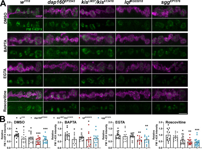
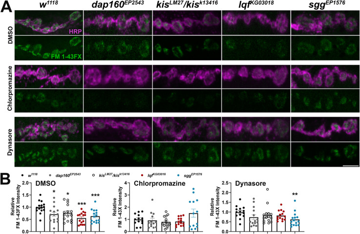
We extended our analyses of CME and ADBE to include additional inhibitors and endocytic mutants as negative controls. In addition to the lqfKG03016 mutant, which exhibits alterations in both CME and ADBE [37, 60], we used dap160EP2543 and sggEP1576 mutants, which have impaired CME [40] and ADBE [44], respectively. Endocytosis was examined using the lipophilic dye, FM 1–43FX, to label newly endocytosed synaptic vesicles [34] after one min stimulation with 90 mM KCl in 1.0 mM Ca2+. Stimulation using high K+ induces both CME and ADBE [61]. This stimulation paradigm resulted in decreased endocytosis, as indicated by a significant reduction in FM 1-43FX internalization, in all four mutants (Fig 4B, left). Next, larvae were pretreated with either 100 μM BAPTA-AM, 25 μM EGTA-AM, or 200 μM Roscovitine before one min stimulation with 90 mM KCl in 1.0 mM Ca2+. Each compound pretreatment led to a reduction in endocytosis in w1118 controls (S3 Fig). Roscovitine inhibits cyclin-dependent kinase 5, which is required for ADBE [62], by competing with ATP for the kinase domain [63]. Mutations in either dap160 or kis did not affect endocytosis when NMJs were pretreated with BAPTA or EGTA (Fig 4A, middle panels). Similar as lqfKG0301 and sggEP1576 mutants, however, kisLM27/kisk13416 mutants exhibited reduced endocytosis after pretreatment with the ADBE inhibitor, Roscovitine (Fig 4B). Given that kis mutants did not exhibit a further reduction in endocytosis when ADBE was inhibited by BAPTA or EGTA, Kis likely functionally influences ADBE.

Fig 4. Kismet mutants exhibit minimal changes in endocytosis when ADBE is inhibited.

Fig 4

Endocytosis was assessed by measuring internalization of the lipophilic dye FM 1-43FX after one min stimulation with 90 mM KCl. Animals were pretreated with either 100 μM BAPTA-AM, 25 μM EGTA-AM, or 200 μM Roscovitine to inhibit ADBE. (A) Panels show high resolution confocal micrographs of terminal presynaptic motor neuron boutons (HRP, magenta) after internalization of the lipophilic dye FM 1-43FX (green). (B) Data for each condition were normalized to w1118 controls. Bars indicate means, points represent individual larvae, and error bars represent SEM. Endocytic and kis mutants show reduced endocytosis. Endocytosis was unchanged in kis mutants pretreated with BAPTA or EGTA but reduced when pretreated with Roscovitine. Scale bar = 5 μm.

We used the same protocol to assess whether Kis also functionally contributes to CME. We again used 100 μM Dynasore to inhibit CME. We also employed Chlorpromazine, which inhibits assembly of clathrin coats [64] possibly by interfering with Dyn-lipid binding [65]. Pretreatment with each compound led to a reduction in endocytosis w1118 controls (S3 Fig). None of the mutants examined exhibited changes in endocytosis after pretreatment with 200 μM Chlorpromazine while only sggEP1576 mutants exhibited a further decrease in endocytosis after pretreatment with 100 μM Dynasore (Fig 5). These data, coupled with our electrophysiological analyses, suggest that Kis functionally contributes to both CME and ADBE.

Fig 5. Kismet mutants do not show changes in endocytosis when CME is inhibited.

Fig 5

Endocytosis was assessed by measuring internalization of the lipophilic dye FM 1-43FX after one min stimulation with 90 mM KCl. Larvae were pretreated with either 200 μM Chlorpromazine or 100 μM Dynasore to inhibit CME. (A) Panels show high resolution confocal micrographs of terminal presynaptic motor neuron boutons (HRP, magenta) after internalization of the lipophilic dye FM 1-43FX (green). (B) Data for each condition were normalized to w1118 controls. Bars indicate means, points represent individual larvae, and error bars represent SEM. Endocytosis was unchanged in kis mutants pretreated with Chlorpromazine or Dynasore. Scale bar = 5 μm.

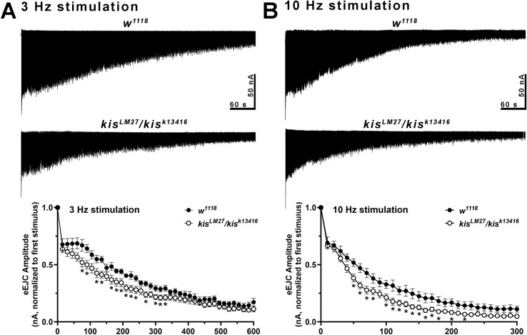
Reduced eEJC amplitudes during LFS and HFS and endocytosis could be a consequence of kis mutant synapses containing fewer synaptic vesicles. To test this possibility, we used the vesicular H+ pump inhibitor, Bafilomycin A1, to block glutamate uptake into vesicles [66]. Because Bafilomycin inhibits the refilling of glutamate into newly endocytosed vesicles, eEJC amplitudes diminish upon successive stimulation as less glutamate is released. eEJC amplitudes were assessed after 20 min incubation with 2 μM Bafilomycin in 1.0 mM Ca2+ during 3 or 10 Hz stimulation. 3 Hz stimulation promotes vesicle release from the readily releasable and recycling vesicle pools while 10 Hz stimulation mobilizes the reserve pool of vesicles [67]. kis mutants exhibited a more pronounced reduction in eEJC amplitudes relative to the first stimuli during both 3 and 10 Hz stimulation compared with w1118 controls (Fig 6). These data indicate kis mutant synapses possess fewer vesicles.

Fig 6. Kismet helps maintain vesicle pools.

Fig 6

Representative responses (top) in controls (n = 9) and kis mutants (n = 10) to 3 (A) or 10 (B) Hz stimulation in the presence of 2 μM Bafilomycin, which inhibits vesicle refilling. Muscles were clamped at -60 mV and recordings were obtained in HL-3 + 1.0 mM Ca2+. 3 Hz stimulation depletes the readily releasable and recycling pool of vesicles while 10 Hz stimulation deplete the reserve pool of vesicles. Vesicle pools are smaller in kis mutants as indicated by reduced eEJCs induced by stimulation. Bottom graphs show mean eEJC amplitudes normalized to the first stimulus for each condition. Error bars represent the SEM.

Kismet is important in postsynaptic muscles for endocytosis

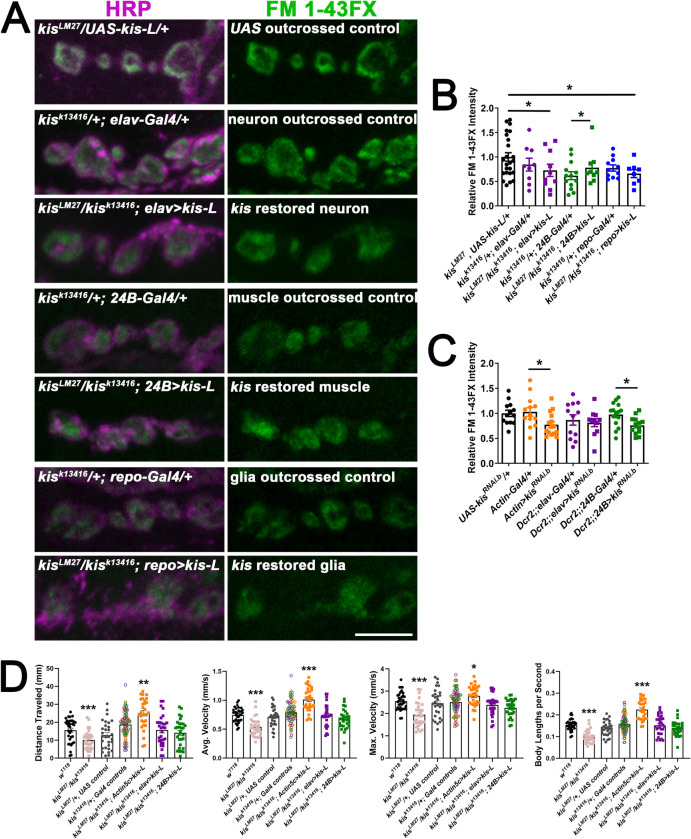
We performed rescue experiments to determine the tissue-specific requirements of Kis for endocytosis and locomotor behavior. We restored kis expression in kisLM27/kisk13416 mutants by expressing UAS-kis-L in all tissues using the Actin5c-Gal4 driver, neurons using the elav-Gal4 driver, postsynaptic muscle using the 24B-Gal4 driver, or glial cells using the repo-Gal4 driver. Endocytosis was induced by one min stimulation with 90 mM KCl in HL-3 containing 1.0 mM Ca2+ [34]. Expression of kis-L in all tissues restored endocytosis (S4A and S4B Fig). There were slight but significant reductions in endocytosis, as indicated by reduced FM 1-43FX internalization, when kis-L was expressed in neurons or glia of kis mutants compared with UAS but not driver-specific outcrossed controls (Fig 7A and 7B). When kis-L was expressed in muscles of kis mutants using the 24B-Gal4 driver, however, endocytosis was increased compared with the muscle-specific outcrossed control. There was no difference in endocytosis between kis mutants expressing kis-L in postsynaptic muscle and UAS-kis-L/+ outcrossed controls (Fig 7A and 7B). These data indicate that Kis specifically functions postsynaptically to promote endocytosis as postsynaptic kis-L expression in kisLM27/kisk13416 mutants partially rescues endocytic deficits.

Fig 7. Kismet is required in postsynaptic muscles for proper endocytosis of presynaptic vesicles.

Fig 7

Tissue-specific drivers were used to restore kis expression in specific tissues of kis mutants including presynaptic motor neurons (using the elav-Gal4 driver), postsynaptic muscle (using the 24B-Gal4 driver), or glial cells (using the repo-Gal4 driver). A) High resolution confocal micrographs of terminal presynaptic motor neuron boutons (HRP, magenta) after internalization of the lipophilic dye FM 1-43FX (green) after one min stimulation with 90 mM KCl in genotypes as listed. Scale bar = 5 μm. B) Quantification of FM 1-43FX fluorescence intensity indicates that when kis is restored in postsynaptic muscles of kis mutants, presynaptic endocytosis returns to control levels. C) Quantification of FM 1-43FX fluorescence intensity after tissue-specific knockdown of Kis. Knockdown of Kis in all tissues using the Actin5c-Gal4 driver or in postsynaptic muscles using the 24B-Gal4 driver resulted in impaired endocytosis. D) Histograms show quantification as determined by wrMTrck of larval crawling behavior on an agar arena for 30 s. Distance traveled, maximum velocity, average velocity, and crawling velocity normalized to body size (body lengths per second). Restoring kis expression in all tissues led to enhanced larval crawling.

To further probe the tissue-specific contributions of Kis, we knocked down Kis in all tissues, neurons, or postsynaptic muscle to determine if we could mimic the reduction in endocytosis in kis mutants. Expression of UAS-kisRNAi.b was previously shown to reduce Kis levels by 90% in third instar wing discs [29] and reduces kis transcripts by 56.2% in all tissues (S4C Fig). When Kis was knocked down in all tissues using the Actin5c-Gal4 driver, the intensity of the FM signal was decreased to approximately 76.7% of control levels. Similarly, knock down of Kis in postsynaptic muscles using the 24B-Gal4 driver, resulted in a 76.8% decrease in endocytosis (Fig 7C). These values are similar to those of kisLM27/kisk13416 mutants, which exhibit a 72.6% decrease in endocytosis. There was no change in endocytosis when Kis was knocked down in neurons (Fig 7C). These data support the hypothesis that postsynaptic Kis regulates presynaptic endocytosis.

Kis mutants show impaired locomotor behaviors with reductions in the velocity of movement and total distance traveled (Fig 7D). Expression of UAS-kis-L in all tissues of kisLM27/kisk13416 mutants using the Actin5c-Gal4 driver resulted in increases in total distance traveled and both the average and maximum velocity of larval movement (Fig 7D). In contrast, expression of UAS-kis-L in neurons or muscles of kisLM27/kisk13416 mutants did not affect larval movement compared with controls (Fig 7D). These data indicate that Kis functions in all tissues to promote proper larval locomotion.

Expression of human CHD7 in kis mutants restores locomotion but not endocytosis

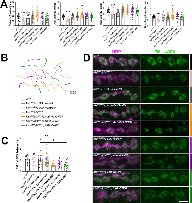
Kis is 63% identical to human CHD7 with notable conservation within all major functional domains [26]. CHD7 regulates the transcription of gene products that promote migration of neural crest cells [68] but is also expressed in human adult cortical neurons [22]. Because CHD7 influences the expression of genes required for cell adhesion, neurotransmission, and synaptic plasticity [69], we sought to determine whether expression of human CHD7 in kis mutants would restore endocytosis and locomotion. We expressed a Drosophila optimized human CHD7 in all tissues, neurons, or postsynaptic muscles in kisLM27/kisk13416 mutants after verifying that CHD7-V5 was expressed in the nuclei of these tissues (S5A Fig). Although there was some extranuclear/cytoplasmic V5 signal, the most intense signal was localized to the nucleus. Transgenic expression of CHD7 was accomplished in all tissues using the Actin5c-Gal4 driver, in neurons using the elav-Gal4 driver, or in postsynaptic muscle using the 24B-Gal4 driver in kisLM27/kisk13416 mutants. Expression of CHD7 in all three tissue types restored the maximum velocity of larval movement and distance traveled (Fig 8A and 8B). CHD7 expression in all tissues produced a significant increase in average velocity of larval movement while CHD7 expression in neurons or postsynaptic muscle did not restore this aspect of behavior (Fig 8A and 8B). Conversely, expression of CHD7 failed to restore endocytosis as indicated by internalization of FM 1-43FX (Fig 8C and 8D). Expression of CHD7 in all tissues or postsynaptic muscle instead resulted in reduced endocytosis compared with the UAS control but not the Gal4 control (Fig 8C and 8D). These data indicate that CHD7 restores locomotion, but not endocytosis, in kisLM27/kisk13416 mutants.

Fig 8. Expression of human CHD7 in kis mutants restores behavior but not endocytosis.

Fig 8

Tissue-specific drivers were used to express human CHD7 in all tissues (using the Actin5c-Gal4 driver), presynaptic motor neurons (using the elav-Gal4 driver), or postsynaptic muscle (using the 24B-Gal4 driver) of kis mutants. A) Histograms show quantification as determined by wrMTrck of larval crawling behavior on an agar arena for 30 s. Distance traveled, maximum velocity, average velocity, and crawling velocity normalized to body size (body lengths per second). B) Representative traces of larval crawling behavior for the genotypes listed. C) Quantification of FM 1-43FX fluorescence intensity indicates that expression of human CHD7 does not restore endocytosis in kis mutants. D) High resolution confocal micrographs of terminal presynaptic motor neuron boutons (HRP, magenta) after internalization of the lipophilic dye FM 1-43FX (green) after 1 min stimulation with 90 mM KCl in genotypes as listed. Scale bar = 5 μm.

Expression of an ATPase deficient Kis in kis mutants rescues endocytosis and behavior

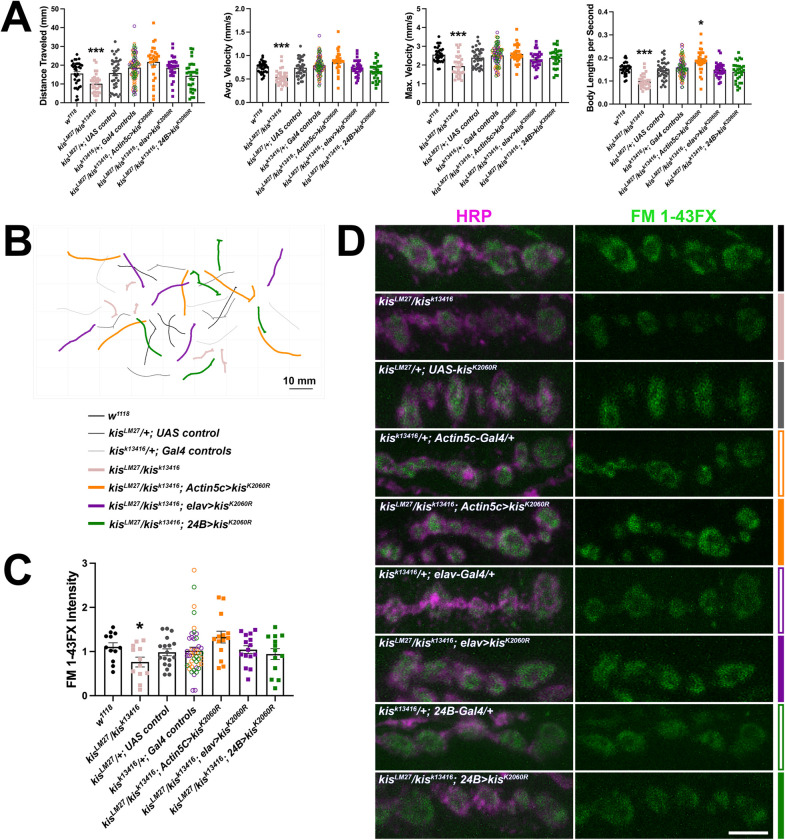
If the chromatin remodeling activity of Kis is important for endocytosis, we would expect that expression of an ATPase deficient Kis in kis mutants would fail to rescue either endocytosis and/or behavior. We mutated the Lys residue within the conserved ATP binding site [70] to an Arg (KisK2060R). Mutation of this Lys residue results in the inability of ATPases, including the mammalian Kis homolog, CHD8 [71], to hydrolyze ATP and remodel chromatin [72, 73]. We expressed UAS-kisK2060R in all tissues, neurons, or postsynaptic muscles in kisLM27/kisk13416 mutants. We verified KisK2060R-V5 was expressed in the nuclei of each tissue (S5B Fig). Similar as CHD7-V5, we noted that there was some extranuclear/cytoplasmic signal, which was most apparent in postsynaptic tissue where KisK2060R-V5 exhibited a punctate nuclear and extranuclear distribution. Notably, this signal was not present in the muscle nuclei of larvae expressing UAS-kisK2060R in neurons. Expression of KisK2060R in all tissues, neurons, or postsynaptic muscles in kisLM27/kisk13416 mutants restored both endocytosis (Fig 9C and 9D) and locomotor behavior (Fig 9A and 9B) as there were no significant differences in locomotion or endocytosis between experimental animals and outcrossed controls. These findings suggest that Kis may have functional implications at the synapse that are, at least partly, independent of its chromatin remodeling activity.

Fig 9. Expression of an ATPase deficient Kis, KisK2060R, in kis mutants restores behavior and endocytosis.

Fig 9

Tissue-specific drivers were used to express KisK2060R in all tissues (using the Actin5c-Gal4 driver), presynaptic motor neurons (using the elav-Gal4 driver), or postsynaptic muscle (using the 24B-Gal4 driver) of kis mutants. A) Histograms show quantification as determined by wrMTrck of larval crawling behavior on an agar arena for 30 s. Distance traveled, maximum velocity, average velocity, and crawling velocity normalized to body size (body lengths per second). B) Representative traces of larval crawling behavior for the genotypes listed. C) Quantification of FM 1-43FX fluorescence intensity indicates that expression of KisK2060R restores endocytosis in kis mutants. D) High resolution confocal micrographs of terminal presynaptic motor neuron boutons (HRP, magenta) after internalization of the lipophilic dye FM 1-43FX (green) after one min stimulation with 90 mM KCl in genotypes as listed. Scale bar = 5 μm.

Discussion

Our data indicate that Kis promotes both CME and ADBE possibly by regulating the expression of gene products that mediate these processes (Fig 1) and maintaining synaptic vesicle pools (Fig 6). Kis is important in postsynaptic muscles for endocytosis as postsynaptic Kis-L expression in kis mutants restores endocytosis (Fig 7). Surprisingly, the ATPase activity of Kis is dispensable for endocytosis and locomotion (Fig 9).

Kis promotes both CME and ADBE

Several forms of endocytosis exist in neurons to collectively internalize nutrients, detect growth and guidance cues, and maintain synaptic membrane homeostasis [13]. While several endocytic mechanisms are thought to replenish synaptic vesicles [11], there is little evidence for kiss-and-run at the Drosophila NMJ [74]. Compensatory endocytosis and ultrafast endocytosis are thought to occur at the NMJ but there are few direct demonstrations of these mechanisms [12]. CME is thought to be the most prevalent form of endocytosis at the synapse [11] but is limited by the duration required to select cargo and activate CME proteins [75]. Other modes of endocytosis, therefore, compensate for these inherent limitations in CME to enable synaptic homeostasis. ADBE, which quickly internalizes large areas of membrane, is utilized during periods of intense neuronal activity [14]. Kis functionally contributes to both forms of endocytosis as evidenced by the reduction in kis mutant eEJC amplitudes during stimulation protocols that induce ADBE (Fig 2) or CME (Fig 3). Further, when ADBE or CME are pharmacologically inhibited, kis mutants do not show further reductions in endocytosis (Figs 4 and 5).

Postsynaptic Kis-L expression in kis mutants rescues endocytosis (Fig 7A and 7B). Similarly, knock down of Kis in postsynaptic muscle and all tissues mimics the kisLM27/kisk13416 mutant phenotype (Fig 7C). These data indicate that postsynaptic Kis regulates presynaptic endocytosis. Although this may seem unexpected, retrograde signaling via cell adhesion molecules (CAMs) and diffusible molecules released postsynaptically regulate many presynaptic processes including the synaptic vesicle cycle [76]. Presynaptic endocytosis is enhanced by production of nitric oxide downstream of N-methyl D-aspartate (NMDA) receptor signaling, which increases production of the membrane lipid phosphatidylinositol 4,5-biphosphate (PIP2) [77]. PIP2 recruits adaptor protein 2 (AP2) to the plasma membrane [78] to enable endocytosis by selecting cargo and binding to clathrin [79]. Transsynaptic crosstalk through CAMs also modulates endocytosis as null mutations in Neuroligin 1 (Nlg1) enhance endocytosis without affecting exocytosis or the size of vesicle pools [80].

Kis restricts the synaptic localization of the CAM FasII [30], the Drosophila ortholog of neural cell adhesion molecule (NCAM). Similarly, loss of function of Chd7 [81] or Chd8 [82] results in differential expression of several CAMs including ncams and nlgs as determined by ChIP-Seq. Kis binds near the regulatory regions of fasII, fasIII, nlg2, and nlg4 in Drosophila intestinal stem cells [83]. NCAM negatively regulates ADBE [84], possibly by activating protein kinase B/Akt [85, 86], and binds directly to AP2 [87]. Therefore, mutations in kis may lead to deficient endocytosis due to the accumulation of CAMs, which might physically restrict invagination of the plasma membrane.

Alternatively, Kis may regulate CME and ADBE by regulating proteins that organize microdomains of the synapse like Rabs. Rab GTPases facilitate directional membrane trafficking between compartments of the endomembrane system [88]. Specifically, Rab11 traffics cargo between recycling endosomes and the plasma membrane [89] to influence the concentration of lipids and membrane-associated proteins [90]. Kis promotes the transcription of rab11 and Rab11 localization to the synapse [31]. Rab11, when constitutively active, increases the number of rat cerebellar granule cell neuron terminals executing ADBE. Nlg2, Nlg3, and NCAM were present in bulk endosomes [91] suggesting that Rab11 controls the localization of CAMs by regulating ADBE. Thus, Kis may functionally influence endocytosis by a combination of mechanisms including, but not limited to, regulating the expression of endocytic gene transcripts, expression and/or localization of endocytic proteins, CAMs, and Rab11.

Kis is 63% identical to human CHD7 [26]. Despite this conservation, expression of human CHD7 in all tissues or postsynaptic muscles of kis mutants did not restore endocytosis (Fig 8C and 8D) as was observed with UAS-kis-L expression in the same tissue types (Fig 7A and 7B). CHD family chromatin remodeling enzymes assemble with DNA binding proteins [92], histone methytransferases [93], and other proteins to form large, multisubunit complexes. Although the organization and identities of the subunits and the mechanism by which chromatin remodeling enzyme complexes recognize nucleosomes is poorly understood [94], it is recognized that the complexes are developmental- and tissue-specific [95, 96]. Therefore, it is possible that overexpression of CHD7 might produce a dominant negative effect in kis mutants where CHD7 sequesters other transcriptional regulators rendering them inactive. This possibility, however, seems unlikely because CHD7 expression in all tissues, neurons, or postsynaptic muscle of kis mutants restored locomotor behaviors (Fig 8A and 8B). It is more plausible that this discrepancy arises because different tissue types are required for locomotion and endocytosis. Locomotor behaviors require broader, multi-circuit coordination compared with endocytosis. Larval locomotion is produced by central nervous system central pattern generators, which are subject to upstream cholinergic control, and peripheral motor neurons. This circuit may be initiated independent of, but is modulated by, sensory feedback [97]. Presynaptic endocytosis is influenced by interactions between pre- and postsynaptic cells and associated glia [98]. CHD7 binds to unique regulatory loci in different tissues including human neural crest cells, induced pluripotent stem cell-derived neuroepithelial cells, and induced pluripotent stem cells. Further, CHD7 preferentially associates with the transcription factors TFAP2A and NR2F1/F2 in neural crest cells but not neuroepithelial cells [99]. Thus, more CHD7 sensitive gene products may be required for locomotion compared with endocytosis.

Kis may transcriptionally regulate endocytosis

CHD7, CHD8, and Kis influence the transcription of thousands of gene products [81, 83, 100, 101]. Therefore, it is not surprising that Kis affects the transcript levels of several endocytic genes including AP2α, endoB [31], PI3K92E, and sgg/GSK3β (Fig 1). In Drosophila adult intestinal stem cells, Kis was bound near regulatory sequences for several endocytic genes including amphiphysin, AP2α, clathrin light and clathrin heavy chain, dyn, draper 1, flower, lqf, and synaptojanin [83]. We did not observe altered transcript levels of clathrin heavy chain (Fig 1), amphiphysin, flower, lqf, or synaptojanin [31] via RT-qPCR. Similarly, we did not detect Kis binding to the promoter or within 200 bp of the transcription start site of dyn even though Kis is enriched at the promoters and transcription start sites for dap160 and endoB [31]. These discrepancies likely reflect developmental- and/or tissue-specific differences in chromatin remodeling complexes as a result of differential expression of specific transcription factors [96, 102].

Kis, CHD7, and CHD8 contain several domains including two chromodomains, which bind to methylated N-terminal histone residues [103, 104], and an ATPase domain [105]. The latter repositions histones relative to DNA to expose transcriptional regulatory sequences [106]. We found that expression of an ATPase-dead Kis, KisK2060R, in kis mutants restored locomotor behaviors and endocytosis (Fig 9). Similarly, expression of KisK2060R partially rescued the size of stem cell clones in the kis10D26 loss of function mutant [83]. Taken together, these data suggest that Kis may function independent of its ATPase domain to form functional regulatory transcriptional complexes. Chromatin remodeling proteins are integral components of large, multisubunit complexes, the composition of which differ in a cell- and developmental-dependent manner [95, 96]. In Drosophila intestinal stem cells, Kis colocalizes with the histone methyltransferase, Trithorax related (Trr), to regulate the transcription of target genes [83]. Kis recruits the histone methyltransferases, Trithorax and Absent, Small or Homeotic Discs 1 (ASH1) to Drosophila salivary polytene chromosomes [93]. Similarly, CHD8 recruits ASH2L, which is part of a lysine methyltransferase complex, to regulate gene expression required for differentiation of mouse oligodendrocyte precursor cells. Notably, differentiation was partially restored by inhibition of KDM5, a demethyltransferase, in conditional Chd8 knock outs [107] indicating that CHD8’s role in oligodendrocyte precursor cell differentiation occurs partly because of its ability to recruit histone methyltransferases. KDM5 interacts with Kis and Trr in Drosophila adult heads [108] indicating these complexes may be similar across species.

Collectively, these data are consistent with clinical findings, which show that mutations throughout CHD proteins perturb their function. Approximately 30% of CHARGE-associated Chd7 mutations lie within functional domains including the ATPase domain [28] and the percentage of Chd8 point mutations within the ATPase domain associated with autism spectrum disorders is 8% [109]. Thus, residues throughout CHD7 and CHD8 contribute to their transcriptional function. Indeed, Chd7 mutations in CHARGE patients produce different phenotypes depending on the location with mutations in the chromodomains resulting in the most severe phenotypes [28]. Similarly, while mutations in the chromodomains of zebrafish chd7 produce the morphological characteristics of CHARGE Syndrome including craniofacial defects, mutations in the ATPase domain do not [110].

Alternatively, the capacity of KisK2060R to restore locomotor behaviors and endocytosis in kis mutants may occur due to a novel cytoplasmic role for Kis. Other components of chromatin remodeling complexes including the MYST family of acetyltransferases [111] and members of the histone deacetyltransferase (HDAC) family [112] are localized in both the nucleus and cytoplasm where they recognize cytoplasmic substrates. CHD1 [113] and CHD9 [114] were detected in the cytoplasm of mitotic cells. We have not detected Kis in the cytoplasm of ventral nerve cord neurons or postsynaptic muscle cells [30]. Given that 1) Kis binds to regulatory regions of the endocytic genes dap160 and endoB [31], 2) chromatin remodeling enzymes are integral components of transcriptional complexes that include additional proteins that influence transcription, and 3) mutations outside the ATPase domain of CHD proteins exert the most severe phenotypes, we favor the hypothesis that Kis may influence transcription independent of its ATPase domain.

Implications for neurodevelopmental disorders and neurodegenerative diseases

The synaptic vesicle cycle, which includes processes that enable release and recycling of vesicles, is disrupted in neurodevelopmental disorders [115] and neurodegenerative diseases [76]. Genes that regulate the synaptic vesicle cycle are differentially expressed in human Alzheimer’s disease patient neurons [116118]. It is unclear, however, whether these genetic changes precede the formation of amyloid β (Aβ) plaques, which are produced by proteolytic cleavage of amyloid precursor protein (APP) [25]. Cellular exposure to Aβ40 and Aβ42 aggregates impairs endocytosis in PC12 and neuroblastoma cells and Aβ40 slows the intracellular trafficking of recycling endosomes [116]. Impaired trafficking of endosomes containing amyloid precursor protein (APP) exacerbates production of Aβ40 and Aβ42 as the amyloidogenic pathway of APP processing is favored [119].

Although it is recognized that chromatin remodeling is a contributing factor to neurodevelopmental disorders including autism spectrum disorders [120], how aberrant chromatin remodeling functionally influences the synaptic vesicle cycle is relatively unexplored. We find that Kis maintains synaptic vesicle pools as kis mutants exhibit reductions in the sizes of the readily releasable, recycling, and reserve pools of vesicles (Fig 6). CME and ADBE both replenish synaptic vesicle pools [12, 13] but the extent to which each contributes is not well understood. Because the synaptic vesicle cycle is intimately linked to the endomembrane system [121], it may be difficult to distinguish discrete pools of vesicles. The readily releasable pool of vesicles was regenerated from CME while the reserve pool was bolstered by ADBE in cultured rat cerebellar neurons [122]. CME replenished 63% of the vesicle pool in cultured hippocampal neurons at 9 DIV but only 39% at 19 DIV indicating ADBE is preferentially used in mature neurons to replenish synaptic vesicles [123].

The reduced size of synaptic vesicle pools in kis mutants may lead to the decrease in evoked potentials [30], failed homeostatic presynaptic plasticity [124], and impaired motor behavior (Figs 7D, 8A and 8B, 9A and 9B) observed in kis mutants. It is not clear whether deficient endocytosis in kis mutants leads to the decrease in the synaptic vesicle pool or whether kis mutants produce fewer synaptic vesicles de novo. Our data begins to uncover correlates between aberrant chromatin remodeling and synaptic processes. Given the altered state of chromatin in neurodevelopmental disorders and neurodegenerative diseases [125], a better understanding of the synaptic correlates affecting cognitive and behavioral processes is needed.

Supporting information

S1 Fig. CME is inhibited by Dynasore but not BAPTA or EGTA.

eEJCs were recorded during 5 Hz stimulation for five minutes in HL-3 + 1.0 mM Ca2+. 100 μM BAPTA (n = 10), 25 μM EGTA (n = 10), 100 μM Dynasore (n = 9), or an equal volume of DMSO (controls, n = 11) were applied for 10 minutes prior to neuronal stimulation. Each eEJC is normalized to the first stimulus for each condition. Points represent mean relative eEJC amplitudes. Error bars represent the SEM.

(TIF)

pone.0300255.s001.tif (336.2KB, tif)
S2 Fig. ADBE is not inhibited by BAPTA, EGTA, or Dynasore.

eEJCs were measured in HL-3 + 1.0 mM Ca2+ for 60 sec of 20 Hz HFS to induce ADBE and during a 50 sec recovery period with 0.2 Hz stimulation. 100 μM BAPTA (n = 10), 25 μM EGTA (n = 9), 100 μM Dynasore (n = 9), or an equal volume of DMSO (controls, n = 10) were applied for 10 minutes prior to neuronal stimulation. Each eEJC is normalized to the first stimulus for each condition. Points represent mean relative eEJC amplitudes. Error bars represent the SEM.

(TIF)

pone.0300255.s002.tif (294.4KB, tif)
S3 Fig. Genotype by genotype comparison of ADBE and CME compounds.

Endocytosis was assessed by measuring internalization of the lipophilic dye FM 1-43FX after one min stimulation with 90 mM KCl. Panels show high resolution confocal micrographs of terminal presynaptic motor neuron boutons (HRP, magenta) after internalization of the lipophilic dye FM 1-43FX (green). Data for each genotype were normalized to the DMSO control condition.

(TIF)

pone.0300255.s003.tif (389.9KB, tif)
S4 Fig. Restoration of Kis in all tissues rescues endocytosis.

The Actin5c-Gal4 driver was used to express UAS-kis-L in all tissues of kis mutants. A) High resolution confocal micrographs of terminal presynaptic motor neuron boutons (HRP, magenta) after internalization of the lipophilic dye FM 1-43FX (green) after one min stimulation with 90 mM KCl in genotypes as listed. Scale bar = 5 μm. B) Quantification of FM 1-43FX fluorescence. C) Relative expression of CNS transcripts was assessed via RT-qPCR. 2-ΔΔC(t) values are indicated. Data includes four biological replicates each including three technical replicates. Technical replicates are represented by the points for the representative genotypes. Bars indicate the SEM.

(TIF)

pone.0300255.s004.tif (3.4MB, tif)
S5 Fig. CHD7-V5 and KisK2060R-V5 are localized to the nuclei of tissues.

Confocal micrographs showing V5 (green) and HRP (magenta, neuron) immunolabeling and DAPI labeling. Left and middle large panels show representative ventral nerve cords with single nuclei depicted in the small panels of kis mutants expressing human CHD7 (A) or the ATPase deficient KisK2060R (B). Scale bar = 5 μm. Right panels show representative muscles with single nuclei depicted in the small panels of kis mutants expressing human CHD7 (A) or the ATPase deficient KisK2060R (B). Scale bar = 5 μm.

(TIF)

pone.0300255.s005.tif (2.3MB, tif)
S1 Table. Statistical comparisons and corresponding p-values for figures.

(XLSX)

pone.0300255.s006.xlsx (15.5KB, xlsx)

Acknowledgments

We thank the Bloomington Drosophila Stock Center for fly stocks (NIH P40OD018537), Carly Gridley for help with the FM 1-43FX rescue experiments, Dave Featherstone for his mentoring, and Southern Illinois University Edwardsville for travel support.

Data Availability

All relevant data are within the manuscript and its Supporting Information files.

Funding Statement

This work was funded by the National Institute of Neurological Disorders and Stroke of the NIH under award numbers 1R15NS101608-01A1 and 2R15NS101608-02A1 (to FL) and by Southern Illinois University Edwardsville’s Competitive Graduate Award (to EH). The funders had no role in study design, data collection and analysis, decision to publish, or preparation of the manuscript.

References

  • 1.Katz B, Miledi R. The timing of calcium action during neuromuscular transmission. The Journal of Physiology. 1967;189(3):535–44. doi: 10.1113/jphysiol.1967.sp008183 [DOI] [PMC free article] [PubMed] [Google Scholar]
  • 2.Südhof Thomas C. Neurotransmitter Release: The Last Millisecond in the Life of a Synaptic Vesicle. Neuron. 2013;80(3):675–90. doi: 10.1016/j.neuron.2013.10.022 [DOI] [PMC free article] [PubMed] [Google Scholar]
  • 3.Van Vactor D, Sigrist SJ. Presynaptic morphogenesis, active zone organization and structural plasticity in Drosophila. Current Opinion in Neurobiology. 2017;43:119–29. Epub 20170405. doi: 10.1016/j.conb.2017.03.003 ; PubMed Central PMCID: PMC5501089. [DOI] [PMC free article] [PubMed] [Google Scholar]
  • 4.Katz B, Miledi R. The role of calcium in neuromuscular facilitation. The Journal of Physiology. 1968;195(2):481–92. doi: 10.1113/jphysiol.1968.sp008469 [DOI] [PMC free article] [PubMed] [Google Scholar]
  • 5.Doussau F, Schmidt H, Dorgans K, Valera AM, Poulain B, Isope P. Frequency-dependent mobilization of heterogeneous pools of synaptic vesicles shapes presynaptic plasticity. eLife. 2017;6:e28935. doi: 10.7554/eLife.28935 [DOI] [PMC free article] [PubMed] [Google Scholar]
  • 6.Rey S, Marra V, Smith C, Staras K. Nanoscale Remodeling of Functional Synaptic Vesicle Pools in Hebbian Plasticity. Cell Reports. 2020;30(6):2006–17.e3. doi: 10.1016/j.celrep.2020.01.051 [DOI] [PMC free article] [PubMed] [Google Scholar]
  • 7.Rosenmund C, Stevens CF. Definition of the Readily Releasable Pool of Vesicles at Hippocampal Synapses. Neuron. 1996;16(6):1197–207. doi: 10.1016/s0896-6273(00)80146-4 [DOI] [PubMed] [Google Scholar]
  • 8.Pieribone VA, Shupliakov O, Brodin L, Hilfiker-Rothenfluh S, Czernik AJ, Greengard P. Distinct pools of synaptic vesicles in neurotransmitter release. Nature. 1995;375(6531):493–7. doi: 10.1038/375493a0 . [DOI] [PubMed] [Google Scholar]
  • 9.Zhang M, Augustine GJ. Synapsins and the Synaptic Vesicle Reserve Pool: Floats or Anchors? Cells. 2021;10(3). Epub 20210316. doi: 10.3390/cells10030658 ; PubMed Central PMCID: PMC8002314. [DOI] [PMC free article] [PubMed] [Google Scholar]
  • 10.Hoopmann P, Punge A, Barysch SV, Westphal V, Bückers J, Opazo F, et al. Endosomal sorting of readily releasable synaptic vesicles. Proceedings of the National Academy of Sciences. 2010;107(44):19055–60. doi: 10.1073/pnas.1007037107 [DOI] [PMC free article] [PubMed] [Google Scholar]
  • 11.Chanaday NL, Cousin MA, Milosevic I, Watanabe S, Morgan JR. The Synaptic Vesicle Cycle Revisited: New Insights into the Modes and Mechanisms. J Neurosci. 2019;39(42):8209–16. doi: 10.1523/JNEUROSCI.1158-19.2019 ; PubMed Central PMCID: PMC6794917. [DOI] [PMC free article] [PubMed] [Google Scholar]
  • 12.Gan Q, Watanabe S. Synaptic Vesicle Endocytosis in Different Model Systems. Frontiers in Cellular Neuroscience. 2018;12:171. Epub 20180628. doi: 10.3389/fncel.2018.00171 ; PubMed Central PMCID: PMC6031744. [DOI] [PMC free article] [PubMed] [Google Scholar]
  • 13.Camblor-Perujo S, Kononenko NL. Brain-specific functions of the endocytic machinery. The FEBS Journal. 2022;289(8):2219–46. doi: 10.1111/febs.15897 [DOI] [PubMed] [Google Scholar]
  • 14.Clayton EL, Cousin MA. The molecular physiology of activity-dependent bulk endocytosis of synaptic vesicles. Journal of Neurochemistry. 2009;111(4):901–14. Epub 20090916. doi: 10.1111/j.1471-4159.2009.06384.x ; PubMed Central PMCID: PMC2871311. [DOI] [PMC free article] [PubMed] [Google Scholar]
  • 15.Bonnycastle K, Davenport EC, Cousin MA. Presynaptic dysfunction in neurodevelopmental disorders: Insights from the synaptic vesicle life cycle. Journal of Neurochemistry. 2021;157(2):179–207. doi: 10.1111/jnc.15035 [DOI] [PubMed] [Google Scholar]
  • 16.Schreij AM, Fon EA, McPherson PS. Endocytic membrane trafficking and neurodegenerative disease. Cell Mol Life Sci. 2016;73(8):1529–45. Epub 20151231. doi: 10.1007/s00018-015-2105-x . [DOI] [PMC free article] [PubMed] [Google Scholar]
  • 17.Sweatt JD. The emerging field of neuroepigenetics. Neuron. 2013;80(3):624–32. doi: 10.1016/j.neuron.2013.10.023 ; PubMed Central PMCID: PMC3878295. [DOI] [PMC free article] [PubMed] [Google Scholar]
  • 18.Kim S, Kaang BK. Epigenetic regulation and chromatin remodeling in learning and memory. Exp Mol Med. 2017;49(1):e281. Epub 20170113. doi: 10.1038/emm.2016.140 ; PubMed Central PMCID: PMC5291841. [DOI] [PMC free article] [PubMed] [Google Scholar]
  • 19.Wilson KD, Porter EG, Garcia BA. Reprogramming of the epigenome in neurodevelopmental disorders. Crit Rev Biochem Mol Biol. 2022;57(1):73–112. Epub 20211002. doi: 10.1080/10409238.2021.1979457 . [DOI] [PMC free article] [PubMed] [Google Scholar]
  • 20.Berson A, Nativio R, Berger SL, Bonini NM. Epigenetic Regulation in Neurodegenerative Diseases. Trends Neurosci. 2018;41(9):587–98. Epub 20180607. doi: 10.1016/j.tins.2018.05.005 ; PubMed Central PMCID: PMC6174532. [DOI] [PMC free article] [PubMed] [Google Scholar]
  • 21.Manning BJ, Yusufzai T. The ATP-dependent chromatin remodeling enzymes CHD6, CHD7, and CHD8 exhibit distinct nucleosome binding and remodeling activities. Journal of Biological Chemistry. 2017;292(28):11927–36. Epub 20170521. doi: 10.1074/jbc.M117.779470 ; PubMed Central PMCID: PMC5512084. [DOI] [PMC free article] [PubMed] [Google Scholar]
  • 22.Weng MK, Zimmer B, Poltl D, Broeg MP, Ivanova V, Gaspar JA, et al. Extensive transcriptional regulation of chromatin modifiers during human neurodevelopment. PLoS One. 2012;7(5):e36708. Epub 20120509. doi: 10.1371/journal.pone.0036708 ; PubMed Central PMCID: PMC3348879. [DOI] [PMC free article] [PubMed] [Google Scholar]
  • 23.Parikshak NN, Gandal MJ, Geschwind DH. Systems biology and gene networks in neurodevelopmental and neurodegenerative disorders. Nat Rev Genet. 2015;16(8):441–58. Epub 20150707. doi: 10.1038/nrg3934 ; PubMed Central PMCID: PMC4699316. [DOI] [PMC free article] [PubMed] [Google Scholar]
  • 24.Restrepo LJ, DePew AT, Moese ER, Tymanskyj SR, Parisi MJ, Aimino MA, et al. gamma-secretase promotes Drosophila postsynaptic development through the cleavage of a Wnt receptor. Dev Cell. 2022;57(13):1643–60 e7. Epub 20220601. doi: 10.1016/j.devcel.2022.05.006 ; PubMed Central PMCID: PMC9283269. [DOI] [PMC free article] [PubMed] [Google Scholar]
  • 25.Scheuner D, Eckman C, Jensen M, Song X, Citron M, Suzuki N, et al. Secreted amyloid beta-protein similar to that in the senile plaques of Alzheimer’s disease is increased in vivo by the presenilin 1 and 2 and APP mutations linked to familial Alzheimer’s disease. Nat Med. 1996;2(8):864–70. doi: 10.1038/nm0896-864 . [DOI] [PubMed] [Google Scholar]
  • 26.Latcheva NK, Ghosh R, Marenda DR. The epigenetics of CHARGE syndrome. Frontiers in Biology. 2016;11(2):85–95. doi: 10.1007/s11515-016-1398-y [DOI] [Google Scholar]
  • 27.Saladi SV, de la Serna IL. ATP dependent chromatin remodeling enzymes in embryonic stem cells. Stem Cell Reviews and Reports. 2010;6(1):62–73. doi: 10.1007/s12015-010-9120-y ; PubMed Central PMCID: PMC2862992. [DOI] [PMC free article] [PubMed] [Google Scholar]
  • 28.Basson MA, van Ravenswaaij-Arts C. Functional Insights into Chromatin Remodelling from Studies on CHARGE Syndrome. Trends in Genetics. 2015;31(10):600–11. Epub 20150924. doi: 10.1016/j.tig.2015.05.009 ; PubMed Central PMCID: PMC4604214. [DOI] [PMC free article] [PubMed] [Google Scholar]
  • 29.Melicharek DJ, Ramirez LC, Singh S, Thompson R, Marenda DR. Kismet/CHD7 regulates axon morphology, memory and locomotion in a Drosophila model of CHARGE syndrome. Human Molecular Genetics. 2010;19(21):4253–64. Epub 20100817. doi: 10.1093/hmg/ddq348 ; PubMed Central PMCID: PMC2951870. [DOI] [PMC free article] [PubMed] [Google Scholar]
  • 30.Ghosh R, Vegesna S, Safi R, Bao H, Zhang B, Marenda DR, et al. Kismet positively regulates glutamate receptor localization and synaptic transmission at the Drosophila neuromuscular junction. PLoS One. 2014;9(11):e113494. Epub 20141120. doi: 10.1371/journal.pone.0113494 ; PubMed Central PMCID: PMC4239079. [DOI] [PMC free article] [PubMed] [Google Scholar]
  • 31.Latcheva NK, Delaney TL, Viveiros JM, Smith RA, Bernard KM, Harsin B, et al. The CHD Protein, Kismet, is Important for the Recycling of Synaptic Vesicles during Endocytosis. Scientific Reports. 2019;9(1):19368. doi: 10.1038/s41598-019-55900-6 [DOI] [PMC free article] [PubMed] [Google Scholar]
  • 32.Melicharek D, Shah A, DiStefano G, Gangemi AJ, Orapallo A, Vrailas-Mortimer AD, et al. Identification of novel regulators of atonal expression in the developing Drosophila retina. Genetics. 2008;180(4):2095–110. Epub 20081001. doi: 10.1534/genetics.108.093302 ; PubMed Central PMCID: PMC2600944. [DOI] [PMC free article] [PubMed] [Google Scholar]
  • 33.Livak KJ, Schmittgen TD. Analysis of relative gene expression data using real-time quantitative PCR and the 2(-Delta Delta C(T)) Method. Methods. 2001;25(4):402–8. doi: 10.1006/meth.2001.1262 . [DOI] [PubMed] [Google Scholar]
  • 34.Verstreken P, Ohyama T, Bellen HJ. FM 1–43 labeling of synaptic vesicle pools at the Drosophila neuromuscular junction. Methods Mol Biol. 2008;440:349–69. doi: 10.1007/978-1-59745-178-9_26 ; PubMed Central PMCID: PMC2712823. [DOI] [PMC free article] [PubMed] [Google Scholar]
  • 35.Schindelin J, Arganda-Carreras I, Frise E, Kaynig V, Longair M, Pietzsch T, et al. Fiji: an open-source platform for biological-image analysis. Nat Methods. 2012;9(7):676–82. Epub 20120628. doi: 10.1038/nmeth.2019 ; PubMed Central PMCID: PMC3855844. [DOI] [PMC free article] [PubMed] [Google Scholar]
  • 36.Nussbaum-Krammer CI, Neto MF, Brielmann RM, Pedersen JS, Morimoto RI. Investigating the Spreading and Toxicity of Prion-like Proteins Using the Metazoan Model Organism C. elegans. JoVE. 2015;(95):e52321. doi: 10.3791/52321 [DOI] [PMC free article] [PubMed] [Google Scholar]
  • 37.Saheki Y, De Camilli P. Synaptic vesicle endocytosis. Cold Spring Harbor Perspectives in Biology. 2012;4(9):a005645. Epub 20120901. doi: 10.1101/cshperspect.a005645 ; PubMed Central PMCID: PMC3428771. [DOI] [PMC free article] [PubMed] [Google Scholar]
  • 38.Alabi AA, Tsien RW. Synaptic vesicle pools and dynamics. Cold Spring Harb Perspect Biol. 2012;4(8):a013680. Epub 20120801. doi: 10.1101/cshperspect.a013680 ; PubMed Central PMCID: PMC3405865. [DOI] [PMC free article] [PubMed] [Google Scholar]
  • 39.Morgan JR, Comstra HS, Cohen M, Faundez V. Presynaptic membrane retrieval and endosome biology: defining molecularly heterogeneous synaptic vesicles. Cold Spring Harb Perspect Biol. 2013;5(10):a016915. Epub 20131001. doi: 10.1101/cshperspect.a016915 ; PubMed Central PMCID: PMC3783047. [DOI] [PMC free article] [PubMed] [Google Scholar]
  • 40.Pechstein A, Bacetic J, Vahedi-Faridi A, Gromova K, Sundborger A, Tomlin N, et al. Regulation of synaptic vesicle recycling by complex formation between intersectin 1 and the clathrin adaptor complex AP2. Proc Natl Acad Sci U S A. 2010;107(9):4206–11. Epub 20100216. doi: 10.1073/pnas.0911073107 ; PubMed Central PMCID: PMC2840162. [DOI] [PMC free article] [PubMed] [Google Scholar]
  • 41.Sundborger A, Soderblom C, Vorontsova O, Evergren E, Hinshaw JE, Shupliakov O. An endophilin-dynamin complex promotes budding of clathrin-coated vesicles during synaptic vesicle recycling. Journal of Cell Science. 2011;124(Pt 1):133–43. doi: 10.1242/jcs.072686 ; PubMed Central PMCID: PMC3001412. [DOI] [PMC free article] [PubMed] [Google Scholar]
  • 42.Bai J, Hu Z, Dittman JS, Pym EC, Kaplan JM. Endophilin functions as a membrane-bending molecule and is delivered to endocytic zones by exocytosis. Cell. 2010;143(3):430–41. doi: 10.1016/j.cell.2010.09.024 ; PubMed Central PMCID: PMC2996235. [DOI] [PMC free article] [PubMed] [Google Scholar]
  • 43.Chou VT, Johnson SA, Van Vactor D. Synapse development and maturation at the drosophila neuromuscular junction. Neural Development. 2020;15(1):11. doi: 10.1186/s13064-020-00147-5 [DOI] [PMC free article] [PubMed] [Google Scholar]
  • 44.Nicholson-Fish JC, Cousin MA, Smillie KJ. Phosphatidylinositol 3-Kinase Couples Localised Calcium Influx to Activation of Akt in Central Nerve Terminals. Neurochem Res. 2016;41(3):534–43. Epub 20150722. doi: 10.1007/s11064-015-1663-5 ; PubMed Central PMCID: PMC4799249. [DOI] [PMC free article] [PubMed] [Google Scholar]
  • 45.Long AA, Mahapatra CT, Woodruff EA, 3rd, Rohrbough J, Leung HT, Shino S, et al. The nonsense-mediated decay pathway maintains synapse architecture and synaptic vesicle cycle efficacy. J Cell Sci. 2010;123(Pt 19):3303–15. Epub 20100907. doi: 10.1242/jcs.069468 ; PubMed Central PMCID: PMC2939802. [DOI] [PMC free article] [PubMed] [Google Scholar]
  • 46.Muller M, Liu KS, Sigrist SJ, Davis GW. RIM controls homeostatic plasticity through modulation of the readily-releasable vesicle pool. J Neurosci. 2012;32(47):16574–85. doi: 10.1523/JNEUROSCI.0981-12.2012 ; PubMed Central PMCID: PMC3523185. [DOI] [PMC free article] [PubMed] [Google Scholar]
  • 47.Heerssen H, Fetter RD, Davis GW. Clathrin dependence of synaptic-vesicle formation at the Drosophila neuromuscular junction. Curr Biol. 2008;18(6):401–9. doi: 10.1016/j.cub.2008.02.055 ; PubMed Central PMCID: PMC2699046. [DOI] [PMC free article] [PubMed] [Google Scholar]
  • 48.Cadavid AL, Ginzel A, Fischer JA. The function of the Drosophila fat facets deubiquitinating enzyme in limiting photoreceptor cell number is intimately associated with endocytosis. Development. 2000;127(8):1727–36. doi: 10.1242/dev.127.8.1727 . [DOI] [PubMed] [Google Scholar]
  • 49.Clayton EL, Evans GJ, Cousin MA. Activity-dependent control of bulk endocytosis by protein dephosphorylation in central nerve terminals. J Physiol. 2007;585(Pt 3):687–91. Epub 20070621. doi: 10.1113/jphysiol.2007.137539 ; PubMed Central PMCID: PMC2375512. [DOI] [PMC free article] [PubMed] [Google Scholar]
  • 50.Zhu Y, Xu J, Heinemann SF. Two pathways of synaptic vesicle retrieval revealed by single-vesicle imaging. Neuron. 2009;61(3):397–411. doi: 10.1016/j.neuron.2008.12.024 ; PubMed Central PMCID: PMC2707496. [DOI] [PMC free article] [PubMed] [Google Scholar]
  • 51.Sun T, Wu XS, Xu J, McNeil BD, Pang ZP, Yang W, et al. The role of calcium/calmodulin-activated calcineurin in rapid and slow endocytosis at central synapses. J Neurosci. 2010;30(35):11838–47. doi: 10.1523/JNEUROSCI.1481-10.2010 ; PubMed Central PMCID: PMC2954493. [DOI] [PMC free article] [PubMed] [Google Scholar]
  • 52.Wu XS, McNeil BD, Xu J, Fan J, Xue L, Melicoff E, et al. Ca(2+) and calmodulin initiate all forms of endocytosis during depolarization at a nerve terminal. Nat Neurosci. 2009;12(8):1003–10. Epub 20090726. doi: 10.1038/nn.2355 ; PubMed Central PMCID: PMC4887276. [DOI] [PMC free article] [PubMed] [Google Scholar]
  • 53.Morton A, Marland JR, Cousin MA. Synaptic vesicle exocytosis and increased cytosolic calcium are both necessary but not sufficient for activity-dependent bulk endocytosis. J Neurochem. 2015;134(3):405–15. Epub 20150514. doi: 10.1111/jnc.13132 ; PubMed Central PMCID: PMC4950031. [DOI] [PMC free article] [PubMed] [Google Scholar]
  • 54.Augustine GJ, Santamaria F, Tanaka K. Local calcium signaling in neurons. Neuron. 2003;40(2):331–46. doi: 10.1016/s0896-6273(03)00639-1 . [DOI] [PubMed] [Google Scholar]
  • 55.Yamashita T. Ca2+-dependent regulation of synaptic vesicle endocytosis. Neurosci Res. 2012;73(1):1–7. Epub 20120303. doi: 10.1016/j.neures.2012.02.012 . [DOI] [PubMed] [Google Scholar]
  • 56.Nakamura Y. EGTA Can Inhibit Vesicular Release in the Nanodomain of Single Ca(2+) Channels. Front Synaptic Neurosci. 2019;11:26. Epub 20191001. doi: 10.3389/fnsyn.2019.00026 ; PubMed Central PMCID: PMC6779814. [DOI] [PMC free article] [PubMed] [Google Scholar]
  • 57.Zhou S, Yuan X, Liu Q, Zhang X, Pan X, Zang L, et al. BAPTA-AM, an intracellular calcium chelator, inhibits RANKL-induced bone marrow macrophages differentiation through MEK/ERK, p38 MAPK and Akt, but not JNK pathways. Cytokine. 2010;52(3):210–4. Epub 20100727. doi: 10.1016/j.cyto.2010.07.003 . [DOI] [PubMed] [Google Scholar]
  • 58.Kirchhausen T, Macia E, Pelish HE. Use of dynasore, the small molecule inhibitor of dynamin, in the regulation of endocytosis. Methods Enzymol. 2008;438:77–93. doi: 10.1016/S0076-6879(07)38006-3 ; PubMed Central PMCID: PMC2796620. [DOI] [PMC free article] [PubMed] [Google Scholar]
  • 59.Macia E, Ehrlich M, Massol R, Boucrot E, Brunner C, Kirchhausen T. Dynasore, a cell-permeable inhibitor of dynamin. Dev Cell. 2006;10(6):839–50. doi: 10.1016/j.devcel.2006.04.002 . [DOI] [PubMed] [Google Scholar]
  • 60.Clayton EL, Anggono V, Smillie KJ, Chau N, Robinson PJ, Cousin MA. The phospho-dependent dynamin-syndapin interaction triggers activity-dependent bulk endocytosis of synaptic vesicles. J Neurosci. 2009;29(24):7706–17. doi: 10.1523/JNEUROSCI.1976-09.2009 ; PubMed Central PMCID: PMC2713864. [DOI] [PMC free article] [PubMed] [Google Scholar]
  • 61.Vijayakrishnan N, Woodruff EA, 3rd, Broadie K. Rolling blackout is required for bulk endocytosis in non-neuronal cells and neuronal synapses. J Cell Sci. 2009;122(Pt 1):114–25. Epub 20081209. doi: 10.1242/jcs.036673 ; PubMed Central PMCID: PMC2714403. [DOI] [PMC free article] [PubMed] [Google Scholar]
  • 62.Evans GJ, Cousin MA. Activity-dependent control of slow synaptic vesicle endocytosis by cyclin-dependent kinase 5. J Neurosci. 2007;27(2):401–11. doi: 10.1523/JNEUROSCI.3809-06.2007 ; PubMed Central PMCID: PMC2099210. [DOI] [PMC free article] [PubMed] [Google Scholar]
  • 63.Pao PC, Tsai LH. Three decades of Cdk5. J Biomed Sci. 2021;28(1):79. Epub 20211123. doi: 10.1186/s12929-021-00774-y ; PubMed Central PMCID: PMC8609871. [DOI] [PMC free article] [PubMed] [Google Scholar]
  • 64.Yao CK, Liu YT, Lee IC, Wang YT, Wu PY. A Ca2+ channel differentially regulates Clathrin-mediated and activity-dependent bulk endocytosis. PLoS Biol. 2017;15(4):e2000931. Epub 20170417. doi: 10.1371/journal.pbio.2000931 ; PubMed Central PMCID: PMC5393565. [DOI] [PMC free article] [PubMed] [Google Scholar]
  • 65.Daniel JA, Chau N, Abdel-Hamid MK, Hu L, von Kleist L, Whiting A, et al. Phenothiazine-derived antipsychotic drugs inhibit dynamin and clathrin-mediated endocytosis. Traffic. 2015;16(6):635–54. Epub 20150409. doi: 10.1111/tra.12272 . [DOI] [PubMed] [Google Scholar]
  • 66.Cavelier P, Attwell D. Neurotransmitter depletion by bafilomycin is promoted by vesicle turnover. Neurosci Lett. 2007;412(2):95–100. Epub 20061122. doi: 10.1016/j.neulet.2006.10.040 ; PubMed Central PMCID: PMC6930131. [DOI] [PMC free article] [PubMed] [Google Scholar]
  • 67.Delgado R, Maureira C, Oliva C, Kidokoro Y, Labarca P. Size of vesicle pools, rates of mobilization, and recycling at neuromuscular synapses of a Drosophila mutant, shibire. Neuron. 2000;28(3):941–53. doi: 10.1016/s0896-6273(00)00165-3 . [DOI] [PubMed] [Google Scholar]
  • 68.Bajpai R, Chen DA, Rada-Iglesias A, Zhang J, Xiong Y, Helms J, et al. CHD7 cooperates with PBAF to control multipotent neural crest formation. Nature. 2010;463(7283):958–62. Epub 20100203. doi: 10.1038/nature08733 ; PubMed Central PMCID: PMC2890258. [DOI] [PMC free article] [PubMed] [Google Scholar]
  • 69.Schulz Y, Freese L, Manz J, Zoll B, Volter C, Brockmann K, et al. CHARGE and Kabuki syndromes: a phenotypic and molecular link. Hum Mol Genet. 2014;23(16):4396–405. Epub 20140404. doi: 10.1093/hmg/ddu156 . [DOI] [PubMed] [Google Scholar]
  • 70.Walker JE, Saraste M, Runswick MJ, Gay NJ. Distantly related sequences in the alpha- and beta-subunits of ATP synthase, myosin, kinases and other ATP-requiring enzymes and a common nucleotide binding fold. EMBO J. 1982;1(8):945–51. doi: 10.1002/j.1460-2075.1982.tb01276.x ; PubMed Central PMCID: PMC553140. [DOI] [PMC free article] [PubMed] [Google Scholar]
  • 71.Thompson BA, Tremblay V, Lin G, Bochar DA. CHD8 is an ATP-dependent chromatin remodeling factor that regulates beta-catenin target genes. Mol Cell Biol. 2008;28(12):3894–904. Epub 20080331. doi: 10.1128/MCB.00322-08 ; PubMed Central PMCID: PMC2423111. [DOI] [PMC free article] [PubMed] [Google Scholar]
  • 72.Corona DF, Langst G, Clapier CR, Bonte EJ, Ferrari S, Tamkun JW, et al. ISWI is an ATP-dependent nucleosome remodeling factor. Mol Cell. 1999;3(2):239–45. doi: 10.1016/s1097-2765(00)80314-7 . [DOI] [PubMed] [Google Scholar]
  • 73.Khavari PA, Peterson CL, Tamkun JW, Mendel DB, Crabtree GR. BRG1 contains a conserved domain of the SWI2/SNF2 family necessary for normal mitotic growth and transcription. Nature. 1993;366(6451):170–4. doi: 10.1038/366170a0 . [DOI] [PubMed] [Google Scholar]
  • 74.Dickman DK, Horne JA, Meinertzhagen IA, Schwarz TL. A slowed classical pathway rather than kiss-and-run mediates endocytosis at synapses lacking synaptojanin and endophilin. Cell. 2005;123(3):521–33. doi: 10.1016/j.cell.2005.09.026 . [DOI] [PubMed] [Google Scholar]
  • 75.Watanabe S, Boucrot E. Fast and ultrafast endocytosis. Curr Opin Cell Biol. 2017;47:64–71. Epub 20170406. doi: 10.1016/j.ceb.2017.02.013 . [DOI] [PubMed] [Google Scholar]
  • 76.Park C, Jung S, Park H. Single vesicle tracking for studying synaptic vesicle dynamics in small central synapses. Curr Opin Neurobiol. 2022;76:102596. Epub 20220706. doi: 10.1016/j.conb.2022.102596 . [DOI] [PubMed] [Google Scholar]
  • 77.Micheva KD, Buchanan J, Holz RW, Smith SJ. Retrograde regulation of synaptic vesicle endocytosis and recycling. Nat Neurosci. 2003;6(9):925–32. doi: 10.1038/nn1114 . [DOI] [PubMed] [Google Scholar]
  • 78.Smith SM, Smith CJ. Capturing the mechanics of clathrin-mediated endocytosis. Curr Opin Struct Biol. 2022;75:102427. Epub 20220721. doi: 10.1016/j.sbi.2022.102427 . [DOI] [PubMed] [Google Scholar]
  • 79.Beacham GM, Partlow EA, Hollopeter G. Conformational regulation of AP1 and AP2 clathrin adaptor complexes. Traffic. 2019;20(10):741–51. Epub 20190806. doi: 10.1111/tra.12677 ; PubMed Central PMCID: PMC6774827. [DOI] [PMC free article] [PubMed] [Google Scholar]
  • 80.Luo JK, Melland H, Nithianantharajah J, Gordon SL. Postsynaptic Neuroligin-1 Mediates Presynaptic Endocytosis During Neuronal Activity. Front Mol Neurosci. 2021;14:744845. Epub 20211008. doi: 10.3389/fnmol.2021.744845 ; PubMed Central PMCID: PMC8531268. [DOI] [PMC free article] [PubMed] [Google Scholar]
  • 81.Schnetz MP, Handoko L, Akhtar-Zaidi B, Bartels CF, Pereira CF, Fisher AG, et al. CHD7 targets active gene enhancer elements to modulate ES cell-specific gene expression. PLoS Genet. 2010;6(7):e1001023. Epub 20100715. doi: 10.1371/journal.pgen.1001023 ; PubMed Central PMCID: PMC2904778. [DOI] [PMC free article] [PubMed] [Google Scholar]
  • 82.Katayama Y, Nishiyama M, Shoji H, Ohkawa Y, Kawamura A, Sato T, et al. CHD8 haploinsufficiency results in autistic-like phenotypes in mice. Nature. 2016;537(7622):675–9. Epub 20160907. doi: 10.1038/nature19357 . [DOI] [PubMed] [Google Scholar]
  • 83.Gervais L, van den Beek M, Josserand M, Salle J, Stefanutti M, Perdigoto CN, et al. Stem Cell Proliferation Is Kept in Check by the Chromatin Regulators Kismet/CHD7/CHD8 and Trr/MLL3/4. Dev Cell. 2019;49(4):556–73 e6. doi: 10.1016/j.devcel.2019.04.033 ; PubMed Central PMCID: PMC6547167. [DOI] [PMC free article] [PubMed] [Google Scholar]
  • 84.Polo-Parada L, Bose CM, Plattner F, Landmesser LT. Distinct roles of different neural cell adhesion molecule (NCAM) isoforms in synaptic maturation revealed by analysis of NCAM 180 kDa isoform-deficient mice. J Neurosci. 2004;24(8):1852–64. doi: 10.1523/JNEUROSCI.4406-03.2004 ; PubMed Central PMCID: PMC6730389. [DOI] [PMC free article] [PubMed] [Google Scholar]
  • 85.Neiiendam JL, Kohler LB, Christensen C, Li S, Pedersen MV, Ditlevsen DK, et al. An NCAM-derived FGF-receptor agonist, the FGL-peptide, induces neurite outgrowth and neuronal survival in primary rat neurons. J Neurochem. 2004;91(4):920–35. doi: 10.1111/j.1471-4159.2004.02779.x . [DOI] [PubMed] [Google Scholar]
  • 86.Smillie KJ, Cousin MA. Akt/PKB controls the activity-dependent bulk endocytosis of synaptic vesicles. Traffic. 2012;13(7):1004–11. Epub 20120429. doi: 10.1111/j.1600-0854.2012.01365.x ; PubMed Central PMCID: PMC3488115. [DOI] [PMC free article] [PubMed] [Google Scholar]
  • 87.Shetty A, Sytnyk V, Leshchyns’ka I, Puchkov D, Haucke V, Schachner M. The neural cell adhesion molecule promotes maturation of the presynaptic endocytotic machinery by switching synaptic vesicle recycling from adaptor protein 3 (AP-3)- to AP-2-dependent mechanisms. J Neurosci. 2013;33(42):16828–45. doi: 10.1523/JNEUROSCI.2192-13.2013 ; PubMed Central PMCID: PMC6618524. [DOI] [PMC free article] [PubMed] [Google Scholar]
  • 88.Kiral FR, Kohrs FE, Jin EJ, Hiesinger PR. Rab GTPases and Membrane Trafficking in Neurodegeneration. Curr Biol. 2018;28(8):R471–R86. doi: 10.1016/j.cub.2018.02.010 ; PubMed Central PMCID: PMC5965285. [DOI] [PMC free article] [PubMed] [Google Scholar]
  • 89.Nieuwenhuis B, Eva R. ARF6 and Rab11 as intrinsic regulators of axon regeneration. Small GTPases. 2020;11(6):392–401. Epub 20180517. doi: 10.1080/21541248.2018.1457914 ; PubMed Central PMCID: PMC6124649. [DOI] [PMC free article] [PubMed] [Google Scholar]
  • 90.Rozes-Salvador V, Gonzalez-Billault C, Conde C. The Recycling Endosome in Nerve Cell Development: One Rab to Rule Them All? Front Cell Dev Biol. 2020;8:603794. Epub 20201210. doi: 10.3389/fcell.2020.603794 ; PubMed Central PMCID: PMC7793921. [DOI] [PMC free article] [PubMed] [Google Scholar]
  • 91.Kokotos AC, Peltier J, Davenport EC, Trost M, Cousin MA. Activity-dependent bulk endocytosis proteome reveals a key presynaptic role for the monomeric GTPase Rab11. Proc Natl Acad Sci U S A. 2018;115(43):E10177–E86. Epub 20181009. doi: 10.1073/pnas.1809189115 ; PubMed Central PMCID: PMC6205440. [DOI] [PMC free article] [PubMed] [Google Scholar]
  • 92.Sokpor G, Castro-Hernandez R, Rosenbusch J, Staiger JF, Tuoc T. ATP-Dependent Chromatin Remodeling During Cortical Neurogenesis. Front Neurosci. 2018;12:226. Epub 20180409. doi: 10.3389/fnins.2018.00226 ; PubMed Central PMCID: PMC5900035. [DOI] [PMC free article] [PubMed] [Google Scholar]
  • 93.Dorighi KM, Tamkun JW. The trithorax group proteins Kismet and ASH1 promote H3K36 dimethylation to counteract Polycomb group repression in Drosophila. Development. 2013;140(20):4182–92. Epub 20130904. doi: 10.1242/dev.095786 ; PubMed Central PMCID: PMC3787758. [DOI] [PMC free article] [PubMed] [Google Scholar]
  • 94.Jiang D, Li T, Guo C, Tang TS, Liu H. Small molecule modulators of chromatin remodeling: from neurodevelopment to neurodegeneration. Cell Biosci. 2023;13(1):10. Epub 20230116. doi: 10.1186/s13578-023-00953-4 ; PubMed Central PMCID: PMC9841685. [DOI] [PMC free article] [PubMed] [Google Scholar]
  • 95.Hargreaves DC, Crabtree GR. ATP-dependent chromatin remodeling: genetics, genomics and mechanisms. Cell Res. 2011;21(3):396–420. Epub 20110301. doi: 10.1038/cr.2011.32 ; PubMed Central PMCID: PMC3110148. [DOI] [PMC free article] [PubMed] [Google Scholar]
  • 96.Meier K, Brehm A. Chromatin regulation: how complex does it get? Epigenetics. 2014;9(11):1485–95. doi: 10.4161/15592294.2014.971580 ; PubMed Central PMCID: PMC4622878. [DOI] [PMC free article] [PubMed] [Google Scholar]
  • 97.Gjorgjieva J, Berni J, Evers JF, Eglen SJ. Neural circuits for peristaltic wave propagation in crawling Drosophila larvae: analysis and modeling. Front Comput Neurosci. 2013;7:24. Epub 20130404. doi: 10.3389/fncom.2013.00024 ; PubMed Central PMCID: PMC3616270. [DOI] [PMC free article] [PubMed] [Google Scholar]
  • 98.Barber CN, Raben DM. Lipid Metabolism Crosstalk in the Brain: Glia and Neurons. Front Cell Neurosci. 2019;13:212. Epub 20190521. doi: 10.3389/fncel.2019.00212 ; PubMed Central PMCID: PMC6536584. [DOI] [PMC free article] [PubMed] [Google Scholar]
  • 99.Sanosaka T, Okuno H, Mizota N, Andoh-Noda T, Sato M, Tomooka R, et al. Chromatin remodeler CHD7 targets active enhancer region to regulate cell type-specific gene expression in human neural crest cells. Sci Rep. 2022;12(1):22648. Epub 20221231. doi: 10.1038/s41598-022-27293-6 ; PubMed Central PMCID: PMC9805427. [DOI] [PMC free article] [PubMed] [Google Scholar]
  • 100.Sugathan A, Biagioli M, Golzio C, Erdin S, Blumenthal I, Manavalan P, et al. CHD8 regulates neurodevelopmental pathways associated with autism spectrum disorder in neural progenitors. Proc Natl Acad Sci U S A. 2014;111(42):E4468–77. Epub 20141007. doi: 10.1073/pnas.1405266111 ; PubMed Central PMCID: PMC4210312. [DOI] [PMC free article] [PubMed] [Google Scholar]
  • 101.Wilkinson B, Grepo N, Thompson BL, Kim J, Wang K, Evgrafov OV, et al. The autism-associated gene chromodomain helicase DNA-binding protein 8 (CHD8) regulates noncoding RNAs and autism-related genes. Transl Psychiatry. 2015;5(5):e568. Epub 20150519. doi: 10.1038/tp.2015.62 ; PubMed Central PMCID: PMC4471293. [DOI] [PMC free article] [PubMed] [Google Scholar]
  • 102.Murawska M, Brehm A. CHD chromatin remodelers and the transcription cycle. Transcription. 2011;2(6):244–53. Epub 20111101. doi: 10.4161/trns.2.6.17840 ; PubMed Central PMCID: PMC3265784. [DOI] [PMC free article] [PubMed] [Google Scholar]
  • 103.Flanagan JF, Mi LZ, Chruszcz M, Cymborowski M, Clines KL, Kim Y, et al. Double chromodomains cooperate to recognize the methylated histone H3 tail. Nature. 2005;438(7071):1181–5. doi: 10.1038/nature04290 . [DOI] [PubMed] [Google Scholar]
  • 104.Srinivasan S, Dorighi KM, Tamkun JW. Drosophila Kismet regulates histone H3 lysine 27 methylation and early elongation by RNA polymerase II. PLoS Genet. 2008;4(10):e1000217. Epub 20081010. doi: 10.1371/journal.pgen.1000217 ; PubMed Central PMCID: PMC2563034. [DOI] [PMC free article] [PubMed] [Google Scholar]
  • 105.Hoffmann A, Spengler D. Chromatin Remodeler CHD8 in Autism and Brain Development. J Clin Med. 2021;10(2). Epub 20210119. doi: 10.3390/jcm10020366 ; PubMed Central PMCID: PMC7835889. [DOI] [PMC free article] [PubMed] [Google Scholar]
  • 106.Bartholomew B. Regulating the chromatin landscape: structural and mechanistic perspectives. Annu Rev Biochem. 2014;83:671–96. Epub 20140305. doi: 10.1146/annurev-biochem-051810-093157 ; PubMed Central PMCID: PMC4332854. [DOI] [PMC free article] [PubMed] [Google Scholar]
  • 107.Zhao C, Dong C, Frah M, Deng Y, Marie C, Zhang F, et al. Dual Requirement of CHD8 for Chromatin Landscape Establishment and Histone Methyltransferase Recruitment to Promote CNS Myelination and Repair. Dev Cell. 2018;45(6):753–68 e8. doi: 10.1016/j.devcel.2018.05.022 ; PubMed Central PMCID: PMC6063525. [DOI] [PMC free article] [PubMed] [Google Scholar]
  • 108.Yheskel M, Sidoli S, Secombe J. Proximity labeling reveals a new in vivo network of interactors for the histone demethylase KDM5. Epigenetics Chromatin. 2023;16(1):8. Epub 20230218. doi: 10.1186/s13072-023-00481-y ; PubMed Central PMCID: PMC9938590. [DOI] [PMC free article] [PubMed] [Google Scholar]
  • 109.An Y, Zhang L, Liu W, Jiang Y, Chen X, Lan X, et al. De novo variants in the Helicase-C domain of CHD8 are associated with severe phenotypes including autism, language disability and overgrowth. Hum Genet. 2020;139(4):499–512. Epub 20200124. doi: 10.1007/s00439-020-02115-9 . [DOI] [PubMed] [Google Scholar]
  • 110.Hodorovich DR, Lindsley PM, Berry AA, Burton DF, Marsden KC. Morphological and sensorimotor phenotypes in a zebrafish CHARGE syndrome model are domain-dependent. Genes Brain Behav. 2023;22(3):e12839. Epub 20230130. doi: 10.1111/gbb.12839 ; PubMed Central PMCID: PMC10242184. [DOI] [PMC free article] [PubMed] [Google Scholar]
  • 111.Sapountzi V, Cote J. MYST-family histone acetyltransferases: beyond chromatin. Cell Mol Life Sci. 2011;68(7):1147–56. Epub 20101204. doi: 10.1007/s00018-010-0599-9 . [DOI] [PMC free article] [PubMed] [Google Scholar]
  • 112.Cho Y, Cavalli V. HDAC signaling in neuronal development and axon regeneration. Curr Opin Neurobiol. 2014;27:118–26. Epub 20140412. doi: 10.1016/j.conb.2014.03.008 ; PubMed Central PMCID: PMC4122610. [DOI] [PMC free article] [PubMed] [Google Scholar]
  • 113.Stokes DG, Perry RP. DNA-binding and chromatin localization properties of CHD1. Mol Cell Biol. 1995;15(5):2745–53. doi: 10.1128/MCB.15.5.2745 ; PubMed Central PMCID: PMC230505. [DOI] [PMC free article] [PubMed] [Google Scholar]
  • 114.Salomon-Kent R, Marom R, John S, Dundr M, Schiltz LR, Gutierrez J, et al. New Face for Chromatin-Related Mesenchymal Modulator: n-CHD9 Localizes to Nucleoli and Interacts With Ribosomal Genes. J Cell Physiol. 2015;230(9):2270–80. doi: 10.1002/jcp.24960 ; PubMed Central PMCID: PMC6363339. [DOI] [PMC free article] [PubMed] [Google Scholar]
  • 115.John A, Ng-Cordell E, Hanna N, Brkic D, Baker K. The neurodevelopmental spectrum of synaptic vesicle cycling disorders. J Neurochem. 2021;157(2):208–28. Epub 20200818. doi: 10.1111/jnc.15135 . [DOI] [PubMed] [Google Scholar]
  • 116.Omtri RS, Thompson KJ, Tang X, Gali CC, Panzenboeck U, Davidson MW, et al. Differential Effects of Alzheimer’s Disease Abeta40 and 42 on Endocytosis and Intraneuronal Trafficking. Neuroscience. 2018;373:159–68. Epub 20180111. doi: 10.1016/j.neuroscience.2018.01.003 . [DOI] [PubMed] [Google Scholar]
  • 117.Seshadri S, Fitzpatrick AL, Ikram MA, DeStefano AL, Gudnason V, Boada M, et al. Genome-wide analysis of genetic loci associated with Alzheimer disease. JAMA. 2010;303(18):1832–40. doi: 10.1001/jama.2010.574 ; PubMed Central PMCID: PMC2989531. [DOI] [PMC free article] [PubMed] [Google Scholar]
  • 118.Talwar P, Silla Y, Grover S, Gupta M, Agarwal R, Kushwaha S, et al. Genomic convergence and network analysis approach to identify candidate genes in Alzheimer’s disease. BMC Genomics. 2014;15(1):199. Epub 20140315. doi: 10.1186/1471-2164-15-199 ; PubMed Central PMCID: PMC4028079. [DOI] [PMC free article] [PubMed] [Google Scholar]
  • 119.Bu G, Cam J, Zerbinatti C. LRP in amyloid-beta production and metabolism. Ann N Y Acad Sci. 2006;1086:35–53. doi: 10.1196/annals.1377.005 . [DOI] [PubMed] [Google Scholar]
  • 120.Sullivan JM, De Rubeis S, Schaefer A. Convergence of spectrums: neuronal gene network states in autism spectrum disorder. Curr Opin Neurobiol. 2019;59:102–11. Epub 20190618. doi: 10.1016/j.conb.2019.04.011 ; PubMed Central PMCID: PMC6888864. [DOI] [PMC free article] [PubMed] [Google Scholar]
  • 121.Ivanova D, Cousin MA. Synaptic Vesicle Recycling and the Endolysosomal System: A Reappraisal of Form and Function. Front Synaptic Neurosci. 2022;14:826098. Epub 20220225. doi: 10.3389/fnsyn.2022.826098 ; PubMed Central PMCID: PMC8916035. [DOI] [PMC free article] [PubMed] [Google Scholar]
  • 122.Cheung G, Jupp OJ, Cousin MA. Activity-dependent bulk endocytosis and clathrin-dependent endocytosis replenish specific synaptic vesicle pools in central nerve terminals. J Neurosci. 2010;30(24):8151–61. doi: 10.1523/JNEUROSCI.0293-10.2010 ; PubMed Central PMCID: PMC2889610. [DOI] [PMC free article] [PubMed] [Google Scholar]
  • 123.Park J, Cho OY, Kim JA, Chang S. Endosome-mediated endocytic mechanism replenishes the majority of synaptic vesicles at mature CNS synapses in an activity-dependent manner. Sci Rep. 2016;6:31807. Epub 20160818. doi: 10.1038/srep31807 ; PubMed Central PMCID: PMC4989163. [DOI] [PMC free article] [PubMed] [Google Scholar]
  • 124.Genc O, An JY, Fetter RD, Kulik Y, Zunino G, Sanders SJ, et al. Homeostatic plasticity fails at the intersection of autism-gene mutations and a novel class of common genetic modifiers. Elife. 2020;9. Epub 20200701. doi: 10.7554/eLife.55775 ; PubMed Central PMCID: PMC7394548. [DOI] [PMC free article] [PubMed] [Google Scholar]
  • 125.Starr JM. Ageing and epigenetics: linking neurodevelopmental and neurodegenerative disorders. Dev Med Child Neurol. 2019;61(10):1134–8. Epub 20190318. doi: 10.1111/dmcn.14210 . [DOI] [PubMed] [Google Scholar]

Decision Letter 0

Alexander G Obukhov

14 Sep 2023

PONE-D-23-22620The CHD family chromatin remodeling enzyme, Kismet, promotes both clathrin-mediated and activity-dependent bulk endocytosisPLOS ONE

Dear Dr. Liebl,

Thank you for submitting your manuscript to PLOS ONE. After careful consideration, we feel that it has merit but does not fully meet PLOS ONE’s publication criteria as it currently stands. Therefore, we invite you to submit a revised version of the manuscript that addresses the points raised during the review process.

Please submit your revised manuscript by Oct 29 2023 11:59PM. If you will need more time than this to complete your revisions, please reply to this message or contact the journal office at plosone@plos.org. When you're ready to submit your revision, log on to https://www.editorialmanager.com/pone/ and select the 'Submissions Needing Revision' folder to locate your manuscript file.

Please include the following items when submitting your revised manuscript:

  • A rebuttal letter that responds to each point raised by the reviewers. You should upload this letter as a separate file labeled 'Response to Reviewers'.

  • A marked-up copy of your manuscript that highlights changes made to the original version. You should upload this as a separate file labeled 'Revised Manuscript with Track Changes'.

  • An unmarked version of your revised paper without tracked changes. You should upload this as a separate file labeled 'Manuscript'.

If you would like to make changes to your financial disclosure, please include your updated statement in your cover letter. Guidelines for resubmitting your figure files are available below the reviewer comments at the end of this letter.

If applicable, we recommend that you deposit your laboratory protocols in protocols.io to enhance the reproducibility of your results. Protocols.io assigns your protocol its own identifier (DOI) so that it can be cited independently in the future. For instructions see: https://journals.plos.org/plosone/s/submission-guidelines#loc-laboratory-protocols. Additionally, PLOS ONE offers an option for publishing peer-reviewed Lab Protocol articles, which describe protocols hosted on protocols.io. Read more information on sharing protocols at https://plos.org/protocols?utm_medium=editorial-email&utm_source=authorletters&utm_campaign=protocols.

We look forward to receiving your revised manuscript.

Kind regards,

Alexander G Obukhov, Ph.D.

Academic Editor

PLOS ONE

Journal Requirements:

When submitting your revision, we need you to address these additional requirements.

1. Please ensure that your manuscript meets PLOS ONE's style requirements, including those for file naming. The PLOS ONE style templates can be found at 

https://journals.plos.org/plosone/s/file?id=wjVg/PLOSOne_formatting_sample_main_body.pdf and 

https://journals.plos.org/plosone/s/file?id=ba62/PLOSOne_formatting_sample_title_authors_affiliations.pdf

2. Thank you for stating the following in the Acknowledgments Section of your manuscript: 

"We thank the Bloomington Drosophila Stock Center for fly stocks (NIH P40OD018537), Carly Gridley for help with the FM 1-43FX rescue experiments, Dave Featherstone for his mentoring, and Southern Illinois University Edwardsville for travel support. This work was funded by the National Institute of Neurological Disorders and Stroke of the NIH under award numbers 1R15NS101608-01A1 and 2R15NS101608-02A1 (to FL) and by Southern Illinois University Edwardsville’s Competitive Graduate Award (to EH). The funders had no role in study design, data collection and analysis, decision to publish, or preparation of the manuscript."

We note that you have provided funding information that is not currently declared in your Funding Statement. However, funding information should not appear in the Acknowledgments section or other areas of your manuscript. We will only publish funding information present in the Funding Statement section of the online submission form. 

Please remove any funding-related text from the manuscript and let us know how you would like to update your Funding Statement. Currently, your Funding Statement reads as follows: 

"This work was funded by the National Institute of Neurological Disorders and Stroke of the NIH under award numbers 1R15NS101608-01A1 and 2R15NS101608-02A1 (to FL) and by Southern Illinois University Edwardsville’s Competitive Graduate Award (to EH). The funders had no role in study design, data collection and analysis, decision to publish, or preparation of the manuscript."

Please include your amended statements within your cover letter; we will change the online submission form on your behalf.

3. We note that you have included the phrase “data not shown” in your manuscript. Unfortunately, this does not meet our data sharing requirements. PLOS does not permit references to inaccessible data. We require that authors provide all relevant data within the paper, Supporting Information files, or in an acceptable, public repository. Please add a citation to support this phrase or upload the data that corresponds with these findings to a stable repository (such as Figshare or Dryad) and provide and URLs, DOIs, or accession numbers that may be used to access these data. Or, if the data are not a core part of the research being presented in your study, we ask that you remove the phrase that refers to these data.

4. Please include complete captions for your Supporting Information files at the end of your manuscript, and update any in-text citations to match accordingly. Please see our Supporting Information guidelines for more information: http://journals.plos.org/plosone/s/supporting-information. 

[Note: HTML markup is below. Please do not edit.]

Reviewers' comments:

Reviewer's Responses to Questions

Comments to the Author

1. Is the manuscript technically sound, and do the data support the conclusions?

The manuscript must describe a technically sound piece of scientific research with data that supports the conclusions. Experiments must have been conducted rigorously, with appropriate controls, replication, and sample sizes. The conclusions must be drawn appropriately based on the data presented.

Reviewer #1: Yes

Reviewer #2: Partly

Reviewer #3: Partly

**********

2. Has the statistical analysis been performed appropriately and rigorously?

Reviewer #1: No

Reviewer #2: No

Reviewer #3: No

**********

3. Have the authors made all data underlying the findings in their manuscript fully available?

The PLOS Data policy requires authors to make all data underlying the findings described in their manuscript fully available without restriction, with rare exception (please refer to the Data Availability Statement in the manuscript PDF file). The data should be provided as part of the manuscript or its supporting information, or deposited to a public repository. For example, in addition to summary statistics, the data points behind means, medians and variance measures should be available. If there are restrictions on publicly sharing data—e.g. participant privacy or use of data from a third party—those must be specified.

Reviewer #1: Yes

Reviewer #2: No

Reviewer #3: Yes

**********

4. Is the manuscript presented in an intelligible fashion and written in standard English?

PLOS ONE does not copyedit accepted manuscripts, so the language in submitted articles must be clear, correct, and unambiguous. Any typographical or grammatical errors should be corrected at revision, so please note any specific errors here.

Reviewer #1: Yes

Reviewer #2: Yes

Reviewer #3: Yes

**********

5. Review Comments to the Author

Please use the space provided to explain your answers to the questions above. You may also include additional comments for the author, including concerns about dual publication, research ethics, or publication ethics. (Please upload your review as an attachment if it exceeds 20,000 characters)

Reviewer #1: In this work, the authors investigate the role of the chromatin remodeling enzyme Kismet in synaptic endocytosis at Drosophila neuromuscular junctions, an excellent and popular system for studying molecular mechanisms of synaptic vesicle endocytosis. The work builds on previous publications by the authors showing that kismet is important for recycling synaptic vesicles during endocytosis.

In extensive experiments using kis mutants and rescue and RNAi approaches, they show that Kismet promotes clathrin-mediated and activity-dependent mass endocytosis of synaptic vesicles. They also show that kismet is important in postsynaptic muscle to promote presynaptic endocytosis as well as locomotion. Interestingly, this function seems to be independent of the ATPase domain.

Although the molecular mechanism of how kismet mediates these processes is still unclear, this work represents an important and novel contribution to the study of the physiological function of kismet. The experiments seem to be carefully executed, however, some points, especially related to evaluation of experiments and statistical analysis, need to be addressed in more detail.

Major points:

1) Experimental details, sample size: Throughout the manuscript, there is no indication of how many animals/samples were used for each experiment. It is only stated in general terms that at least two independent biological experiments were performed. In addition, it is not clear what the sample points shown present. For example, in the endocytosis experiments, does a sample point represent the analysis of a section or a projected z-stack slice? How many sections per larva and how many larvae were used? Electrophysiology - how many experiments were performed with how many larvae? Is the mean value given? What about error bars? All this is of course important for the later statistical analysis and must therefore be indicated in the legends to the figures of the individual experiments.

2) Statistics: The authors state that they used an unpaired t-test when there was only one control. This is not correct, because for experiments with more than two groups, the comparison must be made with a one-way ANOVA and an appropriate post-hoc test. This is independent of the number of control groups. Using an ANOVA you can be confident that any statistically significant result you find is not just due to running many tests. In addition, authors should check for normality distribution.

3) Quantification of endocytosis: as I understand it, the authors measured FM 1-43X intensity. Can the authors rule out an effect of the genetic manipulations/treatments on the size/area of the boutons and thus on the fluorescence intensity of FM 1-43X? Have they also measured HRP fluorescence intensity and is it comparable between NMJs?

4) Figure 1: On what basis were the 10 genes selected to be analyzed by qPCR? Some explanation would be appreciated. While the results for sgg are convincing, three data points that raise the mean dominate the results for PI3K92E and synd, especially for the Kis13416 mutant. What does a data point mean in this context? Does one data point represent one animal/larva? And why is the number of data points different for each gene?

5) Figure S1: Why did BAPTA and EGTA not have an effect on CME or ABDE in control animals? Since they are supposed to inhibit CME and/or ABDE, the finding is surprising.

6) Figure 4 and 5: The presentation of the data (graphs) is very confusing. Here, the untreated and treated genotypes need to be compared to assess how strong the effect of the treatment is in WT and mutants and to judge if the treatments have any effect compared to the respective DMSO control.

7) Figure 7D: The authors conclude from the data shown that Kis functions in all tissues and neurons to promote proper local locomotion. However, reexpression of Kis in postsynaptic muscle increases larval movement while the reexpression in other tissues had no effect. How does that support the conclusion? Also, why is the control group KisLM27/Kisk13416 missing in this graph? It is very confusing to have to jump to the controls of Figures 8 and 9 first to understand Figure 7D.

8) Line 369: UAS-kis-L data should be shown.

9) Line 451: Please show the unpublished data as supplement

Reviewer #2: This paper extends prior work from the lab showing roles for CHD genes in endocytosis by examining the role of the Drosophila kismet in synaptic vesicle recycling. The authors report differences in both clathrin mediated endocytosis and a backup endocytic pathway, activity dependent bulk endocytosis and then use tissue-specific rescue and knock down experiments to try to tease out the where kismet is required for its functions, with results pointing to a role in post-synaptic muscles. Further ATPase- mutants of kismet rescues both endocytosis and movement dysfunction in the mutant whereas human CHD7 only rescues movement functions. Overall, the study adds to our understanding of this large protein and its novel role in synapses. The discussion is thought provoking and contextualizes the work.

Prior to publication, I have major issues that need to be addressed, most important of which is related to Figures 4 and 5 and the use of drugs and their interpretation:

• In figure 4, stars for significance are hard to distinguish from plotted data.

The quantification in the graphs does not appear to match the images shown, which present a much greater than 2-fold reduction in intensity. More representative average images should be shown. Furthermore it is unclear how the ratios are assessed here. They state that they are normalizing all of the samples to w1118 but the exposure cases should be normalize to the vehicle control to determine if the pre-treatment has an effect. In that case, it would appear (from the images provided) that kis is affected by Roscovitine but not EGTA or BAPTA, but dap is only affected by BAPTA and for Iqf and sgg it is unclear because the controls are already very weakly green. The interpretation that this shows kis affects just ADBE seems unjustified. I have similar concerns about the images in Figure 5 and the quantification.

More detail should be provided about the molecular nature of each inhibitor and what it does. It is unclear why different mutants would respond differently to inhibitors of the same process.

• Fig 2. BAPTA and Dynasore are used on the controls, but only the latter on the mutants to show involvement in CME. Both should be tested on the mutants to corroborate the the findings.

• In figure 2A, the curves for kis and Iqf practically overlap but it is shown that only kis mutants are statistically different than control. This is not at all apparent why this would be the case, especially since we do not see error bars for each time point. I think expanding the Y axis in the region from 50-85 would be helpful to see the differences that are being claimed.

Again in figure 7, it seems to me that the statistical differences should be tested between the kis heterozygote and homozygote with the gal4 tissue-specific driver not compared with the UAS-kis-L pan driver. It is also unclear why the kis/+; 24B-GAL4/+ has reduced endocytosis in the first place. It suggests that there is a defect in endocytosis caused by the Gal4 drives itself.

Minor details that should be addressed:

• It should be stated that sgg is on the X chromosome necessitating the use of females since non-fly readers do not know that the first chromosome is the X.

• Significance is not marked in Figure 1. Are any significantly different from controls?

• It is stated that “Recovery from HFS was impaired in kis mutants from 40-50 seconds after HFS 240 and at 40 seconds after HFS in lqf mutants,” but the significance starts show differences at 25s and 30s and respectively.

Line 358, “Similary” should be “Similarly”

Line 426 “for both endocytosis”. Only one item is listed. Remove “both”

Line 437. I think you mean Figs 4,5 not 5,6

Line 480-481: “This circuit may be initiated independent of, but is modulated by, sensory feedback”. needs punctation as marked

Are scale bars the same for all images in Figs 4,5, 7? Were these slides imaged with identical settings?

Reviewer #3: In this study, Hendricks and Liebl assess roles for Drosophila Kismet, a chromodomain family (CHD) protein, in regulating presynaptic endocytosis. Alleles of the mammalian homologs of Kis (CHD7 and CHD8) are linked to autism spectrum disorders and CHARGE syndrome, and are thought to regulate a variety of cell functions including cell adhesion and endocytosis, possibly through chromatin remodeling and/or transcriptional regulation. Using reduction-of-function alleles, the authors report that Kis participates in clathrin-mediated and clathrin-independent endocytosis. Surprisingly, its expression in postsynaptic cells appears to be responsible for its role in presynaptic endocytosis, although the mechanisms are currently unclear.

The study itself presents novel and interesting observations for roles of Kis in endocytosis and, even though they do not yet hint at the mechanism of action, this work should be a relevant beginning point for future work examining relationships between CHD proteins, endocytosis and/or regulation of the synaptic vesicle pool, and neuronal function. In places, the authors report counterintuitive findings but do not provide explanations, and as a result many aspects of this manuscript were difficult to follow. The manner in which data were presented in the figures also created challenges.

Specific comments:

1. Many of graphs were very difficult to interpret when printed at page size. Making the graphs larger or increasing font size of axis labels would be helpful.

2. Throughout the manuscript, the authors distinguish between two forms of endocytosis: clathrin-mediated (CME) and activity-dependent bulk endocytosis (ADBE, which is clathrin-independent). Even in the abstract, the authors state that endocytosis occurs via these two mechanisms, but this is an oversimplification as there are numerous clathrin-independent endocytic pathway. At neurons, ultrafast endocytosis is a clathrin-independent pathway that is distinct from ADBE and which appears to play a prominent role in replenishment of the SV pool. Even if the focus of this study is on CME versus ADBE, the authors should broaden their discussion of endocytic pathways throughout the manuscript to include the likelihood that other pathways are also involved.

3. In Figure 1, the authors describe changes in expression for genes linked to CME, ADBE, or both using a kis hypomorph (k13416) and a loss-of-function (heterozygous LM27/k13416). They report 50% reductions of 150% increases in expression for some genes compared to WT, but the significance (statistical and biological, especially since transcriptionally inactive Kis remains functional for endocytosis and locomotion in their later analyses) is not clear. Statistical analyses should be performed to compare the LM27/k13416 to WT controls and to the k13416 hypomorph.

4. The curves in Figs. 2A and 3A, with accompanying explanation, were confusing. In the text (lines 239-40), the authors state that “recovery from HFS was impaired in kis mutants from 40-50 seconds after HFS and at 40 seconds after HFS in lqf mutants”. Later (lines 253-5), the authors state that kis mutants exhibit a more severe reduction than lqf mutants. In both cases, the wording in the text implies differences between the two mutants, but curves for kis and lqf look essentially identical to one another. At the least, the wording is imprecise and should be modified, but as it stands the data do not support the authors’ conclusions. The curves (as well as those in Figs. 2C, S1 and S2) lack error bars that would add context. Related to this, the fact that Lqf (epsin) is directly involved in CME as an adaptor, and its dephosphorylation triggers ADBE, suggests that it plays roles in both processes. As a result, it is difficult to interpret data using lqf mutants in the context of separating roles in CME versus ADBE.

5. The authors use Dynasore to inhibit dynamin, with the aim of blocking CME. In Figures S1 and S2, they report that Dynasore inhibited CME but not ADBE. This is surprising because there is a solid body of evidence in the literature demonstrating that dynamin is involved in both endocytic pathways [e.g., Winther et al. (2013) J Cell Sci 126:1021-31 and others]. The reported effect on CME (Fig. S1) is surprisingly small and not consistent across time. Without error bars on these plots it is really difficult to assess whether there is or is not a difference, and the small magnitude of change is not convincing.

6. The finding that postsynaptic Kis is required for presynaptic endocytosis is interesting, but counterintuitive. The authors speculate extensively about possible explanations, but do not offer any experimental evidence to support any of these explanations. As a result, these results are descriptive. In the final paragraph of the discussion (lines 533-4), the authors state that “our data begins to uncover potential mechanisms by which aberrant chromatin remodeling affect synaptic processes,” but this study is not mechanistic in nature and this statement should be re-phrased.

7. The methods do not adequately describe how FM1-43 uptake was quantified. Specifically, did the authors correct for size of the regions they measured?

8. The graphs for Fig. 4 are missing labels to identify the samples with blue data points.

9. Wording of the text accompanying Fig. 7 was extremely confusing, and the experiments appear to lack some controls. This section may be difficult to follow for someone unfamiliar with Drosophila genetics. The data shown in Fig. 7 (images and graphs) needs to include WT and kis controls as a frame of reference for the re-expression experiments; moreover, a Kis-restored UAS should be included to show results for whole-organism re-expression. Statistical analyses should be performed relative to WT or kis mutant controls, in addition to the driver-specific controls. Why do the authors see significance compared to the UAS control but not compared to the driver-specific control in Fig. 7B?

10. On lines 357-8, the authors describe Kis knockdown from a previous study in wing discs, but they should also include confirmation and quantification of knockdown in their experimental setup.

11. The finding that transcriptionally-inactive Kis restores endocytosis and locomotor behavior (Fig. 9) is surprising and interesting. Immediately after presenting these results, the first paragraph of the discussion states that “our data indicate that Kis promotes both CME and ADBE, possibly by regulating the expression of gene products that mediate these processes” (lines 424-5). It is true that the authors report changes in gene expression in Fig. 1 (although statistical analyses were not presented as noted above), but this statement disagrees with the data from Fig. 9, which suggest that Kis acts independently of its role in regulating gene expression. There is an entire section of the discussion (lines 487-508) devoted to the possible role of Kis in transcriptional regulation of endocytosis, but this may not be relevant given the authors’ findings.

**********

6. PLOS authors have the option to publish the peer review history of their article (what does this mean?). If published, this will include your full peer review and any attached files.

If you choose “no”, your identity will remain anonymous but your review may still be made public.

Do you want your identity to be public for this peer review? For information about this choice, including consent withdrawal, please see our Privacy Policy.

Reviewer #1: No

Reviewer #2: No

Reviewer #3: No

**********

[NOTE: If reviewer comments were submitted as an attachment file, they will be attached to this email and accessible via the submission site. Please log into your account, locate the manuscript record, and check for the action link "View Attachments". If this link does not appear, there are no attachment files.]

While revising your submission, please upload your figure files to the Preflight Analysis and Conversion Engine (PACE) digital diagnostic tool, https://pacev2.apexcovantage.com/. PACE helps ensure that figures meet PLOS requirements. To use PACE, you must first register as a user. Registration is free. Then, login and navigate to the UPLOAD tab, where you will find detailed instructions on how to use the tool. If you encounter any issues or have any questions when using PACE, please email PLOS at figures@plos.org. Please note that Supporting Information files do not need this step.

PLoS One. 2024 Mar 21;19(3):e0300255. doi: 10.1371/journal.pone.0300255.r002

Author response to Decision Letter 0


25 Oct 2023

We thank the reviewers for the time they dedicated to carefully reviewing our manuscript. We appreciate their insightful comments and are confident that their feedback has improved the manuscript. Reviewer comments are shown in normal text. Our responses are in blue.

Reviewer #1: In this work, the authors investigate the role of the chromatin remodeling enzyme Kismet in synaptic endocytosis at Drosophila neuromuscular junctions, an excellent and popular system for studying molecular mechanisms of synaptic vesicle endocytosis. The work builds on previous publications by the authors showing that kismet is important for recycling synaptic vesicles during endocytosis.

In extensive experiments using kis mutants and rescue and RNAi approaches, they show that Kismet promotes clathrin-mediated and activity-dependent mass endocytosis of synaptic vesicles. They also show that kismet is important in postsynaptic muscle to promote presynaptic endocytosis as well as locomotion. Interestingly, this function seems to be independent of the ATPase domain.

Although the molecular mechanism of how kismet mediates these processes is still unclear, this work represents an important and novel contribution to the study of the physiological function of kismet. The experiments seem to be carefully executed, however, some points, especially related to evaluation of experiments and statistical analysis, need to be addressed in more detail.

Major points:

1) Experimental details, sample size: Throughout the manuscript, there is no indication of how many animals/samples were used for each experiment. It is only stated in general terms that at least two independent biological experiments were performed. In addition, it is not clear what the sample points shown present. For example, in the endocytosis experiments, does a sample point represent the analysis of a section or a projected z-stack slice? How many sections per larva and how many larvae were used? Electrophysiology - how many experiments were performed with how many larvae? Is the mean value given? What about error bars? All this is of course important for the later statistical analysis and must therefore be indicated in the legends to the figures of the individual experiments.

The sample points shown represent the sample size. This information was included in the methods:

Bar graphs in figures show sample sizes. Statistical significance is represented on bar graphs as follows: * = <0.05, ** = <0.01, *** = <0.001 with error bars representing standard error of the mean (SEM). Table S1 shows the statistically significant p - values corresponding to each figure.

However, we have amended the text in the methods to include that sample points for endocytosis experiments represent the mean of the z-stack slices and means are always shown in the graphs. We have also added this information to the figure legends for the figures that show electrophysiological data. For all experiments, the number of Z-sections vary from larva to larva as the depth of the NMJ varies from animal to animal. There was also a supplemental table included to show p-values for all experiments.

2) Statistics: The authors state that they used an unpaired t-test when there was only one control. This is not correct, because for experiments with more than two groups, the comparison must be made with a one-way ANOVA and an appropriate post-hoc test. This is independent of the number of control groups. Using an ANOVA you can be confident that any statistically significant result you find is not just due to running many tests. In addition, authors should check for normality distribution.

Our statistics were performed as the reviewer described. Unpaired t-tests were used, for example, to compare the kis mutant to its w1118 control. For any experiment that included more than one control, and therefore more than two groups, one-way ANOVAs were performed with post hoc analyses. Post hoc tests utilized were determined by the “normality distribution”, which was indicated by Bartlett’s test for homogeneity of variances. Because the reviewer raised this concern, we amended the “Experimental Design and Statistical Analyses” section of the methods to clarify this.

3) Quantification of endocytosis: as I understand it, the authors measured FM 1-43X intensity. Can the authors rule out an effect of the genetic manipulations/treatments on the size/area of the boutons and thus on the fluorescence intensity of FM 1-43X? Have they also measured HRP fluorescence intensity and is it comparable between NMJs?

Although there are not published differences in bouton sizes for any of the genotypes used in our experiments compared to controls, we understand the reviewer’s concern. Fluorescence intensity measurements in Fiji provide the mean pixel intensity normalized for the region of interest/area. Therefore, fluorescence intensity values are not influenced by potential size differences between NMJs. We have not measured HRP fluorescence intensity. Our measurements of FM 1-43FX signal intensity are consistent with other publications 1-3.

4) Figure 1: On what basis were the 10 genes selected to be analyzed by qPCR? Some explanation would be appreciated. While the results for sgg are convincing, three data points that raise the mean dominate the results for PI3K92E and synd, especially for the Kis13416 mutant. What does a data point mean in this context? Does one data point represent one animal/larva? And why is the number of data points different for each gene?

The gene products were selected because of their roles in ADBE and/or CME as described in the results. The data points represent technical replicates and this information has been added to the figure legend. There are 9-12 data points for each gene product. The number of data points/technical replicates vary based on the number of biological replicates (3-4) and whether individual reactions produced product.

5) Figure S1: Why did BAPTA and EGTA not have an effect on CME or ABDE in control animals? Since they are supposed to inhibit CME and/or ABDE, the finding is surprising.

We were also surprised by these results. While BAPTA showed decreased eEJC amplitudes during both low and high frequency stimulation, these reductions were not statistically significant. We followed published protocols for the duration of exposure and these are referenced in the text.

6) Figure 4 and 5: The presentation of the data (graphs) is very confusing. Here, the untreated and treated genotypes need to be compared to assess how strong the effect of the treatment is in WT and mutants and to judge if the treatments have any effect compared to the respective DMSO control.

We agree that there is more than one way to present these data given that both the compounds and genotypes represent the independent variables for these experiments. The experiments were performed one compound at a time instead of one genotype at a time. Therefore, the graphs shown in Figures 4 and 5 quantify mean relative FM 1-43FX intensities for each compound used. The images shown and data obtained for these experiments were performed on the same days. Comparisons of compounds across genotypes were performed on separate days but, given that the same number of controls were used each day, we created a separate set of graphs to compare the results on a compound by compound basis and these data are shown in Fig S3. These data show that all compounds impaired endocytosis in w1118 controls.

7) Figure 7D: The authors conclude from the data shown that Kis functions in all tissues and neurons to promote proper local locomotion. However, reexpression of Kis in postsynaptic muscle increases larval movement while the reexpression in other tissues had no effect. How does that support the conclusion? Also, why is the control group KisLM27/Kisk13416 missing in this graph? It is very confusing to have to jump to the controls of Figures 8 and 9 first to understand Figure 7D.

We thank the reviewer for pointing out this discrepancy in the text. We have amended Figure 7 to include the UAS-Kis-L data (see 8 below) and the kisLM27/kisk13416 mutant along with its control, w1118. We did not include kisLM27/kisk13416 as a control for statistical comparisons, however, because the transheterozygous mutant kisLM27/kisk13416 is not a control for the reexpression experiments. Reexpression of Kis requires introducing the UAS-kis-L transgene in the kisLM27 mutant background and introducing the Gal4 drivers, which are transgenes, in the kisk13416 background. The chromosomal location of the transgenes can affect phenotypes depending on their respective insertion sites in the genome and there can be “leaky” expression of transgenes 4,5. Therefore, given that the presence of transgenes in the genome may produce phenotypes 4,5, outcrossed controls (the kis stocks including either the UAS or the Gal4 crossed with w1118 controls) represent the closest isogenic controls for kis mutants expressing Kis in a specific tissue.

Our data shows that reexpression of Kis in all tissues but not presynaptic motor neurons or postsynaptic muscle increases the distance traveled indicating that kis expression in all tissues is important for larval locomotion. We have amended the text to indicate this and thank the reviewer for catching this error.

8) Line 369: UAS-kis-L data should be shown.

These data have been added to Figure 7.

9) Line 451: Please show the unpublished data as supplement

These data are included in another manuscript and, therefore, cannot be shown here. We amended the text to omit the information that referenced “data not shown”.

Reviewer #2: This paper extends prior work from the lab showing roles for CHD genes in endocytosis by examining the role of the Drosophila kismet in synaptic vesicle recycling. The authors report differences in both clathrin mediated endocytosis and a backup endocytic pathway, activity dependent bulk endocytosis and then use tissue-specific rescue and knock down experiments to try to tease out the where kismet is required for its functions, with results pointing to a role in post-synaptic muscles. Further ATPase- mutants of kismet rescues both endocytosis and movement dysfunction in the mutant whereas human CHD7 only rescues movement functions. Overall, the study adds to our understanding of this large protein and its novel role in synapses. The discussion is thought provoking and contextualizes the work.

Prior to publication, I have major issues that need to be addressed, most important of which is related to Figures 4 and 5 and the use of drugs and their interpretation:

In figure 4, stars for significance are hard to distinguish from plotted data.

We have moved the histograms for Figures 4 and 5 below the panels showing the raw data to address the comment by the third reviewer. We agreed that the histograms were too small making it difficult to discern the stars for significance and read the text of the graphs.

The quantification in the graphs does not appear to match the images shown, which present a much greater than 2-fold reduction in intensity. More representative average images should be shown.

All images to construct the figures were selected because the image was the closest to the mean relative FM 1-43FX intensity. We have amended the figure by choosing different representative images for some genotypes and conditions.

Furthermore it is unclear how the ratios are assessed here. They state that they are normalizing all of the samples to w1118 but the exposure cases should be normalize to the vehicle control to determine if the pre-treatment has an effect. In that case, it would appear (from the images provided) that kis is affected by Roscovitine but not EGTA or BAPTA, but dap is only affected by BAPTA and for Iqf and sgg it is unclear because the controls are already very weakly green. The interpretation that this shows kis affects just ADBE seems unjustified. I have similar concerns about the images in Figure 5 and the quantification.

We included additional graphs to show the data normalized to the vehicle control for each genotype. These data were requested by Reviewer 1 and are shown in Figure S3. Our interpretation is that Kis affects both ADBE and CME as evidenced by kis mutants not exhibiting further reductions in endocytosis when BAPTA and EGTA were used to inhibit ADBE and when Chlorpromazine and Dynasore were used to inhibit CME. These data are now shown in Figures 4, 5 and S3.

More detail should be provided about the molecular nature of each inhibitor and what it does. It is unclear why different mutants would respond differently to inhibitors of the same process.

Although each of these inhibitors were previously used to inhibit ADBE and/or CME, there is no indication in the literature why the inhibitors would differentially affect the same genotypes. We could speculate that the affinity of the inhibitor for its molecular target and the penetrance of the mutant alleles may contribute to these differential affects.

Fig 2. BAPTA and Dynasore are used on the controls, but only the latter on the mutants to show involvement in CME. Both should be tested on the mutants to corroborate the the findings.

We think the reviewer is referring to Fig S2, which shows that none of the compounds used, BAPTA, Dynasore, nor EGTA impair endocytosis in w1118 controls. Endocytosis was induced by high frequency stimulation (20 Hz for one minute) and evoked eEJC amplitudes were recorded. Because none of the compounds impaired endocytosis in controls, they were not used on the mutants.

In figure 2A, the curves for kis and Iqf practically overlap but it is shown that only kis mutants are statistically different than control. This is not at all apparent why this would be the case, especially since we do not see error bars for each time point. I think expanding the Y axis in the region from 50-85 would be helpful to see the differences that are being claimed.

We appreciate this suggestion and made several iterations of this figure prior to submission. Because of the proximity between the curves, adding error bars made it impossible to distinguish one line from another. It was also difficult to expand the Y-axis in the region specified as it distorted the rest of the graph. We revised the Figure so that the 2A panel is larger. This makes it easier to delineate the curves relative to one another.

Again in figure 7, it seems to me that the statistical differences should be tested between the kis heterozygote and homozygote with the gal4 tissue-specific driver not compared with the UAS-kis-L pan driver. It is also unclear why the kis/+; 24B-GAL4/+ has reduced endocytosis in the first place. It suggests that there is a defect in endocytosis caused by the Gal4 drives itself.

We appreciate these suggestions. We did not statistically compare kisLM27/kisk13416 mutants with kisk13416 homozygotes with Gal4 drivers partly because the stocks containing Gal4 drivers are not homozygous for the Gal4 drivers. In addition, both UAS-kis-L and the Gal4 drivers are transgenes. As described above (Reviewer 1 #7), transgene insertion sites can affect phenotypes depending on their respective insertion sites in the genome. Further, there can be “leaky” expression of transgenes 4,5 and these potential influences on phenotypes are not present in kisLM27/kisk13416 mutants. Therefore, given that the presence of transgenes in the genome may produce phenotypes 4,5, outcrossed controls (the kis stocks including either the UAS or the Gal4 crossed with w1118 controls) represent the closest isogenic controls for kisLM27/kisk13416 mutants expressing Kis in a specific tissue.

Minor details that should be addressed:

It should be stated that sgg is on the X chromosome necessitating the use of females since non-fly readers do not know that the first chromosome is the X.

This is a good point. The manuscript includes that information in the first section of the methods.

Significance is not marked in Figure 1. Are any significantly different from controls?

Statistical analyses are problematic with n<5 6. All RT-qPCR experiments included 3-4 biological replicates. Each biological replicate includes 30 central nervous systems. Three technical replicates were performed for each biological replicate. Because all three technical replicates used the same pool of RNA, they aren’t considered independent samples. Therefore, we have not previously published statistical analyses of our RT-qPCR data 2,7 and this is consistent with recent publications that also show RT-qPCR data but do not use t-tests to analyze those data 8,9.

It is stated that “Recovery from HFS was impaired in kis mutants from 40-50 seconds after HFS 240 and at 40 seconds after HFS in lqf mutants,” but the significance starts show differences at 25s and 30s and respectively.

Recovery from high frequency stimulation (HFS) occurs after the HFS stimuli ceased. Thus, the significant differences at 25 and 30 sec occurred during HFS. Once the HFS stimuli ceased, both kis and lqf mutants exhibited impaired recovery as evidenced by diminished evoked eEJCs at 40-50 and 40 sec, respectively, after HFS. We added a phrase in the text to clarify this.

Line 358, “Similary” should be “Similarly”

Line 426 “for both endocytosis”. Only one item is listed. Remove “both”

Line 437. I think you mean Figs 4,5 not 5,6

Line 480-481: “This circuit may be initiated independent of, but is modulated by, sensory feedback”. needs punctation as marked

We thank the reviewer for finding these typos. We have corrected them.

Are scale bars the same for all images in Figs 4,5, 7? Were these slides imaged with identical settings?

Yes, the scale bars indicate 5 μm. Slides/larvae were imaged by taking the mean of settings used for all controls and applying those means to image all experimental animals on the same day. This is described in the methods section under “Immunocytochemistry and FM Labeling”.

Reviewer #3: In this study, Hendricks and Liebl assess roles for Drosophila Kismet, a chromodomain family (CHD) protein, in regulating presynaptic endocytosis. Alleles of the mammalian homologs of Kis (CHD7 and CHD8) are linked to autism spectrum disorders and CHARGE syndrome, and are thought to regulate a variety of cell functions including cell adhesion and endocytosis, possibly through chromatin remodeling and/or transcriptional regulation. Using reduction-of-function alleles, the authors report that Kis participates in clathrin-mediated and clathrin-independent endocytosis. Surprisingly, its expression in postsynaptic cells appears to be responsible for its role in presynaptic endocytosis, although the mechanisms are currently unclear.

The study itself presents novel and interesting observations for roles of Kis in endocytosis and, even though they do not yet hint at the mechanism of action, this work should be a relevant beginning point for future work examining relationships between CHD proteins, endocytosis and/or regulation of the synaptic vesicle pool, and neuronal function. In places, the authors report counterintuitive findings but do not provide explanations, and as a result many aspects of this manuscript were difficult to follow. The manner in which data were presented in the figures also created challenges.

Specific comments:

1. Many of graphs were very difficult to interpret when printed at page size. Making the graphs larger or increasing font size of axis labels would be helpful.

We appreciated this suggestion and revised Figures 4 and 5. We also revised Figures 2, 3, and 7 to make graphs larger.

2. Throughout the manuscript, the authors distinguish between two forms of endocytosis: clathrin-mediated (CME) and activity-dependent bulk endocytosis (ADBE, which is clathrin-independent). Even in the abstract, the authors state that endocytosis occurs via these two mechanisms, but this is an oversimplification as there are numerous clathrin-independent endocytic pathway. At neurons, ultrafast endocytosis is a clathrin-independent pathway that is distinct from ADBE and which appears to play a prominent role in replenishment of the SV pool. Even if the focus of this study is on CME versus ADBE, the authors should broaden their discussion of endocytic pathways throughout the manuscript to include the likelihood that other pathways are also involved.

Although we acknowledge that other forms of endocytosis occur at synapses, we only alluded to this in the introduction and discussion. We have amended these descriptions in both locations and revised the abstract to mention the other forms of endocytosis.

3. In Figure 1, the authors describe changes in expression for genes linked to CME, ADBE, or both using a kis hypomorph (k13416) and a loss-of-function (heterozygous LM27/k13416). They report 50% reductions of 150% increases in expression for some genes compared to WT, but the significance (statistical and biological, especially since transcriptionally inactive Kis remains functional for endocytosis and locomotion in their later analyses) is not clear. Statistical analyses should be performed to compare the LM27/k13416 to WT controls and to the k13416 hypomorph.

We appreciate this suggestion. See our response to reviewer 2 above.

4. The curves in Figs. 2A and 3A, with accompanying explanation, were confusing. In the text (lines 239-40), the authors state that “recovery from HFS was impaired in kis mutants from 40-50 seconds after HFS and at 40 seconds after HFS in lqf mutants”. Later (lines 253-5), the authors state that kis mutants exhibit a more severe reduction than lqf mutants. In both cases, the wording in the text implies differences between the two mutants, but curves for kis and lqf look essentially identical to one another. At the least, the wording is imprecise and should be modified, but as it stands the data do not support the authors’ conclusions. The curves (as well as those in Figs. 2C, S1 and S2) lack error bars that would add context. Related to this, the fact that Lqf (epsin) is directly involved in CME as an adaptor, and its dephosphorylation triggers ADBE, suggests that it plays roles in both processes. As a result, it is difficult to interpret data using lqf mutants in the context of separating roles in CME versus ADBE.

We thank the reviewer for pointing out this wording. We have revised the text so that there are no implied differences between kis and lqf mutant responses to HFS and low frequency stimulation. We chose to use lqf mutants for the very reason the reviewer describes. We hypothesized that Kis may be important for both CME and ADBE. To begin to test this hypothesis, we needed to compare kis mutants to another mutant that also would affect both processes. We chose the lqf mutant because of its roles in CME and ADBE as described in the results. Subsequent experiments using chemical inhibitors and additional mutants were designed to assess either CME or ADBE.

5. The authors use Dynasore to inhibit dynamin, with the aim of blocking CME. In Figures S1 and S2, they report that Dynasore inhibited CME but not ADBE. This is surprising because there is a solid body of evidence in the literature demonstrating that dynamin is involved in both endocytic pathways [e.g., Winther et al. (2013) J Cell Sci 126:1021-31 and others]. The reported effect on CME (Fig. S1) is surprisingly small and not consistent across time. Without error bars on these plots it is really difficult to assess whether there is or is not a difference, and the small magnitude of change is not convincing.

Yes, Dynasore inhibited CME and its effect is quite robust. The most pronounced effect was observed from 160-250 sec. During this time, control eEJCs were 75.6-77.7% of the first response. In contrast, Dynasore treatment produced eEJCs that were 57.8-64.1% of the first response. We added this text to the results. We were also surprised that Dynasore along with BAPTA and EGTA did not affect eEJC amplitudes using a stimulation paradigm that induces ADBE. As described earlier, each of these inhibitors were previously used to inhibit ADBE (see text for references). We used the same pretreatment times and conditions as published protocols but did not observe changes in ADBE.

6. The finding that postsynaptic Kis is required for presynaptic endocytosis is interesting, but counterintuitive. The authors speculate extensively about possible explanations, but do not offer any experimental evidence to support any of these explanations. As a result, these results are descriptive. In the final paragraph of the discussion (lines 533-4), the authors state that “our data begins to uncover potential mechanisms by which aberrant chromatin remodeling affect synaptic processes,” but this study is not mechanistic in nature and this statement should be re-phrased.

We rewrote this sentence to, “Our data begins to uncover potential correlates between aberrant chromatin remodeling and synaptic processes.” We do, however, present experimental evidence to explain why postsynaptic Kis may regulate presynaptic endocytosis. Endocytosis occurs adjacent to cell adhesion molecules 10, which link the cells of the synapse to downstream signaling pathways and their actin cytoskeletons. Kis restricts the localization of the cell adhesion molecule, FasII 11, and this may restrict endocytosis. Further, retrograde signaling impacts many presynaptic processes 12,13.

7. The methods do not adequately describe how FM1-43 uptake was quantified. Specifically, did the authors correct for size of the regions they measured?

See response to Reviewer 1 #3.

8. The graphs for Fig. 4 are missing labels to identify the samples with blue data points.

The labels were present but may have been overlooked due to the size of the graphs as this reviewer pointed out. We have increased the size of the graphs in both Figures 4 and 5.

9. Wording of the text accompanying Fig. 7 was extremely confusing, and the experiments appear to lack some controls. This section may be difficult to follow for someone unfamiliar with Drosophila genetics. The data shown in Fig. 7 (images and graphs) needs to include WT and kis controls as a frame of reference for the re-expression experiments; moreover, a Kis-restored UAS should be included to show results for whole-organism re-expression. Statistical analyses should be performed relative to WT or kis mutant controls, in addition to the driver-specific controls. Why do the authors see significance compared to the UAS control but not compared to the driver-specific control in Fig. 7B?

We also anticipated the challenge for individuals in understanding Drosophila genetics. This is why we included “UAS outcrossed control”, “neuron outcrossed control”, and so on in the panels of the figure. The purpose of the experiments shown in Figure 7 was to identify the tissue-specific requirements for Kis’ role in endocytosis. Given the amount of data already shown in the figure, we chose to omit the restoration of Kis expression in all tissues of kis mutants because those genotypes (one experimental animal and one outcrossed control) do not help identify tissue-specific requirements. w1118 controls and kis mutants are not isogenic controls for kis mutants expressing Kis in specific tissues as described above in the response to Reviewer 1 #7.

10. On lines 357-8, the authors describe Kis knockdown from a previous study in wing discs, but they should also include confirmation and quantification of knockdown in their experimental setup.

We have added the data for knockdown of Kis in all tissues to the manuscript. We don’t have quantification data, however, for neuron-specific, muscle-specific, or glial-specific knockdown. This is because it is not possible to separate neuronal tissue (including glia) from muscle at the Drosophila NMJ because of the structure of the NMJ. The presynaptic motor neurons innervate muscles by forming boutons between muscles and these boutons are buried within muscle tissue.

11. The finding that transcriptionally-inactive Kis restores endocytosis and locomotor behavior (Fig. 9) is surprising and interesting. Immediately after presenting these results, the first paragraph of the discussion states that “our data indicate that Kis promotes both CME and ADBE, possibly by regulating the expression of gene products that mediate these processes” (lines 424-5). It is true that the authors report changes in gene expression in Fig. 1 (although statistical analyses were not presented as noted above), but this statement disagrees with the data from Fig. 9, which suggest that Kis acts independently of its role in regulating gene expression. There is an entire section of the discussion (lines 487-508) devoted to the possible role of Kis in transcriptional regulation of endocytosis, but this may not be relevant given the authors’ findings.

We thank the reviewer for pointing out these sentences in the text. Initially, these sentences may seem contradictory and we have, therefore, revised them. We hypothesize that Kis directly promotes the expression of gene products required for endocytosis while indirectly affecting the expression and/or localization of other gene products required for endocytosis and/or synaptic function. We demonstrated the former by showing that Kis binds both within the promoter and within 200 bp of the transcription start site of dap160 and endoB 7. Kis may indirectly influence the expression and/or localization of other endocytic gene products via several mechanisms, some of which are described in the discussion. First, chromatin remodeling enzymes indirectly influence covalent posttranslational modifications of DNA by assembling in complexes with histone modifying enzymes 14. Second, Kis may influence the localization of some synaptic proteins because it directly affects the expression of scaffolding proteins and cell adhesion molecules 11. Finally, CHD proteins are localized to nucleus, nucleolus, and cytoplasm where they may have context-dependent functions 15.

References

1 Guan, Z., Quinones-Frias, M. C., Akbergenova, Y. & Littleton, J. T. Drosophila Synaptotagmin 7 negatively regulates synaptic vesicle release and replenishment in a dosage-dependent manner. Elife 9 (2020). https://doi.org:10.7554/eLife.55443

2 Hendricks, E. L., Smith, I. R., Prates, B., Barmaleki, F. & Liebl, F. L. W. The CD63 homologs, Tsp42Ee and Tsp42Eg, restrict endocytosis and promote neurotransmission through differential regulation of synaptic vesicle pools. Front Cell Neurosci 16, 957232 (2022). https://doi.org:10.3389/fncel.2022.957232

3 Heo, K. et al. The Rap activator Gef26 regulates synaptic growth and neuronal survival via inhibition of BMP signaling. Mol Brain 10, 62 (2017). https://doi.org:10.1186/s13041-017-0342-7

4 Evangelou, A. et al. Unpredictable Effects of the Genetic Background of Transgenic Lines in Physiological Quantitative Traits. G3 (Bethesda) 9, 3877-3890 (2019). https://doi.org:10.1534/g3.119.400715

5 Haruyama, N., Cho, A. & Kulkarni, A. B. Overview: engineering transgenic constructs and mice. Curr Protoc Cell Biol Chapter 19, Unit 19 10 (2009). https://doi.org:10.1002/0471143030.cb1910s42

6 de Winter, J. C. F. Using the Student's t-test with extremely small sample sizes. Practical Assessment, Research and Validation 18 (2013). https://doi.org:doi.org/10.7275/e4r6-dj05

7 Latcheva, N. K. et al. The CHD Protein, Kismet, is Important for the Recycling of Synaptic Vesicles during Endocytosis. Scientific Reports 9, 19368 (2019). https://doi.org:10.1038/s41598-019-55900-6

8 Borg, R., Herrera, P., Purkiss, A., Cacciottolo, R. & Cauchi, R. J. Reduced levels of ALS gene DCTN1 induce motor defects in Drosophila. Front Neurosci 17, 1164251 (2023). https://doi.org:10.3389/fnins.2023.1164251

9 Dulac, A. et al. A Novel Neuron-Specific Regulator of the V-ATPase in Drosophila. eNeuro 8 (2021). https://doi.org:10.1523/ENEURO.0193-21.2021

10 Grossier, J. P., Xouri, G., Goud, B. & Schauer, K. Cell adhesion defines the topology of endocytosis and signaling. EMBO J 33, 35-45 (2014). https://doi.org:10.1002/embj.201385284

11 Ghosh, R. et al. Kismet positively regulates glutamate receptor localization and synaptic transmission at the Drosophila neuromuscular junction. PLoS One 9, e113494 (2014). https://doi.org:10.1371/journal.pone.0113494

12 Marques, G. & Zhang, B. Retrograde signaling that regulates synaptic development and function at the Drosophila neuromuscular junction. Int Rev Neurobiol 75, 267-285 (2006). https://doi.org:10.1016/S0074-7742(06)75012-7

13 Suvarna, Y., Maity, N. & Shivamurthy, M. C. Emerging Trends in Retrograde Signaling. Mol Neurobiol 53, 2572-2578 (2016). https://doi.org:10.1007/s12035-015-9280-5

14 Jiang, D., Li, T., Guo, C., Tang, T. S. & Liu, H. Small molecule modulators of chromatin remodeling: from neurodevelopment to neurodegeneration. Cell Biosci 13, 10 (2023). https://doi.org:10.1186/s13578-023-00953-4

15 Alendar, A. & Berns, A. Sentinels of chromatin: chromodomain helicase DNA-binding proteins in development and disease. Genes Dev 35, 1403-1430 (2021). https://doi.org:10.1101/gad.348897.121

Attachment

Submitted filename: Response to Reviewers.docx

pone.0300255.s007.docx (62.8KB, docx)

Decision Letter 1

Alexander G Obukhov

20 Nov 2023

PONE-D-23-22620R1The CHD family chromatin remodeling enzyme, Kismet, promotes both clathrin-mediated and activity-dependent bulk endocytosisPLOS ONE

Dear Dr. Liebl,

Thank you for submitting your manuscript to PLOS ONE. The manuscript has been evaluated by the same three experts in the field. Although one reviewer was satisfied with the revisions of the text, two other reviewers indicated that proper controls are still missing in several experiments and that the important statistical analyses are not provided in the manuscript. Therefore, after careful consideration, we feel that the manuscript does not meet PLOS ONE’s publication criteria as it currently stands. However, if you feel that you can do all the requested additional experiments and can perform the missing statistical analyses, you may submit a revised version of the manuscript. Please keep in mind that the same reviewers will be reevaluating your manuscript. Thus, you should focus on carefully addressing all previous and new concerns of Reviewer 2 and Reviewer 3.

Please submit your revised manuscript by Jan 04 2024 11:59PM. If you will need more time than this to complete your revisions, please reply to this message or contact the journal office at plosone@plos.org. When you're ready to submit your revision, log on to https://www.editorialmanager.com/pone/ and select the 'Submissions Needing Revision' folder to locate your manuscript file.

Please include the following items when submitting your revised manuscript:

  • A rebuttal letter that responds to each point raised by the reviewers. You should upload this letter as a separate file labeled 'Response to Reviewers'.

  • A marked-up copy of your manuscript that highlights changes made to the original version. You should upload this as a separate file labeled 'Revised Manuscript with Track Changes'.

  • An unmarked version of your revised paper without tracked changes. You should upload this as a separate file labeled 'Manuscript'.

If you would like to make changes to your financial disclosure, please include your updated statement in your cover letter. Guidelines for resubmitting your figure files are available below the reviewer comments at the end of this letter.

If applicable, we recommend that you deposit your laboratory protocols in protocols.io to enhance the reproducibility of your results. Protocols.io assigns your protocol its own identifier (DOI) so that it can be cited independently in the future. For instructions see: https://journals.plos.org/plosone/s/submission-guidelines#loc-laboratory-protocols. Additionally, PLOS ONE offers an option for publishing peer-reviewed Lab Protocol articles, which describe protocols hosted on protocols.io. Read more information on sharing protocols at https://plos.org/protocols?utm_medium=editorial-email&utm_source=authorletters&utm_campaign=protocols.

With best regards,

Alexander G Obukhov, Ph.D.

Academic Editor

PLOS ONE

[Note: HTML markup is below. Please do not edit.]

Reviewers' comments:

Reviewer's Responses to Questions

Comments to the Author

1. If the authors have adequately addressed your comments raised in a previous round of review and you feel that this manuscript is now acceptable for publication, you may indicate that here to bypass the “Comments to the Author” section, enter your conflict of interest statement in the “Confidential to Editor” section, and submit your "Accept" recommendation.

Reviewer #1: All comments have been addressed

Reviewer #2: (No Response)

Reviewer #3: (No Response)

**********

2. Is the manuscript technically sound, and do the data support the conclusions?

The manuscript must describe a technically sound piece of scientific research with data that supports the conclusions. Experiments must have been conducted rigorously, with appropriate controls, replication, and sample sizes. The conclusions must be drawn appropriately based on the data presented.

Reviewer #1: Yes

Reviewer #2: No

Reviewer #3: Partly

**********

3. Has the statistical analysis been performed appropriately and rigorously?

Reviewer #1: Yes

Reviewer #2: No

Reviewer #3: No

**********

4. Have the authors made all data underlying the findings in their manuscript fully available?

The PLOS Data policy requires authors to make all data underlying the findings described in their manuscript fully available without restriction, with rare exception (please refer to the Data Availability Statement in the manuscript PDF file). The data should be provided as part of the manuscript or its supporting information, or deposited to a public repository. For example, in addition to summary statistics, the data points behind means, medians and variance measures should be available. If there are restrictions on publicly sharing data—e.g. participant privacy or use of data from a third party—those must be specified.

Reviewer #1: Yes

Reviewer #2: Yes

Reviewer #3: Yes

**********

5. Is the manuscript presented in an intelligible fashion and written in standard English?

PLOS ONE does not copyedit accepted manuscripts, so the language in submitted articles must be clear, correct, and unambiguous. Any typographical or grammatical errors should be corrected at revision, so please note any specific errors here.

Reviewer #1: Yes

Reviewer #2: Yes

Reviewer #3: Yes

**********

6. Review Comments to the Author

Please use the space provided to explain your answers to the questions above. You may also include additional comments for the author, including concerns about dual publication, research ethics, or publication ethics. (Please upload your review as an attachment if it exceeds 20,000 characters)

Reviewer #1: (No Response)

Reviewer #2: In general, I feel that the authors minimize a number of the concerns that the reviewers raised initially:

Multiple reviewers bring up the point that dynasore does not affect ADBE. While the authors state that “they are also surprised”, they do nothing to address this concern except state that they followed published protocols. This is not acceptable. They need to resolve why they are getting different results… this aloofness calls into question their experimental findings as a whole.

Rev #1 comment 6. They also do not address the concern here about comparison to the DMSO control.

The response to Rev #2 Fig S2 is not clear. In this case they say there are no effects to ANY of the compounds but Dyasore is used on the mutants. This needs to be clarified.

Rev 2 comment about molecular nature of compounds. These discussion points need to be added to the text.

BoAll reviewers had issues with controls for Fig 7. They must be compare with the Gal4 driver. While the reviewers consider the outcrossed control the most isogenic, their statements about effects of insertion sites of transgenes, further argues that they data must be compared to the Gal4 driver.

In addressing the need for error bars on the qRT-PCR data, the authors now reveal that the results are with the same pool of RNA. This should actually be done as biological and technical replicates.

Reviewer #3: In their revised manuscript, the authors have made a number of substantial improvements in response to the reviewers’ initial comments and concerns. However, many of the concerns raised were not addressed, and significant issues remain:

1. In my original point 3, I expressed concern about analysis of gene expression in Figure 1, and other reviewers expressed concerns about this figure as well. Reviewer 1 asked about differences in the number of data points, which the authors explain as differences in the number of biological replicates (n=3-4). It is not clear why there are differences in the number of biological replicates across samples. To account for day-to-day differences in extraction efficiency and other experimental variables, and to allow comparisons between relative changes, it would seem to make more sense that the analyses be paired and have the same number of biological replicates where each replicate was prepared side-by-side.

Reviewers 2 and 3 requested statistical analysis for the data in Figure 1, which the authors rebutted in saying that statistical analyses are problematic with n<5. A simple solution to addressing this concern is to perform additional replicate experiments so that their n-value is at least 5, which the authors declined to do. Notwithstanding the issue of having different numbers of biological replicates, which in itself is problematic for the reasons described above, the authors should have repeated this experiment or added the necessary additional trials to the existing data, and performed the requested statistical analyses. As it stands, the authors should not be drawing any conclusions about changes in gene expression if those changes are not supported by rigorous analysis.

2. In my original point 4, I expressed concern about the curves shown in Figs. 2A and 3A. One of the original concerns was that there were no error bars provided (as well as in other curves, e.g., Figs. 3C, 6A, 6B, S1, and S2), and these were not included in the revised manuscript. Reviewer 1 also commented on the lack of error bars for these experiments. Part of the importance of including them is that significance is reported for a very limited number of time points, often with surrounding times that have no reported significance. An alternative interpretation of these results might be that overall recovery is “on the cusp” of being statistically significant, where random noise pushes some time points toward being significant (or pushes some time points toward being not significant). This raises questions about whether there are overall changes in the curves. Regardless, error bars or confidence ranges need to be provided for all curves.

3. In my original point 5, I again expressed concern about the lack of error bars in figures S1 and S2, where the close proximity of data points made it difficult to assess the significance of relative changes. This point was not addressed.

4. In my original point 9, I commented that additional control images and quantification needed to be included, but they were not. The authors stated that they chose to omit the data for restoration of Kis expression in all tissues because of the amount of data already included in the figure; some of these could have been placed in a supplemental figure, especially if the authors did not feel that the data helped identify tissue-specific requirements.

5. In my original point 10, I requested confirmation of knockdown in the experiments performed. The authors responded that they added the data for knockdown in all tissues (reported in the text as a 56.2% reduction), but this should be listed as “data not shown” because they did not actually show the result. These data should be added to Figure 7.

**********

7. PLOS authors have the option to publish the peer review history of their article (what does this mean?). If published, this will include your full peer review and any attached files.

If you choose “no”, your identity will remain anonymous but your review may still be made public.

Do you want your identity to be public for this peer review? For information about this choice, including consent withdrawal, please see our Privacy Policy.

Reviewer #1: No

Reviewer #2: No

Reviewer #3: No

**********

[NOTE: If reviewer comments were submitted as an attachment file, they will be attached to this email and accessible via the submission site. Please log into your account, locate the manuscript record, and check for the action link "View Attachments". If this link does not appear, there are no attachment files.]

While revising your submission, please upload your figure files to the Preflight Analysis and Conversion Engine (PACE) digital diagnostic tool, https://pacev2.apexcovantage.com/. PACE helps ensure that figures meet PLOS requirements. To use PACE, you must first register as a user. Registration is free. Then, login and navigate to the UPLOAD tab, where you will find detailed instructions on how to use the tool. If you encounter any issues or have any questions when using PACE, please email PLOS at figures@plos.org. Please note that Supporting Information files do not need this step.

PLoS One. 2024 Mar 21;19(3):e0300255. doi: 10.1371/journal.pone.0300255.r004

Author response to Decision Letter 1


14 Dec 2023

We thank the reviewers for reviewing our manuscript for a second time. We have addressed each of the points below by providing additional explanation, justification based on published literature, and highlight revisions to the manuscript. Reviewer comments are shown in normal text. Our responses are in blue.

Reviewer #2: In general, I feel that the authors minimize a number of the concerns that the reviewers raised initially:

Our intention was not to give the impression that we were minimizing the concerns of the reviewers. Instead, we provided a rationale for the results and representation of data. We often refer to published literature to emphasize that we are not arbitrarily making decisions about our results and representation of data. Instead, we consult the literature to ensure we don’t deviate from recent published standards.

Multiple reviewers bring up the point that dynasore does not affect ADBE. While the authors state that “they are also surprised”, they do nothing to address this concern except state that they followed published protocols. This is not acceptable. They need to resolve why they are getting different results… this aloofness calls into question their experimental findings as a whole.

We did not mean to give the impression that we are lackadaisical but we attributed potential differences to the lack of more literature using Dynasore in flies and/or the challenge of data reproducibility. There are published reports that Dynasore inhibits activity-dependent bulk endocytosis (ADBE) in mammalian in vitro experiments as evidenced by a decrease in dextran import in cultured hippocampal [1] and cerebellar granule neurons [2]. The reports of Dynasore use in flies are largely limited to clathrin-mediated endocytosis [3, 4] but Dynasore was shown to inhibit ADBE in adult fly brains as indicated by inhibited dextran import in surface glia [5]. The challenge of reproducing data has been recognized in the scientific community for over a decade [6]. We strive to mitigate this challenge by ensuring biological replicates represent the variation that is inherent in any phenotype [7].

Rev #1 comment 6. They also do not address the concern here about comparison to the DMSO control.

Reviewer 1’s comment was, “The presentation of the data (graphs) is very confusing. Here, the untreated and treated genotypes need to be compared to assess how strong the effect of the treatment is in WT and mutants and to judge if the treatments have any effect compared to the respective DMSO control.” This comment was made in reference to Figures 4 and 5. Therefore, in response to Reviewer 1’s comment, we added Fig S3, which shows each condition compared to its DMSO control for each genotype. The data corresponding to Figures 4 and 5, then, are shown both on a compound by compound basis (Fig 4, 5) and a genotype by genotype basis (Fig S3).

The response to Rev #2 Fig S2 is not clear. In this case they say there are no effects to ANY of the compounds but Dyasore is used on the mutants. This needs to be clarified.

Fig S2 specifically assesses ADBE, not clathrin mediated endocytosis (CME). Dynasore was only used in experiments to inhibit CME (Fig 3) because we showed in Fig S1 that Dynasore impairs CME induced by 5 Hz stimulation in HL-3 containing 1.0 mM Ca2+. This is specified in the results, “We also used Dynasore, an inhibitor of Dyn GTPase activity [8], to block CME.”

Rev 2 comment about molecular nature of compounds. These discussion points need to be added to the text.

We added additional description of each compound in the text of the results.

BoAll reviewers had issues with controls for Fig 7. They must be compare with the Gal4 driver. While the reviewers consider the outcrossed control the most isogenic, their statements about effects of insertion sites of transgenes, further argues that they data must be compared to the Gal4 driver.

We apologize if we’re misinterpreting the reviewer’s comment but each of the Gal4 driver controls are included in Figure 7. The Gal4 driver controls are compared with the tissue-specific experimental animals where the Gal4 driver is used to express a UAS transgene. Although some researchers only show Gal4 or UAS outcrossed controls, we show both. The latter is consistent with most other Drosophila publications using the Gal4-UAS system (for recent examples see [9-12]) and mitigates the concern of transgene-specific effects. We did not compare the outcrossed controls with w1118 or the wild type Drosophila strains, OR or wt-B, because those comparisons do not answer the experimental questions posed. Further, we could not find any examples of statistical comparisons between Gal4 or UAS outcrossed controls with w1118, OR, or wt-B, in the published literature where the goal of the experiment was to uncover tissue-specific expression requirements.

In addressing the need for error bars on the qRT-PCR data, the authors now reveal that the results are with the same pool of RNA. This should actually be done as biological and technical replicates.

Our response to this point was, “All RT-qPCR experiments included 3-4 biological replicates. Each biological replicate includes 30 central nervous systems. Three technical replicates were performed for each biological replicate. Because all three technical replicates used the same pool of RNA…” Thus, there were both biological (at least three) and technical replicates (three). Only the technical replicates, not the biological replicates, used the same pool of RNA, which were isolated from the central nervous systems of 30 larvae.

Reviewer #3: In their revised manuscript, the authors have made a number of substantial improvements in response to the reviewers’ initial comments and concerns. However, many of the concerns raised were not addressed, and significant issues remain:

1. In my original point 3, I expressed concern about analysis of gene expression in Figure 1, and other reviewers expressed concerns about this figure as well. Reviewer 1 asked about differences in the number of data points, which the authors explain as differences in the number of biological replicates (n=3-4). It is not clear why there are differences in the number of biological replicates across samples. To account for day-to-day differences in extraction efficiency and other experimental variables, and to allow comparisons between relative changes, it would seem to make more sense that the analyses be paired and have the same number of biological replicates where each replicate was prepared side-by-side.

The 2-ΔΔC(t) method of quantifying and representing RT-qPCR data, which is described in the methods, controls for the daily potentially confounding variables the reviewer mentions. This method first subtracts the cycle threshold (C(t)) value of the target transcript reaction from the C(t) value for GAPDH to obtain ΔC(t) for each transcript. Then, the difference between the control, w1118, and kis mutant ΔC(t)s was calculated. Thus, 2-ΔΔC(t) yields the fold change of target gene expression in kis mutants relative to controls, both of which were normalized to the reference transcript, GAPDH. 2-ΔΔC(t) are only calculated using RNA samples isolated the same day with RT-qPCR reactions executed simultaneously, each using 100 ng of RNA. We have added the former information and a reference to the first paper that described the 2-ΔΔC(t) calculation to the methods. We thank the reviewer for pointing out this needed clarification to the methods.

Reviewers 2 and 3 requested statistical analysis for the data in Figure 1, which the authors rebutted in saying that statistical analyses are problematic with n<5. A simple solution to addressing this concern is to perform additional replicate experiments so that their n-value is at least 5, which the authors declined to do. Notwithstanding the issue of having different numbers of biological replicates, which in itself is problematic for the reasons described above, the authors should have repeated this experiment or added the necessary additional trials to the existing data, and performed the requested statistical analyses. As it stands, the authors should not be drawing any conclusions about changes in gene expression if those changes are not supported by rigorous analysis.

We did not perform additional biological replicates because the mean number of biological replicates for RT-qPCR experiments is three. However, we recognized that this mean may have changed since we last published RT-qPCR data and, therefore, searched recent literature for neuronal gene expression RT-qPCR data. Although there are examples in the literature of using two biological replicates (with three technical replicates per biological replicate), these are likely due to the scarcity of vertebrate tissue samples (see for example [13]). All other recent publications we examined used three biological replicates. We did, however, find one paper that used four biological replicates with three technical replicates per biological replicate [14]. Therefore, we added an additional biological replicate to our data and have amended Figure 1 to show this. We also found examples of statistical analyses of RNA fold changes [13, 15] and have added this to Figure 1.

2. In my original point 4, I expressed concern about the curves shown in Figs. 2A and 3A. One of the original concerns was that there were no error bars provided (as well as in other curves, e.g., Figs. 3C, 6A, 6B, S1, and S2), and these were not included in the revised manuscript. Reviewer 1 also commented on the lack of error bars for these experiments. Part of the importance of including them is that significance is reported for a very limited number of time points, often with surrounding times that have no reported significance. An alternative interpretation of these results might be that overall recovery is “on the cusp” of being statistically significant, where random noise pushes some time points toward being significant (or pushes some time points toward being not significant). This raises questions about whether there are overall changes in the curves. Regardless, error bars or confidence ranges need to be provided for all curves.

We apologize for not including error bars in our first revision. As we described, inclusion of the error bars makes it challenging to delineate the different points of the conditions/genotypes. We agree with the reviewer that error bars are important and have revised Figures 2, 3, 6, S1, and S2 to include these data.

3. In my original point 5, I again expressed concern about the lack of error bars in figures S1 and S2, where the close proximity of data points made it difficult to assess the significance of relative changes. This point was not addressed.

We revised Figures S1 and S2 to include error bars.

4. In my original point 9, I commented that additional control images and quantification needed to be included, but they were not. The authors stated that they chose to omit the data for restoration of Kis expression in all tissues because of the amount of data already included in the figure; some of these could have been placed in a supplemental figure, especially if the authors did not feel that the data helped identify tissue-specific requirements.

We agree that these data are ideal for a supplemental figure and thank the reviewer for offering this suggestion. We included the kis mutant phenotype, restoration of Kis in all tissues of kis mutants, and the RT-qPCR data requested in 5 below in a new supplemental figure, S4 Fig.

5. In my original point 10, I requested confirmation of knockdown in the experiments performed. The authors responded that they added the data for knockdown in all tissues (reported in the text as a 56.2% reduction), but this should be listed as “data not shown” because they did not actually show the result. These data should be added to Figure 7.

These data are included in S4 Fig.

1. Li YY, Zhou JX, Fu XW, Bao Y, Xiao Z. Dephospho-dynamin 1 coupled to activity-dependent bulk endocytosis participates in epileptic seizure in primary hippocampal neurons. Epilepsy Res. 2022;182:106915. Epub 20220330. doi: 10.1016/j.eplepsyres.2022.106915. PubMed PMID: 35390701.

2. Clayton EL, Anggono V, Smillie KJ, Chau N, Robinson PJ, Cousin MA. The phospho-dependent dynamin-syndapin interaction triggers activity-dependent bulk endocytosis of synaptic vesicles. J Neurosci. 2009;29(24):7706-17. doi: 10.1523/JNEUROSCI.1976-09.2009. PubMed PMID: 19535582; PubMed Central PMCID: PMCPMC2713864.

3. Gagliardi M, Hernandez A, McGough IJ, Vincent JP. Inhibitors of endocytosis prevent Wnt/Wingless signalling by reducing the level of basal beta-catenin/Armadillo. J Cell Sci. 2014;127(Pt 22):4918-26. Epub 20140918. doi: 10.1242/jcs.155424. PubMed PMID: 25236598; PubMed Central PMCID: PMCPMC4231306.

4. Nemetschke L, Knust E. Drosophila Crumbs prevents ectopic Notch activation in developing wings by inhibiting ligand-independent endocytosis. Development. 2016;143(23):4543-53. doi: 10.1242/dev.141762. PubMed PMID: 27899511.

5. Artiushin G, Zhang SL, Tricoire H, Sehgal A. Endocytosis at the Drosophila blood-brain barrier as a function for sleep. Elife. 2018;7. Epub 20181126. doi: 10.7554/eLife.43326. PubMed PMID: 30475209; PubMed Central PMCID: PMCPMC6255390.

6. Reproducibility and Replicability in Science. Washington (DC)2019.

7. Voelkl B, Altman NS, Forsman A, Forstmeier W, Gurevitch J, Jaric I, et al. Reproducibility of animal research in light of biological variation. Nat Rev Neurosci. 2020;21(7):384-93. Epub 20200602. doi: 10.1038/s41583-020-0313-3. PubMed PMID: 32488205.

8. Kirchhausen T, Macia E, Pelish HE. Use of dynasore, the small molecule inhibitor of dynamin, in the regulation of endocytosis. Methods Enzymol. 2008;438:77-93. doi: 10.1016/S0076-6879(07)38006-3. PubMed PMID: 18413242; PubMed Central PMCID: PMCPMC2796620.

9. Grice SJ, Liu JL. Motor defects in a Drosophila model for spinal muscular atrophy result from SMN depletion during early neurogenesis. PLoS Genet. 2022;18(7):e1010325. Epub 20220725. doi: 10.1371/journal.pgen.1010325. PubMed PMID: 35877682; PubMed Central PMCID: PMCPMC9352204.

10. Walkowicz L, Krzeptowski W, Krzeptowska E, Warzecha K, Salek J, Gorska-Andrzejak J, et al. Glial expression of DmMANF is required for the regulation of activity, sleep and circadian rhythms in the visual system of Drosophila melanogaster. Eur J Neurosci. 2021;54(5):5785-97. Epub 20210317. doi: 10.1111/ejn.15171. PubMed PMID: 33666288.

11. Wang X, Davis RL. Early Mitochondrial Fragmentation and Dysfunction in a Drosophila Model for Alzheimer's Disease. Mol Neurobiol. 2021;58(1):143-55. Epub 20200909. doi: 10.1007/s12035-020-02107-w. PubMed PMID: 32909149; PubMed Central PMCID: PMCPMC7704861.

12. Zhao B, Sun J, Zhang X, Mo H, Niu Y, Li Q, et al. Long-term memory is formed immediately without the need for protein synthesis-dependent consolidation in Drosophila. Nat Commun. 2019;10(1):4550. Epub 20191007. doi: 10.1038/s41467-019-12436-7. PubMed PMID: 31591396; PubMed Central PMCID: PMCPMC6779902.

13. Greguske EA, Maroto AF, Borrajo M, Palou A, Gut M, Esteve-Codina A, et al. Decreased expression of synaptic genes in the vestibular ganglion of rodents following subchronic ototoxic stress. Neurobiol Dis. 2023;182:106134. Epub 20230424. doi: 10.1016/j.nbd.2023.106134. PubMed PMID: 37100209.

14. McSweeney D, Gabriel R, Jin K, Pang ZP, Aronow B, Pak C. CASK loss of function differentially regulates neuronal maturation and synaptic function in human induced cortical excitatory neurons. iScience. 2022;25(10):105187. Epub 20220923. doi: 10.1016/j.isci.2022.105187. PubMed PMID: 36262316; PubMed Central PMCID: PMCPMC9574418.

15. Tan FHP, Azzam G, Najimudin N, Shamsuddin S, Zainuddin A. Behavioural Effects and RNA-seq Analysis of Abeta42-Mediated Toxicity in a Drosophila Alzheimer's Disease Model. Mol Neurobiol. 2023;60(8):4716-30. Epub 20230505. doi: 10.1007/s12035-023-03368-x. PubMed PMID: 37145377.

Attachment

Submitted filename: Response to Reviewers #2.docx

pone.0300255.s008.docx (46.4KB, docx)

Decision Letter 2

Alexander G Obukhov

29 Jan 2024

PONE-D-23-22620R2The CHD family chromatin remodeling enzyme, Kismet, promotes both clathrin-mediated and activity-dependent bulk endocytosisPLOS ONE

Dear Dr. Liebl,

Thank you for submitting your revised manuscript to PLOS ONE. The manuscript has been reevaluated by two previous reviewers. After careful consideration, we feel that it has merit but needs minor revisions before it can be further considered for publication in PLOS ONE. Therefore, we invite you to submit a revised version of the manuscript that addresses the points raised by Reviewer 3.  

Please submit your revised manuscript by Mar 14 2024 11:59PM. If you will need more time than this to complete your revisions, please reply to this message or contact the journal office at plosone@plos.org. When you're ready to submit your revision, log on to https://www.editorialmanager.com/pone/ and select the 'Submissions Needing Revision' folder to locate your manuscript file.

Please include the following items when submitting your revised manuscript:

  • A rebuttal letter that responds to each point raised by the academic editor and reviewer(s). You should upload this letter as a separate file labeled 'Response to Reviewers'.

  • A marked-up copy of your manuscript that highlights changes made to the original version. You should upload this as a separate file labeled 'Revised Manuscript with Track Changes'.

  • An unmarked version of your revised paper without tracked changes. You should upload this as a separate file labeled 'Manuscript'.

If you would like to make changes to your financial disclosure, please include your updated statement in your cover letter. Guidelines for resubmitting your figure files are available below the reviewer comments at the end of this letter.

If applicable, we recommend that you deposit your laboratory protocols in protocols.io to enhance the reproducibility of your results. Protocols.io assigns your protocol its own identifier (DOI) so that it can be cited independently in the future. For instructions see: https://journals.plos.org/plosone/s/submission-guidelines#loc-laboratory-protocols. Additionally, PLOS ONE offers an option for publishing peer-reviewed Lab Protocol articles, which describe protocols hosted on protocols.io. Read more information on sharing protocols at https://plos.org/protocols?utm_medium=editorial-email&utm_source=authorletters&utm_campaign=protocols.

We look forward to receiving your revised manuscript.

Kind regards,

Alexander G Obukhov, Ph.D.

Academic Editor

PLOS ONE

Journal Requirements:

Please review your reference list to ensure that it is complete and correct. If you have cited papers that have been retracted, please include the rationale for doing so in the manuscript text, or remove these references and replace them with relevant current references. Any changes to the reference list should be mentioned in the rebuttal letter that accompanies your revised manuscript. If you need to cite a retracted article, indicate the article’s retracted status in the References list and also include a citation and full reference for the retraction notice.

[Note: HTML markup is below. Please do not edit.]

Reviewers' comments:

Reviewer's Responses to Questions

Comments to the Author

1. If the authors have adequately addressed your comments raised in a previous round of review and you feel that this manuscript is now acceptable for publication, you may indicate that here to bypass the “Comments to the Author” section, enter your conflict of interest statement in the “Confidential to Editor” section, and submit your "Accept" recommendation.

Reviewer #2: All comments have been addressed

Reviewer #3: (No Response)

**********

2. Is the manuscript technically sound, and do the data support the conclusions?

The manuscript must describe a technically sound piece of scientific research with data that supports the conclusions. Experiments must have been conducted rigorously, with appropriate controls, replication, and sample sizes. The conclusions must be drawn appropriately based on the data presented.

Reviewer #2: Yes

Reviewer #3: Partly

**********

3. Has the statistical analysis been performed appropriately and rigorously?

Reviewer #2: Yes

Reviewer #3: Yes

**********

4. Have the authors made all data underlying the findings in their manuscript fully available?

The PLOS Data policy requires authors to make all data underlying the findings described in their manuscript fully available without restriction, with rare exception (please refer to the Data Availability Statement in the manuscript PDF file). The data should be provided as part of the manuscript or its supporting information, or deposited to a public repository. For example, in addition to summary statistics, the data points behind means, medians and variance measures should be available. If there are restrictions on publicly sharing data—e.g. participant privacy or use of data from a third party—those must be specified.

Reviewer #2: Yes

Reviewer #3: Yes

**********

5. Is the manuscript presented in an intelligible fashion and written in standard English?

PLOS ONE does not copyedit accepted manuscripts, so the language in submitted articles must be clear, correct, and unambiguous. Any typographical or grammatical errors should be corrected at revision, so please note any specific errors here.

Reviewer #2: Yes

Reviewer #3: Yes

**********

6. Review Comments to the Author

Please use the space provided to explain your answers to the questions above. You may also include additional comments for the author, including concerns about dual publication, research ethics, or publication ethics. (Please upload your review as an attachment if it exceeds 20,000 characters)

Reviewer #2: (No Response)

Reviewer #3: In this revision, the authors have better addressed the concerns raised by reviewers in the two previous rounds of submission. The data are thus improved and more convincing. I have two minor comments (points 1 and 2 below) that should be addressed based on the latest round of revisions, as well as a concern (point 3 below) about one of the major conclusions drawn (that Kis effects are due to regulation of gene expression). For this conclusion to be convincing, the authors would need to do additional supporting experiments; instead, softening the conclusion and incorporating description/discussion of alternative interpretations of their data, especially with the ATPase-dead mutant of Kis, is necessary.

1. Lines 229-30 are imprecise: as written, the authors state that PI3K92E levels are increased in kis mutants, but this is true only for the LM27/kisk13416 mutant, and not the k13416 hypomorph. The k13416 strain doesn’t seem to be used anywhere else in the paper, so the rationale for its inclusion in this figure is unclear. Importantly, do these two genotypes have the same impact on movement and endocytosis? If so, the fact that only one has a difference in PI3K92E transcript level would imply that the observed change is not important, even if it is statistically significant in LM27/k13416.

2. Line 525: synd should be removed, since the modified data in Figure 1 no longer show changes in its expression.

3. Overall, the fact that ATPase-dead Kis can still correct movement and endocytosis implies that its role in chromatin remodeling (and therefore in regulating transcription) is not required for its regulation of these phenotypes. The authors pointed out in their first rebuttal that Kis localizes to the nucleus, nucleolus, and cytoplasm. Based on these localizations, the conclusion that roles for Kis in endocytosis are due to transcription (e.g., lines 44, 88-90, 522-44) may be true, but cytoplasmic functions cannot be ruled out and indeed may be important given that direct roles of Kis in chromatin remodeling are not required (based on the ATPase-dead mutant results). As written, the conclusion of transcriptional regulation is too strongly stated, especially in the abstract and introduction. It is suitable to speculate on the possibility of gene expression effects in the discussion, but the authors need to make clear that this is one of several possible explanations, and they should expand on alternative interpretations.

**********

7. PLOS authors have the option to publish the peer review history of their article (what does this mean?). If published, this will include your full peer review and any attached files.

If you choose “no”, your identity will remain anonymous but your review may still be made public.

Do you want your identity to be public for this peer review? For information about this choice, including consent withdrawal, please see our Privacy Policy.

Reviewer #2: Yes: Judith L. Yanowitz

Reviewer #3: No

**********

[NOTE: If reviewer comments were submitted as an attachment file, they will be attached to this email and accessible via the submission site. Please log into your account, locate the manuscript record, and check for the action link "View Attachments". If this link does not appear, there are no attachment files.]

While revising your submission, please upload your figure files to the Preflight Analysis and Conversion Engine (PACE) digital diagnostic tool, https://pacev2.apexcovantage.com/. PACE helps ensure that figures meet PLOS requirements. To use PACE, you must first register as a user. Registration is free. Then, login and navigate to the UPLOAD tab, where you will find detailed instructions on how to use the tool. If you encounter any issues or have any questions when using PACE, please email PLOS at figures@plos.org. Please note that Supporting Information files do not need this step.

PLoS One. 2024 Mar 21;19(3):e0300255. doi: 10.1371/journal.pone.0300255.r006

Author response to Decision Letter 2


6 Feb 2024

Response to Reviewer

We thank the reviewer for taking the time to carefully review our manuscript. We believe we have addressed all the concerns raised by the reviewer as described in the points below.

Reviewer #3: In this revision, the authors have better addressed the concerns raised by reviewers in the two previous rounds of submission. The data are thus improved and more convincing. I have two minor comments (points 1 and 2 below) that should be addressed based on the latest round of revisions, as well as a concern (point 3 below) about one of the major conclusions drawn (that Kis effects are due to regulation of gene expression). For this conclusion to be convincing, the authors would need to do additional supporting experiments; instead, softening the conclusion and incorporating description/discussion of alternative interpretations of their data, especially with the ATPase-dead mutant of Kis, is necessary.

1. Lines 229-30 are imprecise: as written, the authors state that PI3K92E levels are increased in kis mutants, but this is true only for the LM27/kisk13416 mutant, and not the k13416 hypomorph. The k13416 strain doesn’t seem to be used anywhere else in the paper, so the rationale for its inclusion in this figure is unclear. Importantly, do these two genotypes have the same impact on movement and endocytosis? If so, the fact that only one has a difference in PI3K92E transcript level would imply that the observed change is not important, even if it is statistically significant in LM27/k13416.

We have amended the text to clarify which specific mutants show significant changes in expression. We also show the kisk13416 data for two reasons. First, kisk13416 is historically the most common kis allele used in publications, possibly because it is homozygous viable. Thus, once published, these data may allow other researchers to draw more direct comparisons with a phenotype of interest. Second, like kisLM27/kisk13416 mutants, kisk13416 mutants also exhibit deficits in endocytosis and reduced synaptic levels of the endocytic proteins Dap160 and EndoB. There are differences, however, in the relative localization of Dynamin (Dyn) to the active zone protein, Bruchpilot (Brp) between kisk13416 and kisLM27/kisk13416 mutants. At rest, Dyn is localized to active zones but is clustered at periactive zones after synaptic stimulation [1]. This relocalization of Dyn does not occur kisk13416 mutants while kisLM27/kisk13416 mutants show a reversal of this relative distribution. Dyn is closer to periactive zones at rest in kisLM27/kisk13416 mutants and then is localized near active zones after stimulation [2]. Collectively, our data indicate that the kisLM27/kisk13416 and kisk13416 mutants have similar phenotypes but there are subtle differences between mutants in other phenotypes.

2. Line 525: synd should be removed, since the modified data in Figure 1 no longer show changes in its expression.

We removed synd. We thank the reviewer for catching this error.

3. Overall, the fact that ATPase-dead Kis can still correct movement and endocytosis implies that its role in chromatin remodeling (and therefore in regulating transcription) is not required for its regulation of these phenotypes. The authors pointed out in their first rebuttal that Kis localizes to the nucleus, nucleolus, and cytoplasm. Based on these localizations, the conclusion that roles for Kis in endocytosis are due to transcription (e.g., lines 44, 88-90, 522-44) may be true, but cytoplasmic functions cannot be ruled out and indeed may be important given that direct roles of Kis in chromatin remodeling are not required (based on the ATPase-dead mutant results). As written, the conclusion of transcriptional regulation is too strongly stated, especially in the abstract and introduction. It is suitable to speculate on the possibility of gene expression effects in the discussion, but the authors need to make clear that this is one of several possible explanations, and they should expand on alternative interpretations.

Chromatin remodeling enzymes, like Kis, regulate transcription through more than just the ATPase domain. We have added two paragraphs to the discussion to better describe transcriptional regulation by CHD7, CHD8, and Kis. We better emphasized the importance of chromatin remodeling enzymes as part of multiprotein transcriptional complexes and the functional importance of CHD protein domains and amino acid residues outside of the ATPase domains. Our conclusions in the abstract (line 44) and last paragraph (lines 88-90) of the introduction are focused on the role of Kis in endocytosis. In both conclusions, we propose that Kis “may” and “possibly” promotes endocytosis through transcriptional mechanisms. Thus, we were careful not to strongly conclude a definitive transcriptional role for Kis in endocytosis. We only have direct evidence for Kis regulating endocytosis via transcriptional regulation, however, as Kis binds to the promoter and transcription start sites of endoB and dap160 [3]. We are also careful to recognize that many of the effects of Kis may be indirect e.g. deficient endocytosis may occur due to increased cell adhesion molecules and/or loss of Rab11 in kis mutants (see paragraphs 4-5 of the discussion).

Although restoration of endocytosis by expressing a Kis lacking the functional ATPase domain in kisLM27/kisk13416 mutants could indicate a cytoplasmic role for Kis, we have no direct evidence for this possibility. Further, there isn’t evidence that can be gleaned from the literature on chromatin remodeling enzymes. Our first rebuttal letter specified that CHD proteins, not Kis, have been detected in nucleus, nucleolus, and the cytoplasm. To our knowledge, there are two published reports of CHD proteins outside the nucleus. CHD9 was detected in the nucleus, nucleolus, and the cytoplasm of osteogenic MBA-15 cells in vitro [4] and CHD1 is found in the cytoplasm of mitotic cells once the nuclear envelope broke down [5]. We have not detected Kis in the cytoplasm and there is little, if any, in the nucleolus of ventral nerve cord cell bodies or postsynaptic muscles [3]. Although some chromatin modifying enzymes also posttranslationally modify cytoplasmic proteins, these enzymes include family members that are primarily localized to the cytoplasm or shuttle between the cytoplasm and nucleus. In contrast, multiple studies demonstrate that point mutations outside the ATPase domain of the Kis homologs, CHD7 and CHD8, produce neuronal transcriptional changes. These data indicate that the transcriptional activity of CHD proteins is not restricted to the ATPase domain. Further, inhibition of KDM5 demethylase activity in Chd8 conditional knock outs partly restores oligodendrocyte precursor cell differentiation [6] indicating that CHD8 partly influences oligodendrocyte precursor cell differentiation by its binding to histone methyltransferases. We have carefully reviewed this information and include additional supporting information in the discussion (see lines 524-569).

1. Winther AM, Vorontsova O, Rees KA, Nareoja T, Sopova E, Jiao W, et al. An Endocytic Scaffolding Protein together with Synapsin Regulates Synaptic Vesicle Clustering in the Drosophila Neuromuscular Junction. J Neurosci. 2015;35(44):14756-70. doi: 10.1523/JNEUROSCI.1675-15.2015. PubMed PMID: 26538647; PubMed Central PMCID: PMCPMC6605226.

2. Latcheva NK, Delaney TL, Viveiros JM, Smith RA, Bernard KM, Harsin B, et al. The CHD Protein, Kismet, is Important for the Recycling of Synaptic Vesicles during Endocytosis. Scientific Reports. 2019;9(1):19368. doi: 10.1038/s41598-019-55900-6.

3. Ghosh R, Vegesna S, Safi R, Bao H, Zhang B, Marenda DR, et al. Kismet positively regulates glutamate receptor localization and synaptic transmission at the Drosophila neuromuscular junction. PLoS One. 2014;9(11):e113494. Epub 20141120. doi: 10.1371/journal.pone.0113494. PubMed PMID: 25412171; PubMed Central PMCID: PMCPMC4239079.

4. Salomon-Kent R, Marom R, John S, Dundr M, Schiltz LR, Gutierrez J, et al. New Face for Chromatin-Related Mesenchymal Modulator: n-CHD9 Localizes to Nucleoli and Interacts With Ribosomal Genes. J Cell Physiol. 2015;230(9):2270-80. doi: 10.1002/jcp.24960. PubMed PMID: 25689118; PubMed Central PMCID: PMCPMC6363339.

5. Stokes DG, Perry RP. DNA-binding and chromatin localization properties of CHD1. Mol Cell Biol. 1995;15(5):2745-53. doi: 10.1128/MCB.15.5.2745. PubMed PMID: 7739555; PubMed Central PMCID: PMCPMC230505.

6. An Y, Zhang L, Liu W, Jiang Y, Chen X, Lan X, et al. De novo variants in the Helicase-C domain of CHD8 are associated with severe phenotypes including autism, language disability and overgrowth. Hum Genet. 2020;139(4):499-512. Epub 20200124. doi: 10.1007/s00439-020-02115-9. PubMed PMID: 31980904.

Attachment

Submitted filename: Response to Reviewer #3.docx

pone.0300255.s009.docx (27.2KB, docx)

Decision Letter 3

Alexander G Obukhov

26 Feb 2024

The CHD family chromatin remodeling enzyme, Kismet, promotes both clathrin-mediated and activity-dependent bulk endocytosis

PONE-D-23-22620R3

Dear Dr. Liebl,

We’re pleased to inform you that your manuscript has been judged scientifically suitable for publication and will be formally accepted for publication once it meets all outstanding technical requirements.

Within one week, you’ll receive an e-mail detailing the required amendments. When these have been addressed, you’ll receive a formal acceptance letter and your manuscript will be scheduled for publication.

An invoice for payment will follow shortly after the formal acceptance. To ensure an efficient process, please log into Editorial Manager at http://www.editorialmanager.com/pone/, click the 'Update My Information' link at the top of the page, and double check that your user information is up-to-date. If you have any billing related questions, please contact our Author Billing department directly at authorbilling@plos.org.

If your institution or institutions have a press office, please notify them about your upcoming paper to help maximize its impact. If they’ll be preparing press materials, please inform our press team as soon as possible -- no later than 48 hours after receiving the formal acceptance. Your manuscript will remain under strict press embargo until 2 pm Eastern Time on the date of publication. For more information, please contact onepress@plos.org.

Kind regards,

Alexander G Obukhov, Ph.D.

Academic Editor

PLOS ONE

Reviewers' comments:

Reviewer's Responses to Questions

Comments to the Author

1. If the authors have adequately addressed your comments raised in a previous round of review and you feel that this manuscript is now acceptable for publication, you may indicate that here to bypass the “Comments to the Author” section, enter your conflict of interest statement in the “Confidential to Editor” section, and submit your "Accept" recommendation.

Reviewer #3: All comments have been addressed

**********

2. Is the manuscript technically sound, and do the data support the conclusions?

The manuscript must describe a technically sound piece of scientific research with data that supports the conclusions. Experiments must have been conducted rigorously, with appropriate controls, replication, and sample sizes. The conclusions must be drawn appropriately based on the data presented.

Reviewer #3: Yes

**********

3. Has the statistical analysis been performed appropriately and rigorously?

Reviewer #3: Yes

**********

4. Have the authors made all data underlying the findings in their manuscript fully available?

The PLOS Data policy requires authors to make all data underlying the findings described in their manuscript fully available without restriction, with rare exception (please refer to the Data Availability Statement in the manuscript PDF file). The data should be provided as part of the manuscript or its supporting information, or deposited to a public repository. For example, in addition to summary statistics, the data points behind means, medians and variance measures should be available. If there are restrictions on publicly sharing data—e.g. participant privacy or use of data from a third party—those must be specified.

Reviewer #3: Yes

**********

5. Is the manuscript presented in an intelligible fashion and written in standard English?

PLOS ONE does not copyedit accepted manuscripts, so the language in submitted articles must be clear, correct, and unambiguous. Any typographical or grammatical errors should be corrected at revision, so please note any specific errors here.

Reviewer #3: Yes

**********

6. Review Comments to the Author

Please use the space provided to explain your answers to the questions above. You may also include additional comments for the author, including concerns about dual publication, research ethics, or publication ethics. (Please upload your review as an attachment if it exceeds 20,000 characters)

Reviewer #3: (No Response)

**********

7. PLOS authors have the option to publish the peer review history of their article (what does this mean?). If published, this will include your full peer review and any attached files.

If you choose “no”, your identity will remain anonymous but your review may still be made public.

Do you want your identity to be public for this peer review? For information about this choice, including consent withdrawal, please see our Privacy Policy.

Reviewer #3: No

**********

Acceptance letter

Alexander G Obukhov

11 Mar 2024

PONE-D-23-22620R3

PLOS ONE

Dear Dr. Liebl,

I'm pleased to inform you that your manuscript has been deemed suitable for publication in PLOS ONE. Congratulations! Your manuscript is now being handed over to our production team.

At this stage, our production department will prepare your paper for publication. This includes ensuring the following:

* All references, tables, and figures are properly cited

* All relevant supporting information is included in the manuscript submission,

* There are no issues that prevent the paper from being properly typeset

If revisions are needed, the production department will contact you directly to resolve them. If no revisions are needed, you will receive an email when the publication date has been set. At this time, we do not offer pre-publication proofs to authors during production of the accepted work. Please keep in mind that we are working through a large volume of accepted articles, so please give us a few weeks to review your paper and let you know the next and final steps.

Lastly, if your institution or institutions have a press office, please let them know about your upcoming paper now to help maximize its impact. If they'll be preparing press materials, please inform our press team within the next 48 hours. Your manuscript will remain under strict press embargo until 2 pm Eastern Time on the date of publication. For more information, please contact onepress@plos.org.

If we can help with anything else, please email us at customercare@plos.org.

Thank you for submitting your work to PLOS ONE and supporting open access.

Kind regards,

PLOS ONE Editorial Office Staff

on behalf of

Dr. Alexander G Obukhov

Academic Editor

PLOS ONE

Associated Data

    This section collects any data citations, data availability statements, or supplementary materials included in this article.

    Supplementary Materials

    S1 Fig. CME is inhibited by Dynasore but not BAPTA or EGTA.

    eEJCs were recorded during 5 Hz stimulation for five minutes in HL-3 + 1.0 mM Ca2+. 100 μM BAPTA (n = 10), 25 μM EGTA (n = 10), 100 μM Dynasore (n = 9), or an equal volume of DMSO (controls, n = 11) were applied for 10 minutes prior to neuronal stimulation. Each eEJC is normalized to the first stimulus for each condition. Points represent mean relative eEJC amplitudes. Error bars represent the SEM.

    (TIF)

    pone.0300255.s001.tif (336.2KB, tif)
    S2 Fig. ADBE is not inhibited by BAPTA, EGTA, or Dynasore.

    eEJCs were measured in HL-3 + 1.0 mM Ca2+ for 60 sec of 20 Hz HFS to induce ADBE and during a 50 sec recovery period with 0.2 Hz stimulation. 100 μM BAPTA (n = 10), 25 μM EGTA (n = 9), 100 μM Dynasore (n = 9), or an equal volume of DMSO (controls, n = 10) were applied for 10 minutes prior to neuronal stimulation. Each eEJC is normalized to the first stimulus for each condition. Points represent mean relative eEJC amplitudes. Error bars represent the SEM.

    (TIF)

    pone.0300255.s002.tif (294.4KB, tif)
    S3 Fig. Genotype by genotype comparison of ADBE and CME compounds.

    Endocytosis was assessed by measuring internalization of the lipophilic dye FM 1-43FX after one min stimulation with 90 mM KCl. Panels show high resolution confocal micrographs of terminal presynaptic motor neuron boutons (HRP, magenta) after internalization of the lipophilic dye FM 1-43FX (green). Data for each genotype were normalized to the DMSO control condition.

    (TIF)

    pone.0300255.s003.tif (389.9KB, tif)
    S4 Fig. Restoration of Kis in all tissues rescues endocytosis.

    The Actin5c-Gal4 driver was used to express UAS-kis-L in all tissues of kis mutants. A) High resolution confocal micrographs of terminal presynaptic motor neuron boutons (HRP, magenta) after internalization of the lipophilic dye FM 1-43FX (green) after one min stimulation with 90 mM KCl in genotypes as listed. Scale bar = 5 μm. B) Quantification of FM 1-43FX fluorescence. C) Relative expression of CNS transcripts was assessed via RT-qPCR. 2-ΔΔC(t) values are indicated. Data includes four biological replicates each including three technical replicates. Technical replicates are represented by the points for the representative genotypes. Bars indicate the SEM.

    (TIF)

    pone.0300255.s004.tif (3.4MB, tif)
    S5 Fig. CHD7-V5 and KisK2060R-V5 are localized to the nuclei of tissues.

    Confocal micrographs showing V5 (green) and HRP (magenta, neuron) immunolabeling and DAPI labeling. Left and middle large panels show representative ventral nerve cords with single nuclei depicted in the small panels of kis mutants expressing human CHD7 (A) or the ATPase deficient KisK2060R (B). Scale bar = 5 μm. Right panels show representative muscles with single nuclei depicted in the small panels of kis mutants expressing human CHD7 (A) or the ATPase deficient KisK2060R (B). Scale bar = 5 μm.

    (TIF)

    pone.0300255.s005.tif (2.3MB, tif)
    S1 Table. Statistical comparisons and corresponding p-values for figures.

    (XLSX)

    pone.0300255.s006.xlsx (15.5KB, xlsx)
    Attachment

    Submitted filename: Response to Reviewers.docx

    pone.0300255.s007.docx (62.8KB, docx)
    Attachment

    Submitted filename: Response to Reviewers #2.docx

    pone.0300255.s008.docx (46.4KB, docx)
    Attachment

    Submitted filename: Response to Reviewer #3.docx

    pone.0300255.s009.docx (27.2KB, docx)

    Data Availability Statement

    All relevant data are within the manuscript and its Supporting Information files.


    Articles from PLOS ONE are provided here courtesy of PLOS

    RESOURCES