Abstract
Purpose
To determine the testability, performance, and test–retest variability (TRV) of visual acuity (VA) assessment using the Freiburg Visual Acuity Test (FrACT) compared to the LEA Symbols Test (LEA) in preschool children.
Methods
In 134 preschool children aged 3.0 to 6.8 years, monocular VA of each eye was measured twice with a four-orientation Landolt C version of the FrACT and once with the LEA. FrACT runs were preceded by a binocular run for explanatory purposes. Test order alternated between subjects. Optotypes were presented on a computer monitor (FrACT) or on cards (LEA) at a distance of 3 m.
Results
Overall, 68% completed the FrACT (91/134 children) and 88% completed the LEA (118/134 children). Testability depended on age: FrACT, 19% (<4 years) and 87% (≥4 years); LEA, 70% (<4 years) and 95% (≥4 years). Mean ± SD VA difference between tests was 0.11 ± 0.19 logarithm of the minimum angle of resolution [logMAR], with LEA reporting better acuity. The difference depended on age (0.27 ± 0.23 logMAR [<4 years], 0.09 ± 0.18 logMAR [≥4 years], P < 0.001) and on test sequence (higher age dependence of FrACT VAs for LEA first, P < 0.001). The 95% limits of agreement for the FrACT TRV were ±0.298 logMAR.
Conclusions
The examiner-independent FrACT, using international reference Landolt C optotypes, can be used to assess VA in preschool children aged ≥4 years, with reliability comparable to other pediatric VA tests.
Translational Relevance
Use of the automated FrACT for VA assessment in preschool children may benefit objectivity and validity as it is a computerized test and employs the international reference Landolt C optotype.
Keywords: FrACT, LEA symbols, visual acuity, test–retest variability, Landolt C
Introduction
Visual acuity (VA) assessment constitutes an essential component in every eye examination, in both children and adults. It has a critical impact on quality of life1 and is the most common primary outcome measure in clinical studies. In literate adults, the ETDRS protocol is recognized as a reference for clinical trials,2 while the eight-orientation Landolt C is the international standard optotype defined by ISO 8596.3 In Germany, the national standard DIN 58220-3, based on the international standard ISO 8596, prescribes the Landolt C optotype for use when VA is tested as part of a medical expert opinion or fitness-to-drive examination.4
There are no such standards for examinations in children, although some testing protocols, such as HOTV, have been proposed, particularly by the PEDIG group (Holmes JM 2001).18 This lack of international standards is due to the age-dependent heterogeneity in visual function and cognitive skills in children. Quantifying visual acuity in early childhood is challenging due to several factors such as illiteracy, limited communication skill, short attention span, lack of compliance, and high examiner dependency, as well as the variable impact of the crowding phenomenon.5–8 Nevertheless, VA assessment in children is of great importance in screening for amblyopia and other eye diseases.
Accordingly, there are a variety of VA tests for children in different age groups. In infants and toddlers, visual evoked potentials9,10 and optokinetic nystagmus11,12 have been used to objectively measure VA. Other methods used in preverbal children include preferential looking tests such as the Teller Acuity Cards13 and the Cardiff Acuity Test.14 Verbal yet preliterate children aged 3 years and older can complete recognition acuity tests, which require naming or matching of pictures/symbols or letters as optotypes (e.g., Allen Cards,15 Wright Figures,16 Kay Pictures,17 the HOTV test,18,19 the Sheridan–Gardiner test,20 and the LEA Symbols Test21). In the same age group, Landolt C and Tumbling E charts are being used as resolution acuity tests.22,23 Older children, who are already familiar with letters, can reliably perform VA test using adult letter charts such as the ETDRS.24–26
For VA tests using differently shaped optotypes (numbers, letters, symbols), there is evidence suggesting that some optotypes are more easily recognizable than others within the same test. This could systematically bias any VA assessment.27 In contrast, Landolt C charts are well standardized and designed to offer equivalence between distinct optotypes, differing only in their respective gap positions.
Such standardized Landolt C optotypes are used in the Freiburg Visual Acuity Test (FrACT), a computer-based, examiner-independent VA test battery complying with ISO 8596. This test has been developed and described in detail by one of the authors,28–30 runs on multiple operating systems, can be downloaded free of charge, and has been validated in various studies.31–33 As VA testing in children is known to be highly examiner dependent, it could benefit from the examiner-independent nature of FrACT.
Therefore, the primary purpose of this study was to investigate testability, performance, and test–retest variability (TRV) of the FrACT as an examiner-independent, automated VA test employing standardized Landolt C optotypes. Another important purpose of this study was to compare the testability and performance of the FrACT with that of a widely used clinical pediatric VA test, the LEA Symbols Test, as routinely used in our pediatric clinic and as recommended in the German guideline concerning amblyopia.34
Materials and Methods
Study Participants
For this observational, cross-sectional study, German preschool children were recruited from the outpatient clinic of the Eye Center of the University of Freiburg Medical Center and from two childcare institutions in the region between March and October 2011.
Inclusion criteria were as follows:
-
•
Age from 3 to 6 years
-
•
Informed consent of the respective legal guardians
Exclusion criteria were a clinical diagnosis of either
-
•
Mental retardation
-
•
Abnormal age-related development (fine/gross motor skills, speech, cognitive skills).
In addition, 19 adolescents and adults (38 eyes) with normal ophthalmologic status aged 14 to 55 years were recruited.
Ethics
Ethics committee approval was obtained (Ethics Committee, University of Freiburg, #366/10). We complied with the Declaration of Helsinki, local laws, and International Council for Harmonisation of Technical Requirements for Pharmaceuticals for Human Use – Good Clinical Practice (ICH-GCP).
VA Testing Protocols
All participants underwent monocular VA testing of each eye with two methods: the FrACT and the LEA Symbols Test (LEA). All tests were performed in an artificially lit room by a single experienced examiner (VJ). The participant was placed alone or on a parent's lap 3 m from the display or LEA chart, respectively (see below). There was no feedback indicating correctness of the responses. The sequence (FrACT or LEA first, no randomization) was alternated between participants. The right eye was always tested first. The other eye was occluded with an adhesive patch. Care was taken to ensure that the study conditions at the Eye Center of the University Medical Center Freiburg and at the childcare institutions were similar. For example, the same computer and display were used for all computer-based testing. The viewing distance (3 m), display brightness (180 cd/m², measured with a Minolta Spotmeter F; Konica Minolta, Osaka), and ambient illuminance (higher than 1% of the display brightness but lower than the display brightness, in accordance with https://michaelbach.de/fract/checklist.html) were kept constant. The dimensions of the rooms where VA testing took place were similar. All VA tests were performed in compliance with ISO 8596.
FrACT
The FrACT is a standardized, validated, computerized visual test developed and described in detail by one of the authors.28,29,35 In short, FrACT tests VA following the Best PEST (parameter estimation by sequential testing) algorithm.36 Here, the VA threshold estimation was performed as previously described.28,29 The Best PEST algorithm calculates the inflection point of the constant fixed slope of the psychometric function of VA. Due to a guessing rate of 25% in our study (four optotypes), the inflection point is where the guessing probability (G) equals 62.5%:
After each trial (Landolt C presentation and participant response), this algorithm calculates the most likely VA based on all previous trials. The corresponding Landolt C size is chosen for the next stimulus presentation. Step sizes take the logarithmic nature of perception into account since the Best PEST algorithm operates on a log(arcmin) scale. Initially, step sizes are quite large (∼3 VA lines) but become smaller the more information on the threshold becomes available via the responses. This results in a smaller number of optotype presentations far away from the patient's VA and consequently a higher number of optotype presentations close to the patient's VA.
In this study, the FrACT was performed at a distance of 3 m, first binocularly as a practice run and then monocularly, twice per eye (in the order OD, OS, OD, OS). With the given monitor size and resolution, VAs from 2.0 to −0.3 logarithm of the minimum angle of resolution (logMAR) could be tested at a distance of 3 m. To increase comprehensibility for children, only four different Landolt C orientations (up, right, down, left) were presented instead of the eight orientations normally used.
Individual standard Landolt C optotypes were displayed on a computer screen as optotypes in black color on a white background. The FrACT uses antialiasing to improve spatial resolution of computer displays in order to assess VA with high resolution without increasing the viewing distance.
Thirty Landolt C rings were shown per run, making a total of 150 Landolt C rings per participant (one binocular test run and two monocular runs per eye). Depending on the child’s preference, a suitable story was invented to promote concentration during the test (e.g., “where can the mouse escape?”). Small breaks (<2 minutes) were taken after each run (i.e., after 30 Landolt Cs). More frequent breaks were given when needed. The children entered the responses via a keypad, where the buttons were spatially arranged in correspondence with the four gap directions of Landolt C rings (e.g., Landolt C with the gap at the top corresponded to the button on the remote control being at the top).
LEA
The LEA is a symbol optotype test (four symbols: circle, heart/apple, square, house) commonly used in preliterate children. The LEA Symbols are based on the same principles as the Bailey–Lovie chart37 and were developed by Lea Hyvärinen et al.21 for better standardization: on each line, there is the same number of optotypes, whose sizes decrease exponentially from line to line. On average, the symbol sizes are 1.5 times larger than the corresponding Snellen E optotypes, so that adult participants achieve the same level of VA. Overall, children show good cooperation on the LEA Symbols test, especially children 3 years and older.38–40
In this study, the LEA test was performed monocularly, once per eye, at a distance of 3 m. We used uncrowded LEA Symbols VA charts (Lighthouse Single Symbol Book, #250600; LEA Test International, LLC, Etters, Pennsylvania, USA). The children’s responses were either naming or matching the symbols, depending on the child's preference. The other symbols on the respective page of the book were covered so that only one symbol was visible at a time. Each VA level contains four symbols/optotypes for each decimal logarithmic step from 2.0 to −0.3 logMAR (24 VA levels total) calibrated to a 3-m test distance. VA was determined using a three-out-of-four criterion following a four-alternative forced-choice procedure. The highest VA at which the participant was able to identify at least three of four optotypes was recorded as the VA. There was 1 trial (= 4 symbols) at each acuity level and therefore a maximum number of 24 trials (= 96 symbols). Trials for individual test levels were not retested.
Statistics
Analysis was carried out using the statistical analysis package R.41 Statistical analyses of VA were performed on the nearly normally distributed log(VAdecimal) = −logMAR scale. A probability value of <0.05 was considered statistically significant. Data values are presented as means ± SDs.
Testability
If the VA test of any eye could not be completed to the last symbol (i.e., 30 of 30 optotypes for the FrACT), that participant’s VA assessment was considered not testable (i.e., successful testability implies successful testing with each eye). Similarly, for participants who could only complete one of the two VA tests (FrACT or LEA), the other test was considered unsuccessful. For the FrACT to be considered testable, only one of the two FrACT runs for an eye had to be successfully completed with each eye. However, there were no cases where one FrACT run was successful and the other was not.
To obtain a measure of dispersion for the testability, 2.5% to 97.5% confidence intervals were calculated in R via bootstrapping, using the “resample” method from the “modelr” package. The results are based on 10,000 samples.
Significance was tested using the Mann–Whitney U test.
VA Depending on Age, Sex, and Method
A mixed analysis of variance (ANOVA) was set up with age and sex as between factors and method (LEA or FrACT) as a within factor using the “aov” procedure of R.
VA Differences Between FrACT and LEA
Only the first FrACT run was taken for comparison with the LEA test. To plot VA differences measured with the comparable LEA and FrACT as a function of age, linear models were fit using the “lm” function of R.
Test–Retest Variability
This could only be tested in the FrACT with its two runs and was quantified via the 95% limits of agreement (LoA)42 analyzed by comparing both test runs performed with each eye. LoA was calculated as follows:
where
SDi = standard deviation of the difference between the first and second test runs per participant.
Results
Study Population
We included a total of 134 children in this study, 76 from the outpatient clinic of the Eye Center of the University Medical Center Freiburg and 58 children from two childcare institutions. Sixty-six children (49%) were male and 68 (51%) female. The children were 3.0 to 6.8 years old (mean age, 4.7 years), with most being between 3.0 and 6.0 years old. Figure 1 displays the age and sex distribution of study participants. Twenty-nine children (22%) had a refractive error corrected with spectacles. Of the 134 participants, 61 (46%) had a preexisting eye disease (see Table). Fifteen (11%) of these conditions were relevant to visual acuity, 11 (8%) of which were related to amblyopia.
Figure 1.
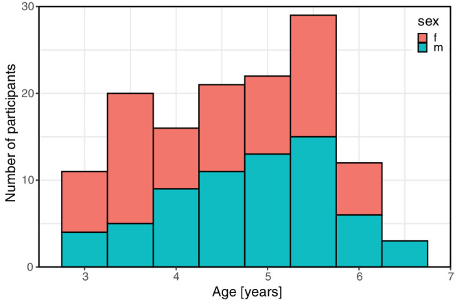
Age and sex distribution of study population. Female participants are shown in red, male participants in blue. Data at age label “3” comprises the interval [3; 3.5) years of age and so on.
Table.
Study Population—Ophthalmologic Diseases
| Ocular Disease | Number of Patients |
|---|---|
| Strabismus without amblyopia | 29 |
| Strabismus with amblyopia | 7 |
| Anisometropia with amblyopia | 3 |
| Chalazion | 3 |
| Aphakia | 2 |
| Duane syndrome | 2 |
| Ptosis without amblyopia | 2 |
| Ptosis with amblyopia | 1 |
| Aniridia | 1 |
| Congenital ocular melanocytosis | 1 |
| History of optic papillitis | 1 |
| Purulent conjunctivitis | 1 |
| Morbus Best | 1 |
| Color vision deficiency | 1 |
| Corneal scar with mild amblyopia | 1 |
| Alternate day squint | 1 |
| Megalopapilla | 1 |
| Chronic blepharoconjunctivitis | 1 |
| Phthisis bulbi after perforating ocular injury | 1 |
| Congenital hyperplasia of the pigment epithelium and epiretinal membrane | 1 |
Preexisting ophthalmologic diseases of participants.
Testability
Figure 2 shows the testability of the methods depending on the age of the study participants. Overall, 118 of 134 children (88% [82–93%] [actual result and bootstrapped 95% confidence interval]) were able to complete the LEA and 91 of 134 children (68% [60–75%]) were able to complete the FrACT (P < .0001).
Figure 2.
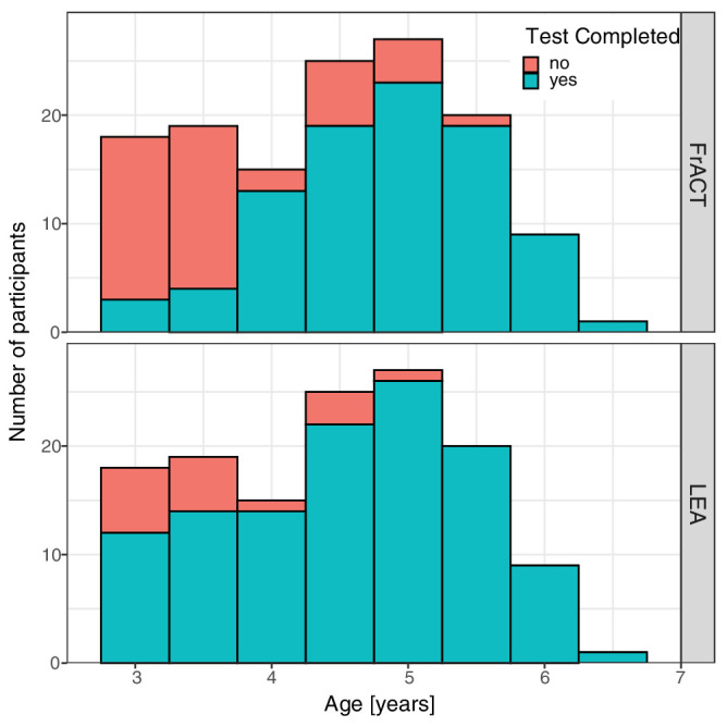
Testability of VA with FrACT (top) and LEA (bottom). For each age group (in 0.5-year steps), the number of patients with successful (blue-green) and failed (red) VA test completion is shown. Successful test completion (“yes”) means test completion in each eye. Age ranges as in Figure 1.
Testability depended on age: the LEA was testable in most participants, including those aged <4 years (<4 years: 70% [54%–84%]; ≥4 years: 95% [90%–99%]). While few participants under 4 years of age were able to complete the FrACT (19% [8%–32%]), 87% [79%–93%] of children aged 4 years and above completed the FrACT successfully. Testability differed significantly between LEA and FrACT below 4.7 years of age (P = 0.033) but not above. Testability was similar in the children examined in the Eye Center of the University Medical Center Freiburg and in the two childcare institutions (segregated data not shown).
Visual Acuity Depending on Age, Sex, Method, and Sequence
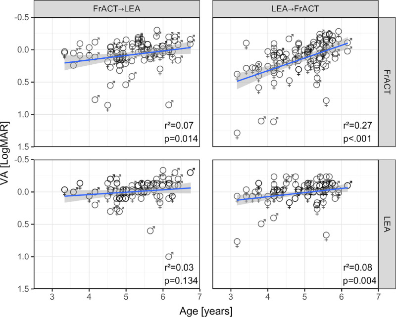
The ANOVA reported significant effects of age (P < 0.0001) and method (P < 0.0001) but no effects of sex (P = 0.37) and sequence (FrACT first or LEA first, P = 0.42). There was also a significant interaction of method × age (P < 0.0001) and age × method × sequence (P = 0.0039). These findings can be graphically appreciated in Figure 3, which plots VA versus age, segregated by method and sequence. As expected, VA improved with age. This age effect differed significantly between methods: the FrACT showed a more pronounced age dependence with a slope of –0.14 logMAR/year (P < 0.0001) compared to the LEA with –0.05 logMAR/year (P = 0.0017). The higher age dependence using the FrACT was due to a sequence effect with higher age dependence in LEA first compared to FrACT first (Fig. 3, compare top left and top right panels).
Figure 3.
- FrACT (FrACT→LEA): VA = −0.07 * age + 0.44, FrACT (LEA→FrACT): VA = −0.20 * age + 1.11
- LEA (FrACT→LEA): VA = −0.04 * age + 0.19, LEA (LEA→FrACT): VA = −0.06 * age + 0.33.
Difference in VA Results Between FrACT and LEA
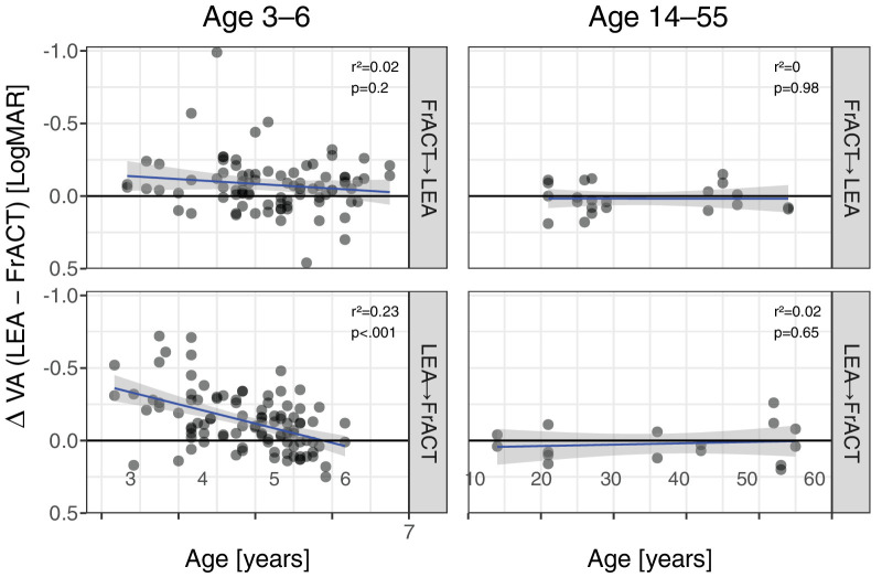
The difference in VA between the FrACT and LEA tests as a function of age and segregated by test sequence is depicted in Figure 4 for the children and an additional control group of 19 older participants (14 to 55 years, mean age: 34.7 years). Whereas the FrACT and LEA tests reported the same mean VA in the older control group regardless of test sequence (mean ± SD; overall: 0.02 ± 0.11 logMAR; FrACT first: 0.02 ± 0.10 logMAR; LEA first: 0.02 ± 0.13 logMAR; P = 0.62), there was an age-dependent trend for LEA to report better VAs in preschool children (overall: 0.11 ± 0.19 logMAR; children aged <4 years: 0.27 ± 0.23 logMAR; children aged ≥4 years: 0.09 ± 0.18 logMAR; P < 0.001). These age-dependent differences were highly dependent on test sequence: for FrACT first, the differences were not significantly age dependent (overall: 0.08 ± 0.19 logMAR; children aged <4 years: 0.12 ± 0.09 logMAR; children aged ≥4 years: 0.07 ± 0.19 logMAR; see top left panel; P = 0.20), whereas LEA first yielded highly age-dependent VA differences (overall: 0.13 ± 0.20 logMAR; children aged <4 years: 0.35 ± 0.24 logMAR; children aged ≥4 years: 0.11 ± 0.18 logMAR; see bottom left panel; P < 0.001). The worse VA values reported by FrACT in LEA first (Fig. 3) explain the larger VA differences in LEA first in Figure 4.
Figure 4.
Difference of VA as reported by LEA minus FrACT versus age. Note the inverted logMAR scale: better acuity up. The thick blue lines represent linear fits with corresponding 95% confidence intervals (gray shaded). The ordinate displays the difference between LEA and FrACT VAs (in logMAR) for eyes of participants who completed both tests. On the abscissa, the age range (in years) was divided in children (left) and older study participants (right). Top row: FrACT first. Bottom row: LEA first. Note that VA differences in children highly depended on test sequence (compare left row top and bottom). P values and r² are given at the top right of each panel.
Test–Retest LoA of the FrACT Results
Test–retest properties were quantified via LoA43 and a Bland–Altman plot (Fig. 5A). We found a negligible bias (0.017 logMAR, better VA for the first test run) and a range of ±0.298 logMAR for the 95% LoAs of TRV (average of all preschool children). Age-specific LoAs were ±0.363 logMAR below 4 years of age and ±0.289 logMAR above. The age dependence of the TRV shows a wide distribution; its slight decrease with age (Fig. 5B, linear regression, r = −0.20, P = 0.0075; LoA = 0.35–0.039 * age) only explains 4% of the variability. In the control population of 19 adults and adolescents, the 95% LoAs of TRV were ±0.154 logMAR.
Figure 5.
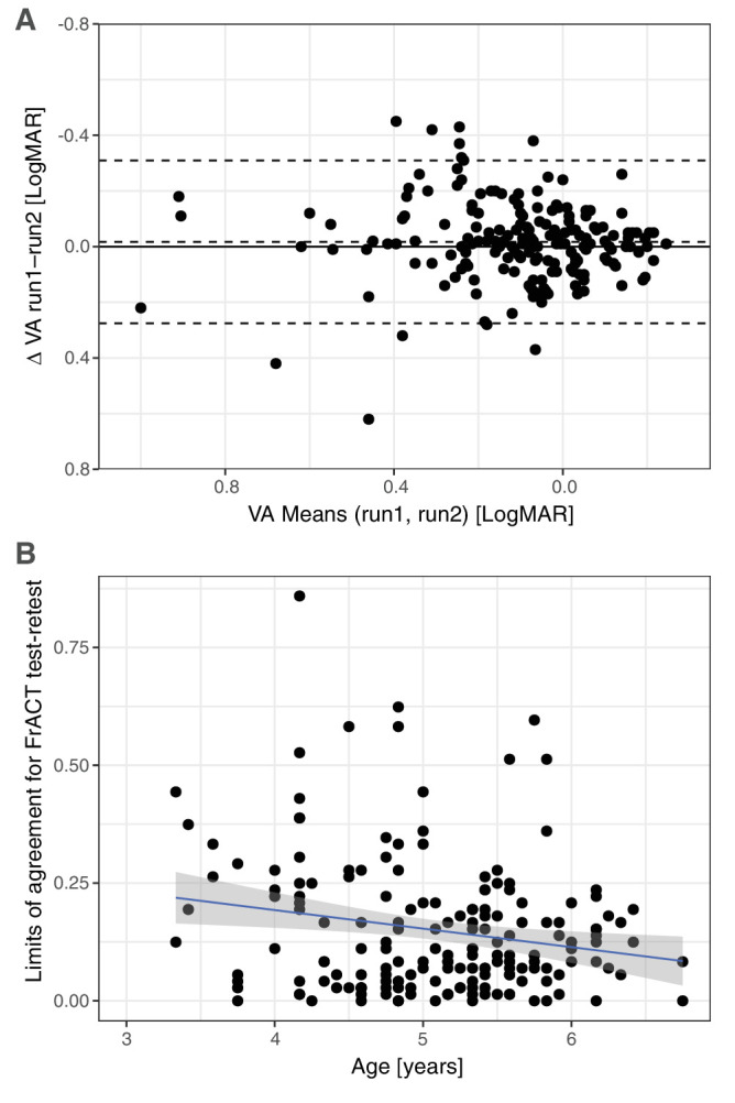
Test–retest variability of FrACT. (A) Bland–Altman plot of FrACT results when two runs were available. The solid line depicts the zero line. Bias (difference between zero line and middle dashed line) is low (−0.02 logMAR). The dashed lines represent ±95% limits of agreement (±0.298 logMAR). (B) Individual limits of agreement versus age, showing wide variability and a small decline with age (r = −0.20, P = 0.0075; LoA = 0.35–0.039 * age): the linear regression explains only 4% of the variance.
Discussion
Differences in Testability Between FrACT and LEA
Testability was age and test dependent. FrACT, for which use in very young preschool children has not been reported previously, showed high testability from the age of 4 years. LEA, which is already widely adopted for use in preschool children,8 was possible in all examined age groups (3–6 years). This agrees with and extends examinations in a German cohort of preschool children (340 children with an average age of 5.1 years) at school enrollment22: in that study, VA assessment using the FrACT and Tumbling E chart was compared. Testability of both FrACT and Tumbling E was 99%, and agreement between FrACT and Tumbling E was very high (approximately ±1 line).
In the present study, completion rates of FrACT and LEA in older preschool children (≥4 years of age) were above 85% (FrACT, 87%; LEA, 95%). Younger preschool children (<4 years of age) showed lower testability with the FrACT (FrACT, 19%; LEA, 70%); the difference in testability was significant below 4.7 years of age. This may reflect the influence of human interaction in VA assessment, particularly at a very young age: Children's responses on the LEA test could be positively biased by human interaction. In contrast, the lack of human interaction in the automated FrACT may lead to reduced compliance.
Visual Acuity Depending on Age and Method
The ANOVA of the VA results revealed a number of expected and unexpected findings: as previously reported,8 we found better results in older children. Besides this, we found that the LEA test yielded better overall VA results, which is in agreement with previous reports.38,44,45 In those studies, VAs measured with Landolt C charts were 0.07 to 0.14 logMAR worse compared to those measured with LEA Symbols—in both children and adults. Better acuity for LEA was also found in comparison to the Tumbling E chart,46 the Bailey–Lovie Letter chart,47 the Patti Pics chart,48 and the ETDRS chart.49 Thus, VA scores achieved in LEA appear to be consistently better than those achieved in other VA tests, especially for the youngest participants. It has been proposed that this discrepancy is due to the fact that LEA Symbols and Landolt rings measure different visual acuity components. In contrast to Landolt rings, LEA Symbols do not differ from each other in only one detail but contain complex spatial information. Thus, while Landolt C rings are supposed to determine the minimum separable or resolution acuity, LEA Symbols actually measure shape recognition acuity, the so-called minimum cognoscible.8,44 This is of importance, since resolution acuity depends more on the quality of the retinal image, whereas recognition acuity demands more cognitive skills. However, the notion that Landolt C optotypes determine resolution acuity has been disputed.6,50
Other possible explanations for the VA differences between LEA and FrACT reported in this study are (1) the high number optotype presentations with the FrACT (see below) and (2) the additional psychomotor hurdle of matching Landolt C directions and pressing the correct button on the remote control in the FrACT.
Effect of Sequence
The ANOVA revealed a highly significant interaction of the factors age, method, and sequence. As illustrated in Figure 3, FrACT VAs showed the greatest age dependence when the LEA test was performed first (i.e., worse VAs in younger children using the FrACT when the LEA test was performed before). We hypothesize that the high number of optotype presentations (FrACT: 5 times 30 optotype presentations [one binocular test run and two monocular runs per eye]; LEA: maximum number of 24 trials per eye [from 2.0 to −0.3 logMAR in steps of 0.1 logMAR] with four optotype presentations each = maximum 192 optotype presentations in total) was too demanding, especially for the youngest children, indicating that our study may overestimate VA differences between LEA and FrACT in children <4 years of age. This also suggests that the FrACT TRVs are possibly overestimated and would improve with a smaller number of trials. Indeed, in a more recent work,22 we found 18 instead of 30 trials for the FrACT to be sufficient.
TRV
In the present study, we determined the TRV of VAs in preschool children and in an older control group using the FrACT (but not the LEA as it was only tested once). The mean TRV of FrACT VAs (given as the 95% LoAs) in preschool children was about ±0.3 logMAR (with some age dependence: ±0.363 logMAR in children <4 years of age, ±0.289 logMAR in children ≥4 years of age). The high TRV found in our study is similar to those reported for other VA testing methods in children: using Landolt C charts in school children aged 6 to 9 years, Schmidt-Bacher et al.51 measured TRVs of ±0.24 to ±0.32 logMAR. Chen et al.52 reported a TRV of ±0.18 logMAR for LEA in amblyopic and healthy children aged 4 to 12 years. Shah et al.53 found TRVs of ±0.14 to ±0.16 logMAR in amblyopic children aged 4 to 15 years, employing printed or computerized crowded Kay Pictures and ETDRS. Others have found similar values.18,54–56 Possible reasons for the high TRV in our study compared to others include differences in testing protocols (test method, mode of optotype presentation mode of response, and as described above especially the high number of optotype presentations) and differences in study populations (younger children in our study).
In adults, TRVs are smaller, ranging from about ±0.1 to ±0.2 logMAR using different VA charts.57–64 The TRV of VAs of adults using the FrACT has been reported as ±0.2 logMAR.29 In the present study, the TRV of FrACT VAs in a control group of 19 adults and adolescents has been ±0.154 logMAR.
Limitations
Our study has some limitations: first, the study design makes a direct comparison between Landolt C optotypes and LEA symbol optotypes difficult, as the testing protocols differed between the FrACT and the LEA: Landolt C optotypes as part of the FrACT were presented on a computer screen and children responded by remote control, whereas LEA Symbols were offered on cards and children responded by naming or matching the recognized symbol on a board. As explained above, the number of optotype presentations in the FrACT was very high. A reduction in the number of presentations might have increased testability without undue loss in precision, especially in younger children. Also, the FrACT and the LEA test differed in their threshold criteria, with the FrACT set at 62.5% guessing probability calculated by the Best PEST algorithm and the LEA at 75% by a three-out-of-four forced-choice procedure. We did not correct for the difference between thresholds. In addition, although care was taken to ensure that the study conditions at the Eye Centre of the University Medical Centre Freiburg and the childcare centers were similar, we cannot exclude a bias due to small differences in setting, for instance, lighting and distractors. Also, the use of a single experienced examiner could have introduced bias toward one test method. Finally, we did not analyze whether LEA or FrACT performs better in children with poor VA since our sample size of such children was too small.
Conclusions
In conclusion, the FrACT, as an examiner-independent tool using the international reference Landolt C optotype, can be used to assess VA in preschool children aged 4 years and older, with reliability comparable to other assessment methods reported in the literature. In this age group, testability (87%) and TRV (±0.29 logMAR) support the use of the FrACT for VA assessment in preschool children. However, the FrACT has limited utility in children under 4 years of age, where the LEA test showed higher testability and a three-line better VA. Given the limitations of our study concerning design and methodology (e.g., number of optotype presentations), further investigations are needed to validate the FrACT against pediatric VA tests and in children with visual impairment.
Acknowledgments
The authors thank the preschool children and their families who generously volunteered their time to participate in this study, are grateful to the staff at the participating childcare institutions for their assistance with coordinating testing sessions, and acknowledge support by the Open Access Publication Fund of the University of Freiburg.
Disclosure: N. Farassat, None; V. Jehle, None; S.P. Heinrich, None; W.A. Lagrèze, None; M. Bach, None
References
- 1. Brown GC. Vision and quality-of-life. Trans Am Ophthalmol Soc. 1999; 97: 473–511. [DOI] [PubMed] [Google Scholar]
- 2. Ferris FL, Bailey I.. Standardizing the measurement of visual acuity for clinical research studies: guidelines from the Eye Care Technology Forum. Ophthalmology. 1996; 103(1): 181–182. [DOI] [PubMed] [Google Scholar]
- 3. ISO. ISO/TC 172/SC 7 Ophthalmic optics and instruments. ISO 8596:2017. ISO. Published 2017, https://www.iso.org/standard/69042.html. Accessed February 15, 2023.
- 4. DIN Deutsches Institut für Normung e.V. DIN EN ISO 8596, Sehschärfenprüfung. Das Normsehzeichen Und Seine Darbietung . Berlin: Beuth Verlag; 1996. [Google Scholar]
- 5. Chandna A. Natural history of the development of visual acuity in infants. Eye (Basingstoke). 1991; 5(1): 20–26. [DOI] [PubMed] [Google Scholar]
- 6. Leat SJ, Yadav NK, Irving EL.. Development of visual acuity and contrast sensitivity in children. J Optom. 2009; 2(1): 19–26. [Google Scholar]
- 7. Brémond-Gignac D, Copin H, Lapillonne A, Milazzo S; European Network of Study. Visual development in infants: physiological and pathological mechanisms. Curr Opin Ophthalmol. 2011; 22(suppl): S1–S8. [DOI] [PubMed] [Google Scholar]
- 8. Anstice NS, Thompson B.. The measurement of visual acuity in children: an evidence-based update. Clin Exp Optom. 2014; 97(1): 3–11. [DOI] [PubMed] [Google Scholar]
- 9. Zheng X, Xu G, Zhang K, et al.. Assessment of human visual acuity using visual evoked potential: a review. Sensors (Switzerland). 2020; 20(19): 1–26. [DOI] [PMC free article] [PubMed] [Google Scholar]
- 10. Hamilton R, Bach M, Heinrich SP, et al.. VEP estimation of visual acuity: a systematic review. Doc Ophthalmol. 2021; 142(1): 25–74. [DOI] [PMC free article] [PubMed] [Google Scholar]
- 11. Hyon JY, Yeo HE, Seo JM, Lee IB, Lee JH, Hwang JM.. Objective measurement of distance visual acuity determined by computerized optokinetic nystagmus test. Invest Ophthalmol Vis Sci. 2010; 51(2): 752–757. [DOI] [PubMed] [Google Scholar]
- 12. Sangi M, Thompson B, Turuwhenua J.. An optokinetic nystagmus detection method for use with young children. IEEE J Transl Eng Health Med. 2015; 3: 1–10. [DOI] [PMC free article] [PubMed] [Google Scholar]
- 13. Teller DY, Morse R, Borton R, Regal D.. Visual acuity for vertical and diagonal gratings in human infants. Vis Res. 1974; 14(12): 1433–1439. [DOI] [PubMed] [Google Scholar]
- 14. Adoh TO, Woodhouse JM, Oduwaiye KA.. The Cardiff Test: a new visual acuity test for toddlers and children with intellectual impairment. A preliminary report. Optom Vis Sci. 1992; 69(6): 427–432. [DOI] [PubMed] [Google Scholar]
- 15. Allen HF. A new picture series for preschool vision testing. Am J Ophthalmol. 1957; 44(1): 38–41. [DOI] [PubMed] [Google Scholar]
- 16. Mocan MC, Najera-Covarrubias M, Wright KW.. Comparison of visual acuity levels in pediatric patients with amblyopia using Wright figures, Allen optotypes, and Snellen letters. J AAPOS. 2005; 9(1): 48–52. [DOI] [PubMed] [Google Scholar]
- 17. Kay H. New method of assessing visual acuity with pictures. Br J Ophthalmol. 1983; 67(2): 131–133. [DOI] [PMC free article] [PubMed] [Google Scholar]
- 18. Holmes JM, Beck RW, Repka MX, et al.. The amblyopia treatment study visual acuity testing protocol. Arch Ophthalmol. 2001; 119(9): 1345–1353. [DOI] [PubMed] [Google Scholar]
- 19. Kupl MT, Dobson V, Peskin E, Quinn G, Schmidt P; Vision in Preschoolers Study Group. The electronic visual acuity tester: testability in preschool children. Optom Vis Sci. 2004; 81(4): 238–244. [DOI] [PubMed] [Google Scholar]
- 20. Sheridan MD. Sheridan-Gardiner test for visual acuity. BMJ. 1970; 2(5701): 108–109. [DOI] [PMC free article] [PubMed] [Google Scholar]
- 21. Hyvärinen L, Näsänen R, Laurinen P.. New visual acuity test for pre-school children. Acta Ophthalmol. 1980; 58(4): 507–511. [DOI] [PubMed] [Google Scholar]
- 22. Bach M, Reuter M, Lagrèze WA.. Vergleich zweier Visustests in der Einschulungsuntersuchung—E-Haken-Einblickgerät versus Freiburger Visustest. Ophthalmologe. 2016; 113(8): 684–689. [DOI] [PubMed] [Google Scholar]
- 23. Lai YH, Wu HJ, Chang SJ.. A reassessment and comparison of the Landolt C and Tumbling E charts in managing amblyopia. Sci Rep. 2021; 11(1): 1–7. [DOI] [PMC free article] [PubMed] [Google Scholar]
- 24. Laidlaw DAH, Abbott A, Rosser DA.. Development of a clinically feasible logMAR alternative to the Snellen chart: performance of the “compact reduced logMAR” visual acuity chart in amblyopic children. Br J Ophthalmol. 2003; 87(10): 1232–1234. [DOI] [PMC free article] [PubMed] [Google Scholar]
- 25. Manny RE, Hussein M, Gwiazda J, Marsh-Tootle W.. Repeatability of ETDRS visual acuity in children. Invest Ophthalmol Vis Sci. 2003; 44(8): 3294–3300. [DOI] [PubMed] [Google Scholar]
- 26. Cotter SA, Chu RH, Chandler DL, et al.. Reliability of the electronic early treatment diabetic retinopathy study testing protocol in children 7 to <13 years old. Am J Ophthalmol. 2003; 136(4): 655–661. [DOI] [PubMed] [Google Scholar]
- 27. Candy TR, Mishoulam SR, Nosofsky RM, Dobson V.. Adult discrimination performance for pediatric acuity test optotypes. Invest Ophthalmol Vis Sci. 2011; 52(7): 4307–4313. [DOI] [PMC free article] [PubMed] [Google Scholar]
- 28. Bach M. The Freiburg Visual Acuity Test—automatic measurement of visual acuity. Optom Vis Sci. 1996; 73(1): 49–53. [DOI] [PubMed] [Google Scholar]
- 29. Bach M. The Freiburg Visual Acuity Test—variability unchanged by post-hoc re-analysis. Graefes Arch Clin Exp Ophthalmol. 2007; 245(7): 965–971. [DOI] [PubMed] [Google Scholar]
- 30. Bach M, Schäfer K.. Visual acuity testing: feedback affects neither outcome nor reproducibility, but leaves participants happier. PLoS ONE. 2016; 11(1): e0147803. [DOI] [PMC free article] [PubMed] [Google Scholar]
- 31. Kurtenbach A, Langrová H, Messias A, Zrenner E, Jägle H.. A comparison of the performance of three visual evoked potential-based methods to estimate visual acuity. Doc Ophthalmol. 2013; 126(1): 45–56. [DOI] [PubMed] [Google Scholar]
- 32. Kollbaum PS, Jansen ME, Kollbaum EJ, Bullimore MA.. Validation of an iPad test of letter contrast sensitivity. Optom Vis Sci. 2014; 91(3): 291–296. [DOI] [PubMed] [Google Scholar]
- 33. Jolly JK, Gray JM, Salvetti AP, Han RC, MacLaren RE.. A randomized crossover study to assess the usability of two new vision tests in patients with low vision. Optom Vis Sci. 2019; 96(6): 443–452. [DOI] [PubMed] [Google Scholar]
- 34. Berufsverband der Augenärzte Deutschlands e.V., Deutsche Ophthalmologische Gesellschaft e.V. Leitlinie 26 a Amblyopie, https://www.dog.org/wp-content/uploads/2009/09/LL-26-Amb-2010-12-29-mit-Inhaltsverz-+Interessenkonfl-Endversion.pdf. Accessed September 7, 2023.
- 35. Bach M. Freiburg Vision Test ('FrACT’). Published 2021, http://michaelbach.de/fract/. Accessed June 3, 2021. [Google Scholar]
- 36. Lieberman HR, Pentland AP.. Microcomputer-based estimation of psychophysical thresholds: the best PEST. Behav Res Methods Instrument. 1982; 14: 21–25. [Google Scholar]
- 37. Bailey IL, Lovie JE.. New design principles for visual acuity letter charts. Am J Optom Physiol Opt. 1976; 53(11): 740–745. [DOI] [PubMed] [Google Scholar]
- 38. Becker R, Hübsch S, Gräf MH, Kaufmann H.. Examination of young children with Lea symbols. Br J Ophthalmol. 2002; 86(5): 513–516. [DOI] [PMC free article] [PubMed] [Google Scholar]
- 39. Bertuzzi F, Orsoni JG, Porta MR, Paliaga GP, Miglior S.. Sensitivity and specificity of a visual acuity screening protocol performed with the Lea Symbols 15-line folding distance chart in preschool children. Acta Ophthalmol Scand. 2006; 84(6): 807–811. [DOI] [PubMed] [Google Scholar]
- 40. Vision in Preschoolers (VIP) Study Group. Effect of age using Lea Symbols or HOTV for preschool vision screening. Optom Vis Sci. 2010; 87(2): 87–95. [DOI] [PMC free article] [PubMed] [Google Scholar]
- 41. R Core Team. R: A Language and Environment for Statistical Computing. Published online 2020, http://www.R-project.org. Accessed August 18, 2014. [Google Scholar]
- 42. Bland JM, Altman DG.. Statistical methods for assessing agreement between two methods of clinical measurement. Lancet. 1986; 1(8476): 307–310. [PubMed] [Google Scholar]
- 43. Bland JM, Altman DG.. Measuring agreement in method comparison studies. Stat Methods Med Res. 1999; 8(2): 135–160. [DOI] [PubMed] [Google Scholar]
- 44. Gräf M, Becker R.. Determining visual acuity with LH symbols and Landolt rings [in German]. Klin Monatsbl Augenheilkd. 1999; 215(2): 86–90. [DOI] [PubMed] [Google Scholar]
- 45. Gräf MH, Becker R, Kaufmann H.. Lea symbols: visual acuity assessment and detection of amblyopia. Graefes Arch Clin Exp Ophthalmol. 2000; 238(1): 53–58. [DOI] [PubMed] [Google Scholar]
- 46. Sanker N, Dhirani S, Bhakat P.. Comparison of visual acuity results in preschool children with lea symbols and Bailey-Lovie E chart. Middle East Afr J Ophthalmol. 2013; 20(4): 345–348. [DOI] [PMC free article] [PubMed] [Google Scholar]
- 47. Dobson V, Maguire M, Orel-Bixler D, Quinn G, Ying GS; Vision in Preschoolers (VIP) Study Group. Visual acuity results in school-aged children and adults: Lea Symbols chart versus Bailey-Lovie chart. Optom Vis Sci. 2003; 80(9): 650–654. [DOI] [PubMed] [Google Scholar]
- 48. Mercer ME, Drover JR, Penney KJ, Courage ML, Adams RJ.. Comparison of Patti Pics and Lea Symbols optotypes in children and adults. Optom Vis Sci. 2013; 90(3): 236–241. [DOI] [PubMed] [Google Scholar]
- 49. Dobson V, Clifford-Donaldson CE, Miller JM, Garvey KA, Harvey EM.. A comparison of Lea Symbol vs ETDRS letter distance visual acuity in a population of young children with a high prevalence of astigmatism. J Am Assoc Pediatr Ophthalmol Strabismus. 2009; 13(3): 253–257. [DOI] [PMC free article] [PubMed] [Google Scholar]
- 50. Heinrich SP, Bach M.. Resolution acuity versus recognition acuity with Landolt-style optotypes. Graefes Arch Clin Exp Ophthalmol. 2013; 251(9): 2235–2241. [DOI] [PubMed] [Google Scholar]
- 51. Schmidt-Bacher A, Pritsch M, Kolling G.. Reliability of three different visual acuity testing procedures in school children. Strabismus. 2007; 15(1): 39–43. [DOI] [PubMed] [Google Scholar]
- 52. Chen SI, Chandna A, Norcia AM, Pettet M, Stone D.. The repeatability of best corrected acuity in normal and amblyopic children 4 to 12 years of age. Invest Ophthalmol Vis Sci. 2006; 47(2): 614–619. [DOI] [PubMed] [Google Scholar]
- 53. Shah N, Laidlaw DAH, Rashid S, Hysi P.. Validation of printed and computerised crowded Kay picture logMAR tests against gold standard ETDRS acuity test chart measurements in adult and amblyopic paediatric subjects. Eye. 2012; 26(4): 593–600. [DOI] [PMC free article] [PubMed] [Google Scholar]
- 54. Kheterpal S, Jones HS, Auld R, Moseley MJ.. Reliability of visual acuity in children with reduced vision. Ophthalmic Physiol Opt. 1996; 16(5): 447–449. [PubMed] [Google Scholar]
- 55. McGraw PV, Winn B, Gray LS, Elliott DB.. Improving the reliability of visual acuity measures in young children. Ophthalmic Physiol Opt. 2000; 20(3): 173–184. [PubMed] [Google Scholar]
- 56. Moke PS, Turpin AH, Beck RW, et al.. Computerized method of visual acuity testing: adaptation of the amblyopia treatment study visual acuity testing protocol. Am J Ophthalmol. 2001; 132(6): 903–909. [DOI] [PubMed] [Google Scholar]
- 57. Arditi A, Cagenello R.. On the statistical reliability of letter-chart visual acuity measurements. Invest Ophthalmol Vis Sci. 1993; 34(1): 120–129. [PubMed] [Google Scholar]
- 58. Siderov J, Tiu AL.. Variability of measurements of visual acuity in a large eye clinic. Acta Ophthalmol Scand. 1999; 77(6): 673–676. [DOI] [PubMed] [Google Scholar]
- 59. Rosser DA, Laidlaw DAH, Murdoch IE.. The development of a “reduced logMAR” visual acuity chart for use in routine clinical practice. Br J Ophthalmol. 2001; 85(4): 432–436. [DOI] [PMC free article] [PubMed] [Google Scholar]
- 60. Rosser DA, Murdoch IE, Fitzke FW, Laidlaw DAH.. Improving on ETDRS acuities: design and results for a computerised thresholding device. Eye. 2003; 17(6): 701–706. [DOI] [PubMed] [Google Scholar]
- 61. Ruamviboonsuk P, Tiensuwan M, Kunawut C, Masayaanon P.. Repeatability of an automated Landolt C test, compared with the early treatment of diabetic retinopathy study (ETDRS) chart testing. Am J Ophthalmol. 2003; 136(4): 662–669. [DOI] [PubMed] [Google Scholar]
- 62. Beck RW, Moke PS, Turpin AH, et al.. A computerized method of visual acuity testing: adaptation of the early treatment of diabetic retinopathy study testing protocol. Am J Ophthalmol. 2003; 135(2): 194–205. [DOI] [PubMed] [Google Scholar]
- 63. Pointer JS. Recognition versus resolution: a comparison of visual acuity results using two alternative test chart optotype. J Optom. 2008; 1(2): 65–70. [Google Scholar]
- 64. Chaikitmongkol V, Nanegrungsunk O, Patikulsila D, Ruamviboonsuk P, Bressler NM.. Repeatability and agreement of visual acuity using the ETDRS number chart, Landolt C chart, or ETDRS alphabet chart in eyes with or without sight-threatening diseases. JAMA Ophthalmol. 2018; 136(3): 286–290. [DOI] [PMC free article] [PubMed] [Google Scholar]


