PLOS ONE logoLink to PLOS ONE
. 2024 Mar 22;19(3):e0300981. doi: 10.1371/journal.pone.0300981

Chemical range recognized by the ligand-binding domain in a representative amino acid-sensing taste receptor, T1r2a/T1r3, from medaka fish

Hikaru Ishida 1, Norihisa Yasui 1, Atsuko Yamashita 1,*
Editor: Israel Silman2
PMCID: PMC10959364  PMID: 38517842

Abstract

Taste receptor type 1 (T1r) proteins are responsible for recognizing nutrient chemicals in foods. In humans, T1r2/T1r3 and T1r1/T1r3 heterodimers serve as the sweet and umami receptors that recognize sugars or amino acids and nucleotides, respectively. T1rs are conserved among vertebrates, and T1r2a/T1r3 from medaka fish is currently the only member for which the structure of the ligand-binding domain (LBD) has been solved. T1r2a/T1r3 is an amino acid receptor that recognizes various l-amino acids in its LBD as observed with other T1rs exhibiting broad substrate specificities. Nevertheless, the range of chemicals that are recognized by T1r2a/T1r3LBD has not been extensively explored. In the present study, the binding of various chemicals to medaka T1r2a/T1r3LBD was analyzed. A binding assay for amino acid derivatives verified the specificity of this protein to l-α-amino acids and the importance of α-amino and carboxy groups for receptor recognition. The results further indicated the significance of the α-hydrogen for recognition as replacing it with a methyl group resulted in a substantially decreased affinity. The binding ability to the protein was not limited to proteinogenic amino acids, but also to non-proteinogenic amino acids, such as metabolic intermediates. Besides l-α-amino acids, no other chemicals showed significant binding to the protein. These results indicate that all of the common structural groups of α-amino acids and their geometry in the l-configuration are recognized by the protein, whereas a wide variety of α-substituents can be accommodated in the ligand binding sites of the LBDs.

Introduction

Animals sense a wide variety of chemicals in foods via taste receptors in their oral cavity. Numerous taste substances can be categorized into five basic taste modalities: sweet, umami, bitter, salty, and sour. The chemicals in each modality are recognized by the specific taste receptors [1]. For example, nutrient substances, such as sugars, amino acids, and nucleotides, are recognized by taste receptor type 1 (T1r) proteins. In humans and mice, the T1r1/T1r3 heterodimer serves as an umami receptor responsible for detecting amino acids and nucleotides, whereas the T1r2/T1r3 heterodimer serves as a sweet receptor responsible for detecting sugars [24]. The ligand specificities of T1r proteins are not strictly conserved among animals and there are some varieties. For example, all T1rs in zebrafish and medaka fish recognize amino acids but not sugars [5].

T1rs belong to the class C G protein-coupled receptor (GPCR) family, which is characterized by the presence of a large ligand-binding domain (LBD) in the extracellular region [6, 7]. The LBD contains a binding site for intrinsic receptor agonists, known as the orthosteric ligand-binding site, in the middle cavity of the domain with a bi-lobal architecture. The intrinsic agonists of T1rs (i.e., sugars, amino acids, and nucleotides) are also recognized at the orthosteric sites in T1rs [812]. Nevertheless, the orthosteric sites in T1rs have distinctive properties compared with those in other class C GPCRs. The former is amenable to accommodate a certain range of chemicals, whereas the latter shows specificity for limited compounds. For example, the human sweet receptor T1r2/T1r3 responds to sucrose as well as artificial sweeteners, such as aspartame, saccharin, and acesulfame K, all of which are recognized by the orthosteric binding site in T1r2-LBD [9, 10]. The mouse T1r1/T1r3 also recognizes a wide array of l-amino acids at the orthosteric binding site of T1r1LBD [11]. These properties are in contrast with those of other class C GPCRs responsible for recognizing signaling molecules, such as metabotropic glutamate receptors, which exhibit strict specificity for l-glutamate [13]. Nevertheless, the broad substrate specificities associated with T1rs correspond to their physiological functions in recognizing various chemicals in the environment using a limited set of receptors. However, these characteristics raise a question of how broadly a range of chemicals can be recognized by the orthosteric site of a particular T1r.

Recent studies have indicated that T1rs are expressed, not only in the oral cavity, but also in many other organs in the body, such as the brain, gastrointestinal system, and reproductive organs [14, 15]. Therefore, these receptors are likely responsible for various physiological functions in these tissues. However, only a fraction of them has been elucidated thus far. Determining the range of substrates for T1rs will provide clues for understanding the physiological functions of T1rs in the body, as well as in taste sensation.

In this study, we examined ligands for T1r2a/T1r3LBD from medaka (Oryizias latipes) as a case study for determining the substrate specificity of a T1r. Medaka T1r2a/T1r3 is an amino acid receptor and the sole T1r protein for which ligand binding and structural analyses using a heterodimeric recombinant protein have been achieved [12, 16, 17]. Medaka T1r2a/T1r3LBD reportedly accommodates a wide range of l-amino acids in orthosteric binding sites [5, 12, 17] and is considered a suitable target protein to address the broad substrate specificities of T1rs. To assess the ligand-binding activity of this protein, we used differential scanning fluorimetry (DSF). In this method, binding is evaluated by the extent of ligand-dependent thermal stabilization of the target protein, which is judged by a high-temperature shift of the melting curves of thermal denaturation detected using an environmentally sensitive fluorescent dye as an indicator [18]. We previously verified that the results of ligand-binding to T1r2a/T1r3LBD analyzed using DSF were consistent with those obtained using other biophysical methodologies [17]. In this study, we performed an assay on an extended range of chemicals using this method and clarified the specificity of this protein.

Materials and methods

Protein samples were prepared as previously described [12, 16]. Briefly, the heterodimer of the C-terminal FLAG-tagged T1r2aLBD and T1r3LBD [12, 19] was stably expressed in Drosophila S2 cells (Invitrogen) and purified from the culture medium using ANTI-FLAG M2 affinity gel (SIGMA).

The ligand binding assay by differential scanning fluorimetry (DSF) and data analysis were performed as described previously [17]. Briefly, the purified protein was dialyzed against the assay buffer (20 mM Tris-HCl, 300 mM NaCl, 2 mM CaCl2, pH 8.0). The sample protein was then mixed with Protein Thermal Shift Dye (Applied Biosystems) and the ligands in the dialysis buffer used for the final dialysis step. Specifically, 1 μg of protein, 1× Dye, and ligands at a concentration for each condition were mixed in 20 μL of reaction mix. Prior to mixing, the ligands, except for steroids, were dissolved in the same solution and the pH was adjusted to 8.0 through the addition of either NaOH or HCl. Because of the low solubilities in aqueous solution, the steroids were first dissolved in dimethyl sulfoxide (DMSO) and added to the reaction mixture at a final DMSO concentration of 10%. Fluorescence intensity changes accompanying the thermal denaturation of the protein were measured using the StepOne Real-Time PCR System (Applied Biosystems) with heating of the sample at the temperature from 25°C to 99°C at a rate of 0.022°C/s. The apparent melting temperature (Tm) of each sample was determined using the maximum of the derivatives of the melting curve (dFluorescence/dT). To ensure sample activity, data from the sample that did not meet the criteria that the Tm in the absence of a ligand was within 51–55°C and the Tm increase (ΔTm) in the presence of 10 mM l-alanine was within 9–11°C were excluded.

The apparent dissociation constant (Kd-app) of the ligand was estimated by fitting the Tm values at different ligand concentrations to an equation based on the thermodynamic folding model proposed by Schellman [20] using KaleidaGraph (Synergy Software) as described previously [17].

Results

Binding of amino acids and their derivatives

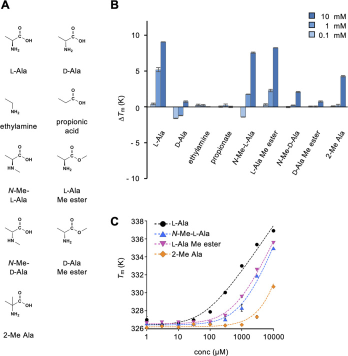
We previously reported that medaka T1r2a/T1r3LBD binds to a variety of l-amino acids [12, 17]. To further address amino acid specificity, the binding of alanine and its derivatives (Fig 1A) to T1r2a/T1r3LBD was examined.

Fig 1. Binding analysis of amino acids and their derivatives to T1r2a/T1r3LBD using differential scanning fluorimetry.

Fig 1

(A) Chemical structures of amino acids and their derivatives tested. (B) ΔTm resulting from the addition of 0.1, 1, and 10 mM amino acids and their derivatives. (C) Dose-dependent Tm changes caused by the addition of l-alanine and its derivatives. Data represent the mean ± standard error of the mean (SEM) of four technical replicates. The fitted line drawn for the 2-methylalanine data assumed the Kd-app as 15 mM, although accurate Kd-app determination was impractical owing to a large fitting error.

As observed previously [17], T1r2a/T1r3LBD showed thermal stabilization, as evidenced by an increase in TmTm), which was dependent on the l-alanine concentration, whereas no significant ΔTm change was observed with the addition of d-alanine (Fig 1B and S1 Fig). These results indicated substantial binding of the former and low or negligible binding of the latter, confirming the l-amino acid specificity of this receptor. Next, we addressed the requirement of each amino acid functional group by testing chemicals devoid of the α-carboxy or amino group of alanine, namely ethylamine and propionate. These results indicated that they did not induce thermal stabilization of the protein, indicating no significant binding. Therefore, both the α-carboxy and the amino group in l-amino acids are essential for recognition by T1r2a/T1r3LBD.

We also addressed the recognition stringency of each amino acid functional group. The l-alanine derivatives, in which the α-carboxy and the amino group are modified [i.e., N-methyl-l-alanine (N-Me-l-Ala) and l-alanine methyl ester (l-Ala Me ester; Fig 1A)], still showed substantial binding signals, whereas their d-enantiomers exhibited little signal (Fig 1B). In addition, 2-methylalanine (2-Me Ala), an achiral alanine derivative in which the hydrogen at the Cα is substituted with a methyl group (Fig 1A), exhibited weaker binding signals. The apparent Kd (Kd-app) of these derivatives derived from a dose-thermal stabilization analysis (Fig 1C) revealed that modification at the α-carboxy or amino group (N-Me-l-Ala and l-Ala Me ester) increased the Kd-app by approximately 10-fold (∼600–1100 μM) compared to the non-modified l-alanine (Kd-app: ∼60 μM), whereas modification at the Cα (2-Me Ala) resulted in a >100-fold increase (Kd-app: > 10 mM; Fig 1C, Table 1). These results suggest that the α-carboxy and the amino groups of amino acids are important for recognition by T1r2a/T1r3LBD, and that their modification impairs recognition to some extent, but not critically. Nevertheless, the modification at Cα results in severe consequences for binding compared with those at the α-carboxy and the amino groups, indicating the significance of the α-carbon and/or hydrogen for T1r2a/T1r3LBD recognition.

Table 1. Apparent dissociation constants (Kd-app) of l-alanine derivatives to medaka T1r2a/T1r3LBD as determined by differential scanning fluorimetry.

ligands Kd-app (μM)
l-alanine 63.9 ± 29.0
N-methyl-l-alanine 1140 ± 407
l-alanine methyl ester 615 ± 154
2-methylalanine ND

Binding of ligands for the other T1rs or class C GPCRs

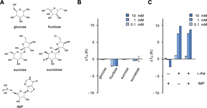
We examined the commonality in ligand specificities among T1rs and other class C GPCRs. A recent study reported that fish T1r2 and T1r3 are not orthologs but paralogs of mammalian Tlr receptors [21]. We examined the binding of sugars (Fig 2A), which are substrates of the mammalian sweet receptors T1r2/T1r3 [2, 4], to medaka T1r2a/T1r3LBD. Glucose, fructose, and sucrose as well as an artificial sweetener sucralose, as a sugar analog, did not exhibit significant binding (Fig 2B). It should be noted that it is not feasible to assess the specific binding of sugars at high concentrations, such as those inducing responses of mammalian T1r2/T1r3 (~300 mM) [2, 4], using DSF, because they intrinsically induce thermal stabilization of a protein, even without specific interactions [22]. Nevertheless, the observation in this study, which showed no noticeable thermal stabilization by sugars up to at least 10 mM, is consistent with the results of a previous study that reported no responses of this receptor to sugars (at 150 mM) [5]. Next, we examined the binding of a nucleotide, inosine monophosphate (IMP), because the mammalian amino acid receptors T1r1/T1r3 recognize IMP as a substrate and exhibit enhanced responses to amino acids in its presence [3, 8]. However, in medaka T1r2a/T1r3LBD, IMP did not bind, and its presence did not enhance alanine binding (Fig 2B).

Fig 2. Binding analysis of typical ligands for T1rs to T1r2a/T1r3LBD.

Fig 2

(A) Chemical structures of the T1r ligands tested. (B) ΔTm resulting from the addition of 0.1, 1, and 10 mM sugars. (C) ΔTm resulting from the addition of 0.1, 1, and 10 mM IMP (left), l-alanine (middle), and l-alanine in the presence of 1 mM IMP (right). Data represent the mean ± SEM of four technical replicates.

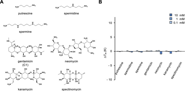
One of the class C GPCRs, calcium sensing receptor (CaSR) responds to polyamines and aminoglycoside antibiotics [23] (Fig 3A). We examined the binding of putrescine, spermidine, and spermine as polyamines and gentamicin, neomycin, kanamycin, and spectinomycin as aminoglycoside antibiotics to medaka T1r2a/T1r3LBD; however, none of these compounds exhibited binding signals (Fig 3B).

Fig 3. Binding analysis of typical CaSR ligands to T1r2a/T1r3LBD.

Fig 3

(A) Chemical structures of the CaSR ligands tested. The structure of the C1 component of the gentamicin complex is shown in the panel, whereas the complex reagent was used for the analysis. (B) ΔTm resulting from the addition of 0.1, 1, and 10 mM CaSR ligands. Data represent the mean ± SEM of four technical replicates.

In summary, we could not find any ligands of medaka T1r2a/T1r3 common to the other T1rs and class C GPCRs other than l-amino acids.

Binding of bioactive compounds

To explore the physiological functions of T1rs in the body, we examined the binding of various bioactive compounds that may serve as substrates in organs other than the oral cavity.

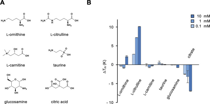
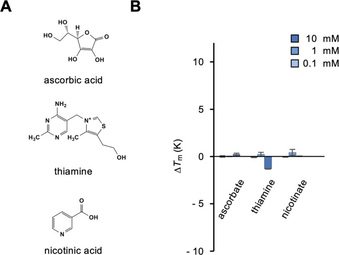
We tested several amino acid-derived metabolites, including l-ornithine, l-citrulline, l-carnitine, and taurine (Fig 4A). l-citrulline and, albeit weakly, l-ornithine, which are metabolites of the urea cycle, showed obvious binding to medaka T1r2a/T1r3LBD (Fig 4B). The results indicated that α-amino acid metabolites serve as ligands for T1r2a/T1r3LBD, even if they are non-proteinogenic amino acids. l-carnitine and taurine, which are biosynthesized from l-amino acids but do not have α-amino acid structures, exhibited no obvious binding to the protein (Fig 4B), again indicating the significance of the α-amino acid structure required for recognition. Other non-amino acid metabolites or biosynthetic precursors, such as glucosamine and citrate, did not induce thermal stabilization of T1r2a/T1r3LBD (Fig 4A and 4B). Other nutrient chemicals were tested, including several water-soluble vitamins, such as ascorbate, thiamine, and nicotinate (Fig 5A); however, none exhibited binding to medaka T1r2a/T1r3LBD (Fig 5B).

Fig 4. Binding analysis of representative amino acid and non-amino acid metabolites and biosynthetic precursors to T1r2a/T1r3LBD.

Fig 4

(A) Chemical structures of the metabolites and biosynthetic precursors tested. (B) ΔTm resulting from the addition of 0.1, 1, and 10 mM metabolites or biosynthetic precursors. Data represent the mean ± SEM of four technical replicates.

Fig 5. Binding analysis of representative water-soluble vitamins to T1r2a/T1r3LBD.

Fig 5

(A) Chemical structures of the vitamins tested. (B) ΔTm resulting from the addition of 0.1, 1, and 10 mM vitamins. Data represent the mean ± SEM of four technical replicates.

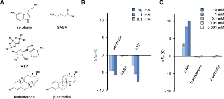
Finally, we tested small signaling molecules, such as neurotransmitters and hormones, considering the fact that T1rs are expressed in the brain and reproductive organs [24, 25] (Fig 6A). Thermal stabilization of T1r2a/T1r3LBD was not observed following the addition of representative neurotransmitters, such as serotonin as a representative of monoamines, γ-aminobutyric acid (GABA), and adenosine triphosphate (ATP) (Fig 6B). The finding that GABA was not recognized by T1r2a/T1r3LBD confirms the specificity of the protein to α-amino acids, but not γ-amino acids. We also examined the binding of representative steroids, testosterone and β-estradiol (Fig 6A). Because steroids exhibited low solubility in the assay buffer, before testing the binding of the steroids in a 10% DMSO buffer, we first confirmed that T1r2a/T1r3LBD showed similar thermal stabilization with the addition of l-alanine in the presence of 10% DMSO to that observed without DMSO (Fig 6C compared with Fig 1B; S1 Fig). Despite testing concentrations up to 0.1 mM, which is the highest soluble concentration for these chemicals in 10% DMSO, no significant binding of these steroids was observed (Fig 6C).

Fig 6. Binding analysis of representative neurotransmitters and hormones to T1r2a/T1r3LBD.

Fig 6

(A) Chemical structures of the neurotransmitters and hormones tested. (B) ΔTm resulting from the addition of 0.1, 1, and 10 mM neurotransmitters. (C) ΔTm resulting from the addition of 0.001, 0.01, and 0.1 mM hormones in 10% DMSO. ΔTm values resulting from the addition of 0.1, 1, and 10 mM l-alanine in 10% DMSO are also shown for comparison. Data represent the mean ± SEM of four technical replicates.

Taken together, among the tested compounds, no bioactive compounds other than l-α-amino acids were identified as ligands for medaka T1r2a/T1r3LBD. Nevertheless, the results also indicate that the l-α-amino acid specificity of T1r2a/T1r3LBD is not limited to the proteinogenic amino acids examined thus far but also to non-proteinogenic amino acids, such as metabolic intermediates and products.

Discussion

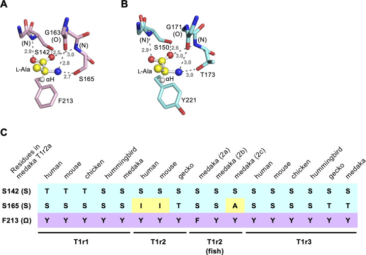
In this study, we examined the ligand specificity of medaka T1r2a/T1r3LBD. The binding assay confirmed the specificity of l-α-amino acid binding to this protein, whereas no significant binding of other chemicals lacking the l-α-amino acid architecture was observed. More specifically, the results indicated the importance of α-amino and carboxy groups in amino acids for recognition by the receptor as observed in the crystallographic structure [12], although small modifications, such as methylation, were found to reduce the affinities but still resulted in binding (Fig 1). Furthermore, the results of the present study indicate the significance of the α-hydrogen for the recognition of amino acids, as replacing this hydrogen with a methyl group resulted in decreased affinity (Fig 1). Because the α-hydrogen in the bound amino acid points toward the aromatic residues just below the substrate, Phe213 in T1r2a and Tyr221 in T1r3 (Fig 7A and 7B), the results indicate that not only the hydrophobic interaction between the ligand and the aromatic residue but also the CH-π interaction between the α-hydrogen of the ligand and the side-chain aromatic ring in the protein is significant for recognition. The manner of recognition explains the l-α-amino acid specificity of the receptor: all of the common structural components of α-amino acids, α-hydrogen, amino, and carboxy groups, and their geometry in the l-configuration, are recognized by the protein. If these groups are present, medaka T1r2a/T1r3 can accommodate a variety of α-substituents in its large ligand-binding pocket [12]. Indeed, we found that the binding ability was not limited to proteinogenic amino acids [17] but also to non-proteinogenic amino acids, such as metabolic intermediates and products (Fig 4). Docking simulations showed that these compounds could be accommodated in the ligand-binding site in a manner similar to the that of known ligands (e.g., l-alanine) without steric clashes (S2 Fig).

Fig 7. Residues important for the interaction with l-amino acids at the ligand-binding sites in T1r2a/T1r3LBD.

Fig 7

(A, B) Close-up views of the ligand-binding sites in T1r2a (A) and T1r3 (B) in the crystal structure of l-alanine-bound T1r2a/T1r3LBD (PDB ID: 5X2N) [12]. Bound l-alanine is shown as a ball-and-stick model. The dashed lines depict the hydrogen bonds labeled with their distances in Å. (C) The “SSΩ” residues corresponding to Ser142, Ser165, and Phe213 in T1r2a for various T1rs. Representative T1rs, in which responses have been reported, are shown [25, 11, 12, 26, 27].

The manner of amino acid recognition in medaka T1r2a/T1r3 is likely conserved among T1rs that share amino acid-responding functions (Fig 7C). In medaka T1r2a, the side-chain hydroxyl groups of Ser142 and Ser165, together with the main-chain amide and carbonyl groups as well as that in another residue (Gly163 in T1r2a), form a hydrogen-bonding network with the α-amino and carboxy groups in the ligand amino acid (Fig 7A). These two Ser/Thr (S) residues, as well as the aromatic residue (Ω; Phe213 in T1r2a) that forms a CH-π interaction with the α-hydrogen in the ligand, are conserved among most T1rs (Fig 7B and 7C). Therefore, the ability to recognize l-α-amino acids may be common to T1rs that share these three “SSΩ” residues at the orthosteric binding site. An exception without strict conservation of the “SSΩ” residues was observed for mammalian T1r2, which has an isoleucine at the residue corresponding to Ser165 [12]. This may be related to the fact that human and mouse sweet receptors T1r2/T1r3 respond to sugars and d-amino acids, but not to l-amino acids [2, 4]. Another exception is medaka T1r2c, which contains an alanine at the same residue. This may be related to the fact that medakaT1r2c/T1r3 specifically responds to l-proline, a secondary amino acid, but not to other l-amino acids with a primary amino group [5, 28]. Conversely, hummingbird T1r1 and gecko T1r2 share “SSΩ” residues, whereas hummingbird T1r1/T1r3 and gecko T1r2/T1r3 exhibit sugar responses [26, 27]. This suggests that the “SSΩ” residues are compatible with the sugar responding function. Nonetheless, the actual receptor functions need to be examined experimentally.

In contrast to the recognition of common groups in l-α-amino acids, the manner of recognition of the α-substituent group likely varies among T1rs, as evidenced by the different substrate specificities observed. For example, human T1r1/T1r3 specifically responds to acidic amino acids, such as l-glutamate and aspartate, whereas mouse T1r1/T1r3 responds to a wide array of l-amino acids except acidic amino acids [3, 4, 11]. Therefore, the range of amino acid specificity for T1r is likely determined by the physicochemical properties of the ligand-binding pocket surrounding the α-substituent group in each receptor. For T1rs sharing broad ligand specificity, such as mouse T1r1/T1r3 and medaka T1r2a/T1r3, the ligands may not be limited to proteinogenic amino acids, but also non-proteinogenic amino acids, as observed for medaka T1r2a/T1r3. This warrants further investigation by binding assays that involve a wider range of chemicals.

Besides l-α-amino acids, no other chemicals tested in this study exhibited significant binding to medaka T1r2a/T1r3LBD. Although the addition of several compounds such as IMP, citric acid, thiamine, serotonin, and ATP provided negative ΔTm values, their actions on the protein are unlikely related to the binding that induces receptor responses, but probably destabilization of the protein due to their acidic/basic properties, because the correlation between the positive ΔTm values and the receptor responses has been reported [17]. On this account, a representative monoamine neurotransmitter serotonin did not show binding expected to induce receptor responses (Fig 6B), which is consistent with the results that T1r2a/T1r3 prefers α-amino acids and not monoamines (ethylamine; Fig 1). Similarly, chemicals sharing chemical structures that are distinct from those of amino acids, such as steroids, did not show obvious binding. However, because the assay was performed solely using the LBD and not the full-length receptor because of methodological limitations, the possibility that these molecules act on other regions in the receptor cannot be excluded. On the other hand, the newly identified l-α-amino acid ligands, l-citrulline and l-ornithine, are cytosolic metabolic intermediates in the urea cycle, whereas they exhibit binding to the extracellular LBD of T1r2a/T1r3. Therefore, it is unclear whether they act as intrinsic ligands for the receptor in vivo. Moreover, the actions of these chemicals on T1r2a/T1r3 as agonists or antagonists are also unclear, although we verified that l-α-amino acid binding analyzed by DSF correlates with the receptor response induced by the ligand [17]. Nonetheless, our results indicate the possibility that a wider range of chemicals beyond those considered may be significant for gustation and could serve as ligands for T1rs. Their physiological relevance should be examined in future studies.

Supporting information

S1 Fig. Thermal melting curves of T1r2a/T1r3LBD.

(A) Representative melting curves in the assay buffer. The Tm values derived from four technical replicated measurements represent no ligand, 53.0 ± 0.16°C; 0.1 mM l-alanine added, 53.4 ± 0.12°C (ΔTm, 0.4°C); 1 mM l-alanine added, 58.2 ± 0.29°C (ΔTm, 5.2°C); 10 mM l-alanine added; 62.0 ± 0.02°C (ΔTm, 9.0°C). (B) Representative melting curves in the assay buffer containing 10% DMSO. The Tm values derived from four technical replicated measurements represent no ligand, 47.3 ± 0.23°C 0.1 mM l-alanine added, 50.9 ± 0.50°C (ΔTm, 3.6°C); 1 mM l-alanine added, 55.9 ± 0.16°C (ΔTm, 8.6°C); 10 mM l-alanine added; 57.5 ± 0.02°C (ΔTm, 10.2°C).

(TIF)

pone.0300981.s001.tif (200.9KB, tif)
S2 Fig. Docking simulations of the compounds revealed binding to T1r2a/3LBD in this study.

(A) A close-up view of the ligand-binding site in T1r2a in the crystal structure of the l-alanine-bound form (PDB ID: 5X2N). (B–D) Representative docking poses of N-methyl-l-alanine (N-Me-l-Ala; ZINC 901468; B), l-alanine methyl ester (l-Ala Me ester; ZINC 34702232; C), and l-citrulline (ZINC 1532614; D) in the ligand-binding site of T1r2a. In panels B–D, the docking simulations were performed using SwissDock (Grosdidier et al. Nucleic Acids Res. 39, W270, 2011), using the coordinates of T1r2aLBD (PDB ID: 5X2N, chain A) without ligands as a target.

(TIF)

S1 File. The file archive including all the data reported in the main text and the Supporting information.

SourceData.xlsx, the data for reproducing the figures in the main text and S1 Fig; 5x2n_A_nolig.pdb, the coordinates of T1r2aLBD used as the input for docking simulations; LNMA.pdb, the coordinates for a representative docked model of N-methyl-l-alanine, shown in S2B Fig; LAME.pdb, the coordinates for a representative docked model of l-alanine methyl ester, shown in S2C Fig; Lcit.pdb, the coordinates for a representative docked model of l-citrulline, shown in S2D Fig.

(ZIP)

pone.0300981.s003.zip (238.9KB, zip)

Acknowledgments

We thank Junya Nitta, Rakuto Mizoguchi, and Mayu Kudo for their help with the protein preparation.

Data Availability

All relevant data are within the manuscript and its Supporting Information files.

Funding Statement

This work was financially supported by JSPS KAKENHI Grant Numbers JP20H03195, JP20H04778, JP21H05524, and JP23H02424, and the Takeda Science Foundation (to A.Y.). The funders had no role in study design, data collection and analysis, decision to publish, or preparation of the manuscript.

References

  • 1.Taruno A, Nomura K, Kusakizako T, Ma Z, Nureki O, Foskett JK. Taste transduction and channel synapses in taste buds. Pflugers Arch. 2021;473:3–13. doi: 10.1007/s00424-020-02464-4 [DOI] [PMC free article] [PubMed] [Google Scholar]
  • 2.Nelson G, Hoon MA, Chandrashekar J, Zhang Y, Ryba NJ, Zuker CS. Mammalian sweet taste receptors. Cell. 2001;106:381–90. doi: 10.1016/s0092-8674(01)00451-2 [DOI] [PubMed] [Google Scholar]
  • 3.Nelson G, Chandrashekar J, Hoon MA, Feng L, Zhao G, Ryba NJ, et al. An amino-acid taste receptor. Nature. 2002;416:199–202. doi: 10.1038/nature726 [DOI] [PubMed] [Google Scholar]
  • 4.Li X, Staszewski L, Xu H, Durick K, Zoller M, Adler E. Human receptors for sweet and umami taste. Proc Natl Acad Sci U S A. 2002;99:4692–6. doi: 10.1073/pnas.072090199 [DOI] [PMC free article] [PubMed] [Google Scholar]
  • 5.Oike H, Nagai T, Furuyama A, Okada S, Aihara Y, Ishimaru Y, et al. Characterization of ligands for fish taste receptors. J Neurosci. 2007;27:5584–92. doi: 10.1523/JNEUROSCI.0651-07.2007 [DOI] [PMC free article] [PubMed] [Google Scholar]
  • 6.Ellaithy A, Gonzalez-Maeso J, Logothetis DA, Levitz J. Structural and Biophysical Mechanisms of Class C G Protein-Coupled Receptor Function. Trends Biochem Sci. 2020;45:1049–64. doi: 10.1016/j.tibs.2020.07.008 [DOI] [PMC free article] [PubMed] [Google Scholar]
  • 7.Yang L, Cui M, Liu B. Current Progress in Understanding the Structure and Function of Sweet Taste Receptor. J Mol Neurosci. 2021;71:234–44. doi: 10.1007/s12031-020-01642-4 [DOI] [PubMed] [Google Scholar]
  • 8.Zhang F, Klebansky B, Fine RM, Xu H, Pronin A, Liu H, et al. Molecular mechanism for the umami taste synergism. Proc Natl Acad Sci U S A. 2008;105:20930–4. doi: 10.1073/pnas.0810174106 [DOI] [PMC free article] [PubMed] [Google Scholar]
  • 9.Zhang F, Klebansky B, Fine RM, Liu H, Xu H, Servant G, et al. Molecular mechanism of the sweet taste enhancers. Proc Natl Acad Sci U S A. 2010;107:4752–7. doi: 10.1073/pnas.0911660107 [DOI] [PMC free article] [PubMed] [Google Scholar]
  • 10.Masuda K, Koizumi A, Nakajima K, Tanaka T, Abe K, Misaka T, et al. Characterization of the modes of binding between human sweet taste receptor and low-molecular-weight sweet compounds. PLoS One. 2012;7:e35380. doi: 10.1371/journal.pone.0035380 [DOI] [PMC free article] [PubMed] [Google Scholar]
  • 11.Toda Y, Nakagita T, Hayakawa T, Okada S, Narukawa M, Imai H, et al. Two distinct determinants of ligand specificity in T1R1/T1R3 (the umami taste receptor). J Biol Chem. 2013;288:36863–77. doi: 10.1074/jbc.M113.494443 [DOI] [PMC free article] [PubMed] [Google Scholar]
  • 12.Nuemket N, Yasui N, Kusakabe Y, Nomura Y, Atsumi N, Akiyama S, et al. Structural basis for perception of diverse chemical substances by T1r taste receptors. Nat Commun. 2017;8:15530. doi: 10.1038/ncomms15530 [DOI] [PMC free article] [PubMed] [Google Scholar]
  • 13.Kang HJ, Menlove K, Ma J, Wilkins A, Lichtarge O, Wensel TG. Selectivity and evolutionary divergence of metabotropic glutamate receptors for endogenous ligands and G proteins coupled to phospholipase C or TRP channels. J Biol Chem. 2014;289:29961–74. doi: 10.1074/jbc.M114.574483 [DOI] [PMC free article] [PubMed] [Google Scholar]
  • 14.Foster SR, Roura E, Thomas WG. Extrasensory perception: odorant and taste receptors beyond the nose and mouth. Pharmacol Ther. 2014;142:41–61. doi: 10.1016/j.pharmthera.2013.11.004 [DOI] [PubMed] [Google Scholar]
  • 15.Behrens M, Meyerhof W. A role for taste receptors in (neuro)endocrinology? J Neuroendocrinol. 2019;31:e12691. doi: 10.1111/jne.12691 [DOI] [PubMed] [Google Scholar]
  • 16.Nango E, Akiyama S, Maki-Yonekura S, Ashikawa Y, Kusakabe Y, Krayukhina E, et al. Taste substance binding elicits conformational change of taste receptor T1r heterodimer extracellular domains. Sci Rep. 2016;6:25745. doi: 10.1038/srep25745 [DOI] [PMC free article] [PubMed] [Google Scholar]
  • 17.Yoshida T, Yasui N, Kusakabe Y, Ito C, Akamatsu M, Yamashita A. Differential scanning fluorimetric analysis of the amino-acid binding to taste receptor using a model receptor protein, the ligand-binding domain of fish T1r2a/T1r3. PLoS One. 2019;14:e0218909. doi: 10.1371/journal.pone.0218909 [DOI] [PMC free article] [PubMed] [Google Scholar]
  • 18.Niesen FH, Berglund H, Vedadi M. The use of differential scanning fluorimetry to detect ligand interactions that promote protein stability. Nat Protoc. 2007;2:2212–21. doi: 10.1038/nprot.2007.321 [DOI] [PubMed] [Google Scholar]
  • 19.Yamashita A, Nango E, Ashikawa Y. A large-scale expression strategy for multimeric extracellular protein complexes using Drosophila S2 cells and its application to the recombinant expression of heterodimeric ligand-binding domains of taste receptor. Protein Sci. 2017;26:2291–301. doi: 10.1002/pro.3271 [DOI] [PMC free article] [PubMed] [Google Scholar]
  • 20.Schellman JA. Maclomolecular binding. Biopolymers. 1975;14:999–1018. [Google Scholar]
  • 21.Nishihara. Latent taste diversity revealed by a vertebrate-wide catalogue of T1R receptors. doi: 10.1101/2023.04.08.532961 [DOI] [PMC free article] [PubMed] [Google Scholar]
  • 22.Ajito S, Iwase H, Takata SI, Hirai M. Sugar-Mediated Stabilization of Protein against Chemical or Thermal Denaturation. J Phys Chem B. 2018;122:8685–97. doi: 10.1021/acs.jpcb.8b06572 [DOI] [PubMed] [Google Scholar]
  • 23.Leach K, Hannan FM, Josephs TM, Keller AN, Moller TC, Ward DT, et al. International Union of Basic and Clinical Pharmacology. CVIII. Calcium-Sensing Receptor Nomenclature, Pharmacology, and Function. Pharmacol Rev. 2020;72:558–604. doi: 10.1124/pr.119.018531 [DOI] [PMC free article] [PubMed] [Google Scholar]
  • 24.Ren X, Zhou L, Terwilliger R, Newton SS, de Araujo IE. Sweet taste signaling functions as a hypothalamic glucose sensor. Front Integr Neurosci. 2009;3:12. doi: 10.3389/neuro.07.012.2009 [DOI] [PMC free article] [PubMed] [Google Scholar]
  • 25.Mosinger B, Redding KM, Parker MR, Yevshayeva V, Yee KK, Dyomina K, et al. Genetic loss or pharmacological blockade of testes-expressed taste genes causes male sterility. Proc Natl Acad Sci U S A. 2013;110:12319–24. doi: 10.1073/pnas.1302827110 [DOI] [PMC free article] [PubMed] [Google Scholar]
  • 26.Baldwin MW, Toda Y, Nakagita T, O’Connell MJ, Klasing KC, Misaka T, et al. Sensory biology. Evolution of sweet taste perception in hummingbirds by transformation of the ancestral umami receptor. Science. 2014;345:929–33. doi: 10.1126/science.1255097 [DOI] [PMC free article] [PubMed] [Google Scholar]
  • 27.Liang Q, Ko MC, Ng NSR, Reh B, Lee JGH, Yamashita A, et al. T1R2-mediated sweet sensing in a lizard. Curr Biol. 2022;32:R1302–R3. doi: 10.1016/j.cub.2022.10.061 [DOI] [PubMed] [Google Scholar]
  • 28.Goda R, Watanabe S, Misaka T. Zebrafish and medaka T1R (taste receptor type 1) proteins mediate highly sensitive recognition of l-proline. FEBS Open Bio. 2023;13:468–77. doi: 10.1002/2211-5463.13558 [DOI] [PMC free article] [PubMed] [Google Scholar]

Decision Letter 0

Israel Silman

23 Jan 2024

PONE-D-23-32338Chemical range recognized by the ligand-binding domain in a representative amino acid-sensing taste receptor, T1r2a/T1r3, from medaka fishPLOS ONE

Dear Dr. Yamashita,

Thank you for submitting your manuscript to PLOS ONE. After careful consideration, we feel that it has merit but does not fully meet PLOS ONE’s publication criteria as it currently stands. Therefore, we invite you to submit a revised version of the manuscript that addresses the points raised during the review process.

Overall, the three reviewers assessed your manuscript favorably. But Reviewer 2 expressed some minor concerns that you are requested to address in full in your revised manuscript.

Please submit your revised manuscript by Mar 08 2024 11:59PM. If you will need more time than this to complete your revisions, please reply to this message or contact the journal office at plosone@plos.org. When you're ready to submit your revision, log on to https://www.editorialmanager.com/pone/ and select the 'Submissions Needing Revision' folder to locate your manuscript file.

Please include the following items when submitting your revised manuscript:

  • A rebuttal letter that responds to each point raised by the academic editor and reviewer(s). You should upload this letter as a separate file labeled 'Response to Reviewers'.

  • A marked-up copy of your manuscript that highlights changes made to the original version. You should upload this as a separate file labeled 'Revised Manuscript with Track Changes'.

  • An unmarked version of your revised paper without tracked changes. You should upload this as a separate file labeled 'Manuscript'.

If you would like to make changes to your financial disclosure, please include your updated statement in your cover letter. Guidelines for resubmitting your figure files are available below the reviewer comments at the end of this letter.

If applicable, we recommend that you deposit your laboratory protocols in protocols.io to enhance the reproducibility of your results. Protocols.io assigns your protocol its own identifier (DOI) so that it can be cited independently in the future. For instructions see: https://journals.plos.org/plosone/s/submission-guidelines#loc-laboratory-protocols. Additionally, PLOS ONE offers an option for publishing peer-reviewed Lab Protocol articles, which describe protocols hosted on protocols.io. Read more information on sharing protocols at https://plos.org/protocols?utm_medium=editorial-email&utm_source=authorletters&utm_campaign=protocols.

We look forward to receiving your revised manuscript.

Kind regards,

Israel Silman

Academic Editor

PLOS ONE

Journal Requirements:

When submitting your revision, we need you to address these additional requirements.

1. Please ensure that your manuscript meets PLOS ONE's style requirements, including those for file naming. The PLOS ONE style templates can be found at 

https://journals.plos.org/plosone/s/file?id=wjVg/PLOSOne_formatting_sample_main_body.pdf and 

https://journals.plos.org/plosone/s/file?id=ba62/PLOSOne_formatting_sample_title_authors_affiliations.pdf

2. Note from Emily Chenette, Editor in Chief of PLOS ONE, and Iain Hrynaszkiewicz, Director of Open Research Solutions at PLOS: Did you know that depositing data in a repository is associated with up to a 25% citation advantage (https://doi.org/10.1371/journal.pone.0230416)? If you’ve not already done so, consider depositing your raw data in a repository to ensure your work is read, appreciated and cited by the largest possible audience. You’ll also earn an Accessible Data icon on your published paper if you deposit your data in any participating repository (https://plos.org/open-science/open-data/#accessible-data).

3. We suggest you thoroughly copyedit your manuscript for language usage, spelling, and grammar. If you do not know anyone who can help you do this, you may wish to consider employing a professional scientific editing service.  

Whilst you may use any professional scientific editing service of your choice, PLOS has partnered with both American Journal Experts (AJE) and Editage to provide discounted services to PLOS authors. Both organizations have experience helping authors meet PLOS guidelines and can provide language editing, translation, manuscript formatting, and figure formatting to ensure your manuscript meets our submission guidelines. To take advantage of our partnership with AJE, visit the AJE website (http://learn.aje.com/plos/) for a 15% discount off AJE services. To take advantage of our partnership with Editage, visit the Editage website (www.editage.com) and enter referral code PLOSEDIT for a 15% discount off Editage services.  If the PLOS editorial team finds any language issues in text that either AJE or Editage has edited, the service provider will re-edit the text for free.

Upon resubmission, please provide the following: 

● The name of the colleague or the details of the professional service that edited your manuscript

● A copy of your manuscript showing your changes by either highlighting them or using track changes (uploaded as a *supporting information* file)

● A clean copy of the edited manuscript (uploaded as the new *manuscript* file)

4. Thank you for stating the following financial disclosure: "This work was financially supported by JSPS KAKENHI Grant Numbers JP20H03195, JP20H04778, JP21H05524, and JP23H02424 (to A.Y.)."  

Please state what role the funders took in the study.  If the funders had no role, please state: ""The funders had no role in study design, data collection and analysis, decision to publish, or preparation of the manuscript."" 

If this statement is not correct you must amend it as needed. 

Please include this amended Role of Funder statement in your cover letter; we will change the online submission form on your behalf.

5. Thank you for stating the following in the Acknowledgments Section of your manuscript: "This work was financially supported by JSPS KAKENHI Grant Numbers JP20H03195, JP20H04778, JP21H05524, and JP23H02424 (to A.Y.)." 

We note that you have provided funding information that is not currently declared in your Funding Statement. However, funding information should not appear in the Acknowledgments section or other areas of your manuscript. We will only publish funding information present in the Funding Statement section of the online submission form. 

Please remove any funding-related text from the manuscript and let us know how you would like to update your Funding Statement. Currently, your Funding Statement reads as follows: "This work was financially supported by JSPS KAKENHI Grant Numbers JP20H03195, JP20H04778, JP21H05524, and JP23H02424 (to A.Y.)." 

Please include your amended statements within your cover letter; we will change the online submission form on your behalf.

6. Please review your reference list to ensure that it is complete and correct. If you have cited papers that have been retracted, please include the rationale for doing so in the manuscript text, or remove these references and replace them with relevant current references. Any changes to the reference list should be mentioned in the rebuttal letter that accompanies your revised manuscript. If you need to cite a retracted article, indicate the article’s retracted status in the References list and also include a citation and full reference for the retraction notice.

[Note: HTML markup is below. Please do not edit.]

Reviewers' comments:

Reviewer's Responses to Questions

Comments to the Author

1. Is the manuscript technically sound, and do the data support the conclusions?

The manuscript must describe a technically sound piece of scientific research with data that supports the conclusions. Experiments must have been conducted rigorously, with appropriate controls, replication, and sample sizes. The conclusions must be drawn appropriately based on the data presented.

Reviewer #1: Yes

Reviewer #2: Yes

Reviewer #3: Yes

**********

2. Has the statistical analysis been performed appropriately and rigorously?

Reviewer #1: Yes

Reviewer #2: N/A

Reviewer #3: Yes

**********

3. Have the authors made all data underlying the findings in their manuscript fully available?

The PLOS Data policy requires authors to make all data underlying the findings described in their manuscript fully available without restriction, with rare exception (please refer to the Data Availability Statement in the manuscript PDF file). The data should be provided as part of the manuscript or its supporting information, or deposited to a public repository. For example, in addition to summary statistics, the data points behind means, medians and variance measures should be available. If there are restrictions on publicly sharing data—e.g. participant privacy or use of data from a third party—those must be specified.

Reviewer #1: Yes

Reviewer #2: Yes

Reviewer #3: Yes

**********

4. Is the manuscript presented in an intelligible fashion and written in standard English?

PLOS ONE does not copyedit accepted manuscripts, so the language in submitted articles must be clear, correct, and unambiguous. Any typographical or grammatical errors should be corrected at revision, so please note any specific errors here.

Reviewer #1: Yes

Reviewer #2: Yes

Reviewer #3: Yes

**********

5. Review Comments to the Author

Please use the space provided to explain your answers to the questions above. You may also include additional comments for the author, including concerns about dual publication, research ethics, or publication ethics. (Please upload your review as an attachment if it exceeds 20,000 characters)

Reviewer #1: Ishida et al reports on binding assays aimed at determining the ligand binding specificity of the medaka fish T1r2a/T1r3 ligand binding domain (LBD). The crystallographic structure of this dimer has been solved, which enables more educated inferences from this study. The study is carefully conducted, and they use a variety of structurally diverse alanine derivatives, non-proteogenic amino acids, and structurally unrelated sugars, neurotransmitters, hormones, and vitamins. They conclude that only L amino acids are capable of binding to the LBD, and that substitutions to the carboxyl and amino groups are more tolerated compared to the substitution of the hydrogen at the alpha carbon. The paper is well written, and the conclusions are well supported by the experiments. My only comment is that they have used very low concentrations of glucose and sucrose. Although these are not natural ligands of fish T1rs, sucrose is known to generate some nerve response at a high concentration of around 300 mM.

Line 171-2: do the authors mean ‘or enhance alanine binding’?

Reviewer #2: The manuscript by Ishida et al. studied the range of chemicals recognized by T1r2a/T1r3, one of the taste receptor type I. Although the structure of the ligand binding domain of the T1r2a/T1r3 (T1r2a/T1r3LBD) has already been determined by the authors, the ligand recognition mechanism by the T1r2a/T1r3LBD has not yet been fully explored. The authors analyzed the binding of various chemicals to medaka T1r2a/T1r3LBDs using their developed assay system with purified medaka T1r2a/T1r3LBD. The authors validated the specificity of this protein for L-α-amino acids and the importance of the α-amino and carboxy groups in receptor recognition through a series of binding assays for amino acid derivatives. In addition to that, they discovered that the binding ability to the receptor is not limited to proteinogenic amino acids, but also to non-proteinogenic amino acids such as metabolic intermediates. In summary, I believe that this work would be an excellent contribution to PLOS ONE. My comments to improve the manuscript are below.

MAIN CONCERN

1. Although the authors have measured the binding affinities for many chemicals, they did not refer to their determined structure except L-Ala in Fig. 7. I believe that it would become a better paper if they could use the structure to discuss all chemicals. For example, can the all chemicals they measured fit into the ligand-binding pocket of the T1r2a/T1r3LBD or not? I suspect that IMP (Fig. 2A) does not fit into the pocket.

2. The authors performed differential scanning fluorimetry (DSF) to measure the binding affinity of the chemicals. Although they described that their assays were performed as described previously (ref 17), when I checked ref 17, there was no detailed description of the method (e.g. protein concentration, dye concentration, etc.). Since this manuscript is primarily concerned with measurements of the binding of chemicals with DSF, the methods should be accurately described in the manuscript.

3. In the case of IMP (Fig. 2C), citric acid (Fig. 4B), thiamine (Fig. 5B), serotonin (Fig. 6B), and ATP (Fig. 6B), ΔTm values were negative. However, there is no description or discussion of the effects of these chemicals on ΔTm. Especially, the effects of citric acid, serotonin, and ATP are so large that they should not be ignored and should be discussed.

4. The authors showed in Fig. 2A that the addition of IMP enhanced the binding affinity to L-Ala. Although this phenomenon has been described in the manuscript, there is no discussion as to why this effect occurred. Perhaps IMP has an allosteric effect on T1r2a/T1r3LBDs, but in any case, some discussions should be needed.

Minor concern

In Fig. 7, dotted lines are drawn as hydrogen bonds, but the length of each bond should be described to show that they are hydrogen bonds.

Reviewer #3: This is a continuation of a line of research that is focused on the structural and functional analysis of taste receptor in the Yamashita lab. In this manuscript, they further characterize the ligand specificity of the medaka T1r2a/T1r3 receptor. As expected, the receptor show specificity toward l-a-amino acids and the authors have demonstrated the importance of a-amino and carboxy groups for ligand recognition. Although a large portion of data shows negative results, such as they could not find any ligands of medaka T1r2/T1r3 common to the other T1rs and class C GPCRs other than L-amino acids, it still adds certain value to the literature. The manuscript is technically sound, and the manuscript is well written and easy to follow.

**********

6. PLOS authors have the option to publish the peer review history of their article (what does this mean?). If published, this will include your full peer review and any attached files.

If you choose “no”, your identity will remain anonymous but your review may still be made public.

Do you want your identity to be public for this peer review? For information about this choice, including consent withdrawal, please see our Privacy Policy.

Reviewer #1: No

Reviewer #2: No

Reviewer #3: Yes: Peihua Jiang

**********

[NOTE: If reviewer comments were submitted as an attachment file, they will be attached to this email and accessible via the submission site. Please log into your account, locate the manuscript record, and check for the action link "View Attachments". If this link does not appear, there are no attachment files.]

While revising your submission, please upload your figure files to the Preflight Analysis and Conversion Engine (PACE) digital diagnostic tool, https://pacev2.apexcovantage.com/. PACE helps ensure that figures meet PLOS requirements. To use PACE, you must first register as a user. Registration is free. Then, login and navigate to the UPLOAD tab, where you will find detailed instructions on how to use the tool. If you encounter any issues or have any questions when using PACE, please email PLOS at figures@plos.org. Please note that Supporting Information files do not need this step.

PLoS One. 2024 Mar 22;19(3):e0300981. doi: 10.1371/journal.pone.0300981.r002

Author response to Decision Letter 0


4 Mar 2024

Reviewer #1:

Ishida et al reports on binding assays aimed at determining the ligand binding specificity of the medaka fish T1r2a/T1r3 ligand binding domain (LBD). The crystallographic structure of this dimer has been solved, which enables more educated inferences from this study. The study is carefully conducted, and they use a variety of structurally diverse alanine derivatives, non-proteogenic amino acids, and structurally unrelated sugars, neurotransmitters, hormones, and vitamins. They conclude that only L amino acids are capable of binding to the LBD, and that substitutions to the carboxyl and amino groups are more tolerated compared to the substitution of the hydrogen at the alpha carbon. The paper is well written, and the conclusions are well supported by the experiments.

Reply:

The authors thank the reviewer for their positive and useful comments on the manuscript.

My only comment is that they have used very low concentrations of glucose and sucrose. Although these are not natural ligands of fish T1rs, sucrose is known to generate some nerve response at a high concentration of around 300 mM.

Reply:

We thank the reviewer for their comment based on the taste physiology. Because the current methodology measures protein stabilization induced by ligand-binding, it is difficult to analyze the specific interactions of sugars at high concentrations, which intrinsically induces protein stabilization even without any specific interaction with the protein. We clarified this point in the section “Binding of ligands for the other T1rs or class C GPCRs” in the Results sections as follows:

(l.170–176, p.8)

“It should be noted that it is not feasible to assess the specific binding of sugars at high concentrations, such as those inducing responses of mammalian T1r2/T1r3 (~300 mM) [2, 4], using DSF, because they intrinsically induce thermal stabilization of a protein, even without specific interactions [22]. Nevertheless, the observation in this study, which showed no noticeable thermal stabilization by sugars up to at least 10 mM, is consistent with the results of a previous study that reported no responses of this receptor to sugars (at 150 mM) [5].”

Related to this change, we have added the following reference to the revised manuscript.

“22. Ajito S, Iwase H, Takata SI, Hirai M. Sugar-Mediated Stabilization of Protein against Chemical or Thermal Denaturation. J Phys Chem B. 2018;122:8685-97. doi: 10.1021/acs.jpcb.8b06572.”

Line 171-2: do the authors mean ‘or enhance alanine binding’?

Reply:

We thank the reviewer for pointing out that the original description was confusing. We intended to mean that IMP DID NOT enhance alanine binding, and revised the sentence as follows:

(l.178-179, p.8)

“However, in medaka T1r2a/T1r3LBD, IMP did not bind, and its presence did not enhance alanine binding (Fig 2B).”

Reviewer #2:

The manuscript by Ishida et al. studied the range of chemicals recognized by T1r2a/T1r3, one of the taste receptor type I. Although the structure of the ligand binding domain of the T1r2a/T1r3 (T1r2a/T1r3LBD) has already been determined by the authors, the ligand recognition mechanism by the T1r2a/T1r3LBD has not yet been fully explored. The authors analyzed the binding of various chemicals to medaka T1r2a/T1r3LBDs using their developed assay system with purified medaka T1r2a/T1r3LBD. The authors validated the specificity of this protein for L-α-amino acids and the importance of the α-amino and carboxy groups in receptor recognition through a series of binding assays for amino acid derivatives. In addition to that, they discovered that the binding ability to the receptor is not limited to proteinogenic amino acids, but also to non-proteinogenic amino acids such as metabolic intermediates. In summary, I believe that this work would be an excellent contribution to PLOS ONE. My comments to improve the manuscript are below.

Reply:

The authors thank the reviewer for their positive comments on the study. We appreciate the comments below, which were useful for revising the manuscript.

MAIN CONCERN

1. Although the authors have measured the binding affinities for many chemicals, they did not refer to their determined structure except L-Ala in Fig. 7. I believe that it would become a better paper if they could use the structure to discuss all chemicals. For example, can the all chemicals they measured fit into the ligand-binding pocket of the T1r2a/T1r3LBD or not? I suspect that IMP (Fig. 2A) does not fit into the pocket.

Reply:

The authors thank the reviewer for their valuable suggestion. We performed the docking simulations for the compounds of which we found the obvious binding to T1r2a/T1r3LBD in this study. We have added the results as S2 Fig and referred to them in the Discussion section of the main text as follows:

“S2 Fig. Docking simulations of the compounds revealed binding to T1r2a/3LBD in this study. (A) A close-up view of the ligand-binding site in T1r2a in the crystal structure of the L-alanine-bound form (PDB ID: 5X2N). (B–D) Representative docking poses of N-methyl-L-alanine (N-Me-L-Ala; ZINC 901468; B), L-alanine methyl ester (L-Ala Me ester; ZINC 34702232; C), and L-citrulline (ZINC 1532614; D) in the ligand-binding site of T1r2a. In panels B–D, the docking simulations were performed using SwissDock (Grosdidier et al. Nucleic Acids Res. 39, W270, 2011), using the coordinates of T1r2aLBD (PDB ID: 5X2N, chain A) without ligands as a target.”

(l.275–277, p.12)

“Docking simulations showed that these compounds could be accommodated in the ligand-binding site in a manner similar to the that of known ligands (e.g., L-alanine) without steric clashes (S2 Fig).”

For the reviewer’s information, we also performed a docking simulation for IMP and found that no docking poses were observed in the ligand-binding pocket, at least under the same conditions as those used in the simulation in S2 Fig, in which the protein model was treated as a rigid body.

Figure. Overlay of all docking poses of IMP (shown in yellow) on T1r2aLBD. The position of the ligand-binding pocket was indicated with a box in dashed lines.

Nevertheless, docking simulations often provide false docking poses for any chemical, even for those that do not show specific binding. Conversely, a simulation under current conditions does not provide a true docking pose if the binding of the ligand requires a conformational change from the input structure. Therefore, we refrained from showing the results of docking simulations for all chemicals and only showed that the compounds that showed binding in this study were capable of binding without a conformational change in the protein taking the conformation in complex with the known ligands.

2. The authors performed differential scanning fluorimetry (DSF) to measure the binding affinity of the chemicals. Although they described that their assays were performed as described previously (ref 17), when I checked ref 17, there was no detailed description of the method (e.g. protein concentration, dye concentration, etc.). Since this manuscript is primarily concerned with measurements of the binding of chemicals with DSF, the methods should be accurately described in the manuscript.

Reply:

We thank the reviewer for pointing out the lack of information. We have added this information to the Materials and Methods section as follows:

(l.99–102, p.5)

“The sample protein was then mixed with Protein Thermal Shift Dye (Applied Biosystems) and the ligands in the dialysis buffer used for the final dialysis step. Specifically, 1 µg of protein, 1× Dye, and ligands at a concentration for each condition were mixed in 20 µL of reaction mix.”

3. In the case of IMP (Fig. 2C), citric acid (Fig. 4B), thiamine (Fig. 5B), serotonin (Fig. 6B), and ATP (Fig. 6B), ΔTm values were negative. However, there is no description or discussion of the effects of these chemicals on ΔTm. Especially, the effects of citric acid, serotonin, and ATP are so large that they should not be ignored and should be discussed.

Reply:

We appreciate the reviewer’s important suggestion. We added the discussion about the relationship between the ΔTm values and the actions on the receptor in the Discussion section as follows:

(l.318–325, p.14)

“Although the addition of several compounds such as IMP, citric acid, thiamine, serotonin, and ATP provided negative ΔTm values, their actions on the protein are unlikely related to the binding that induces receptor responses, but probably destabilization of the protein due to their acidic/basic properties, because the correlation between the positive ΔTm values and the receptor responses has been reported [17]. On this account, a representative monoamine neurotransmitter serotonin did not show binding expected to induce receptor responses (Fig 6B), which is consistent with the results that T1r2a/T1r3 prefers α-amino acids and not monoamines (ethylamine; Fig 1).”

4. The authors showed in Fig. 2A that the addition of IMP enhanced the binding affinity to L-Ala. Although this phenomenon has been described in the manuscript, there is no discussion as to why this effect occurred. Perhaps IMP has an allosteric effect on T1r2a/T1r3LBDs, but in any case, some discussions should be needed.

Reply:

Thank you for pointing this out. We realized that the original description caused confusion. We meant to state that IMP DID NOT enhance alanine binding. To avoid confusion, we revised the sentence as follows:

(l.178-179, p.8)

“However, in medaka T1r2a/T1r3LBD, IMP did not bind, and its presence did not enhance alanine binding (Fig 2B).”

Minor concern

In Fig. 7, dotted lines are drawn as hydrogen bonds, but the length of each bond should be described to show that they are hydrogen bonds.

Reply:

We appreciate the reviewer’s valuable suggestion. We have revised the Fig. 7A and 7B in accordance with their suggestion. Accordingly, the figure legends have been revised as follows:

(l.282–283, p.12)

“The dashed lines depict the hydrogen bonds labeled with their distances in Å.”

Reviewer #3:

This is a continuation of a line of research that is focused on the structural and functional analysis of taste receptor in the Yamashita lab. In this manuscript, they further characterize the ligand specificity of the medaka T1r2a/T1r3 receptor. As expected, the receptor show specificity toward l-a-amino acids and the authors have demonstrated the importance of a-amino and carboxy groups for ligand recognition. Although a large portion of data shows negative results, such as they could not find any ligands of medaka T1r2/T1r3 common to the other T1rs and class C GPCRs other than L-amino acids, it still adds certain value to the literature. The manuscript is technically sound, and the manuscript is well written and easy to follow.

Reply:

The authors thank the reviewer for the positive evaluation of the study.

Attachment

Submitted filename: Responses_to_reviewers_240304_fin.docx

pone.0300981.s004.docx (212.3KB, docx)

Decision Letter 1

Israel Silman

8 Mar 2024

Chemical range recognized by the ligand-binding domain in a representative amino acid-sensing taste receptor, T1r2a/T1r3, from medaka fish

PONE-D-23-32338R1

Dear Dr. Yamashita,

We’re pleased to inform you that your manuscript has been judged scientifically suitable for publication and will be formally accepted for publication once it meets all outstanding technical requirements.

Within one week, you’ll receive an e-mail detailing the required amendments. When these have been addressed, you’ll receive a formal acceptance letter and your manuscript will be scheduled for publication.

An invoice will be generated when your article is formally accepted. Please note, if your institution has a publishing partnership with PLOS and your article meets the relevant criteria, all or part of your publication costs will be covered. Please make sure your user information is up-to-date by logging into Editorial Manager at http://www.editorialmanager.com/pone/ and clicking the ‘Update My Information' link at the top of the page. If you have any questions relating to publication charges, please contact our Author Billing department directly at authorbilling@plos.org.

If your institution or institutions have a press office, please notify them about your upcoming paper to help maximize its impact. If they’ll be preparing press materials, please inform our press team as soon as possible -- no later than 48 hours after receiving the formal acceptance. Your manuscript will remain under strict press embargo until 2 pm Eastern Time on the date of publication. For more information, please contact onepress@plos.org.

Kind regards,

Israel Silman

Academic Editor

PLOS ONE

Additional Editor Comments (optional):

Reviewers' comments:

Reviewer's Responses to Questions

Comments to the Author

1. If the authors have adequately addressed your comments raised in a previous round of review and you feel that this manuscript is now acceptable for publication, you may indicate that here to bypass the “Comments to the Author” section, enter your conflict of interest statement in the “Confidential to Editor” section, and submit your "Accept" recommendation.

Reviewer #2: All comments have been addressed

**********

2. Is the manuscript technically sound, and do the data support the conclusions?

The manuscript must describe a technically sound piece of scientific research with data that supports the conclusions. Experiments must have been conducted rigorously, with appropriate controls, replication, and sample sizes. The conclusions must be drawn appropriately based on the data presented.

Reviewer #2: Yes

**********

3. Has the statistical analysis been performed appropriately and rigorously?

Reviewer #2: Yes

**********

4. Have the authors made all data underlying the findings in their manuscript fully available?

The PLOS Data policy requires authors to make all data underlying the findings described in their manuscript fully available without restriction, with rare exception (please refer to the Data Availability Statement in the manuscript PDF file). The data should be provided as part of the manuscript or its supporting information, or deposited to a public repository. For example, in addition to summary statistics, the data points behind means, medians and variance measures should be available. If there are restrictions on publicly sharing data—e.g. participant privacy or use of data from a third party—those must be specified.

Reviewer #2: Yes

**********

5. Is the manuscript presented in an intelligible fashion and written in standard English?

PLOS ONE does not copyedit accepted manuscripts, so the language in submitted articles must be clear, correct, and unambiguous. Any typographical or grammatical errors should be corrected at revision, so please note any specific errors here.

Reviewer #2: Yes

**********

6. Review Comments to the Author

Please use the space provided to explain your answers to the questions above. You may also include additional comments for the author, including concerns about dual publication, research ethics, or publication ethics. (Please upload your review as an attachment if it exceeds 20,000 characters)

Reviewer #2: The author responded very well to my four main concerns and one minor one, so I have no other specific comments on the revised manuscript.

**********

7. PLOS authors have the option to publish the peer review history of their article (what does this mean?). If published, this will include your full peer review and any attached files.

If you choose “no”, your identity will remain anonymous but your review may still be made public.

Do you want your identity to be public for this peer review? For information about this choice, including consent withdrawal, please see our Privacy Policy.

Reviewer #2: Yes: Haruo Ogawa

**********

Acceptance letter

Israel Silman

13 Mar 2024

PONE-D-23-32338R1

PLOS ONE

Dear Dr. Yamashita,

I'm pleased to inform you that your manuscript has been deemed suitable for publication in PLOS ONE. Congratulations! Your manuscript is now being handed over to our production team.

At this stage, our production department will prepare your paper for publication. This includes ensuring the following:

* All references, tables, and figures are properly cited

* All relevant supporting information is included in the manuscript submission,

* There are no issues that prevent the paper from being properly typeset

If revisions are needed, the production department will contact you directly to resolve them. If no revisions are needed, you will receive an email when the publication date has been set. At this time, we do not offer pre-publication proofs to authors during production of the accepted work. Please keep in mind that we are working through a large volume of accepted articles, so please give us a few weeks to review your paper and let you know the next and final steps.

Lastly, if your institution or institutions have a press office, please let them know about your upcoming paper now to help maximize its impact. If they'll be preparing press materials, please inform our press team within the next 48 hours. Your manuscript will remain under strict press embargo until 2 pm Eastern Time on the date of publication. For more information, please contact onepress@plos.org.

If we can help with anything else, please email us at customercare@plos.org.

Thank you for submitting your work to PLOS ONE and supporting open access.

Kind regards,

PLOS ONE Editorial Office Staff

on behalf of

Prof. Israel Silman

Academic Editor

PLOS ONE

Associated Data

    This section collects any data citations, data availability statements, or supplementary materials included in this article.

    Supplementary Materials

    S1 Fig. Thermal melting curves of T1r2a/T1r3LBD.

    (A) Representative melting curves in the assay buffer. The Tm values derived from four technical replicated measurements represent no ligand, 53.0 ± 0.16°C; 0.1 mM l-alanine added, 53.4 ± 0.12°C (ΔTm, 0.4°C); 1 mM l-alanine added, 58.2 ± 0.29°C (ΔTm, 5.2°C); 10 mM l-alanine added; 62.0 ± 0.02°C (ΔTm, 9.0°C). (B) Representative melting curves in the assay buffer containing 10% DMSO. The Tm values derived from four technical replicated measurements represent no ligand, 47.3 ± 0.23°C 0.1 mM l-alanine added, 50.9 ± 0.50°C (ΔTm, 3.6°C); 1 mM l-alanine added, 55.9 ± 0.16°C (ΔTm, 8.6°C); 10 mM l-alanine added; 57.5 ± 0.02°C (ΔTm, 10.2°C).

    (TIF)

    pone.0300981.s001.tif (200.9KB, tif)
    S2 Fig. Docking simulations of the compounds revealed binding to T1r2a/3LBD in this study.

    (A) A close-up view of the ligand-binding site in T1r2a in the crystal structure of the l-alanine-bound form (PDB ID: 5X2N). (B–D) Representative docking poses of N-methyl-l-alanine (N-Me-l-Ala; ZINC 901468; B), l-alanine methyl ester (l-Ala Me ester; ZINC 34702232; C), and l-citrulline (ZINC 1532614; D) in the ligand-binding site of T1r2a. In panels B–D, the docking simulations were performed using SwissDock (Grosdidier et al. Nucleic Acids Res. 39, W270, 2011), using the coordinates of T1r2aLBD (PDB ID: 5X2N, chain A) without ligands as a target.

    (TIF)

    S1 File. The file archive including all the data reported in the main text and the Supporting information.

    SourceData.xlsx, the data for reproducing the figures in the main text and S1 Fig; 5x2n_A_nolig.pdb, the coordinates of T1r2aLBD used as the input for docking simulations; LNMA.pdb, the coordinates for a representative docked model of N-methyl-l-alanine, shown in S2B Fig; LAME.pdb, the coordinates for a representative docked model of l-alanine methyl ester, shown in S2C Fig; Lcit.pdb, the coordinates for a representative docked model of l-citrulline, shown in S2D Fig.

    (ZIP)

    pone.0300981.s003.zip (238.9KB, zip)
    Attachment

    Submitted filename: Responses_to_reviewers_240304_fin.docx

    pone.0300981.s004.docx (212.3KB, docx)

    Data Availability Statement

    All relevant data are within the manuscript and its Supporting Information files.


    Articles from PLOS ONE are provided here courtesy of PLOS

    RESOURCES