Abstract
The bovine herpesvirus 1 (BHV-1) recombinants BHV-1/eGori and BHV-1/eGsyn were isolated after insertion of expression cassettes which contained either a genomic RNA-derived cDNA fragment (BHV-1/eGori) or a modified, chemically synthesized open reading frame (ORF) (BHV-1/eGsyn), which both encode the attachment glycoprotein G of bovine respiratory syncytial virus (BRSV), a class II membrane glycoprotein. Northern blot analyses and nuclear runoff transcription experiments indicated that transcripts encompassing the authentic BRSV G ORF were unstable in the nucleus of BHV-1/eGori-infected cells. In contrast, high levels of BRSV G RNA were detected in BHV-1/eGsyn-infected cells. Immunoblots showed that the BHV-1/eGsyn-expressed BRSV G glycoprotein contains N- and O-linked carbohydrates and that it is incorporated into the membrane of infected cells and into the envelope of BHV-1/eGsyn virions. The latter was also demonstrated by neutralization of BHV-1/eGsyn infectivity by monoclonal antibodies or polyclonal anti-BRSV G antisera and complement. Our results show that expression of the BRSV G glycoprotein by BHV-1 was dependent on the modification of the BRSV G ORF and indicate that incorporation of class II membrane glycoproteins into BHV-1 virions does not necessarily require BHV-1-specific signals. This raises the possibility of targeting heterologous polypeptides to the viral envelope, which might enable the construction of BHV-1 recombinants with new biological properties and the development of improved BHV-1-based live and inactivated vector vaccines.
Bovine herpesvirus 1 (BHV-1), a member of the subfamily Alphaherpesvirinae with a double-stranded DNA genome of approximately 136 kbp, causes infectious rhinotracheitis and infectious pustular vulvovaginitis as the most common clinical symptoms in cattle (27, 34, 39). Vaccination with attenuated live viruses or inactivated virions is widely used to control the disease and to reduce the concomitant financial losses. As with other large DNA viruses, interest exists in the use of recombinant BHV-1 as an improved live vaccine against BHV-1 infection (1, 21, 42) or as a vector for bi- or multivalent vaccines against BHV-1 and additional bovine pathogens (17, 18). To date, incorporation of heterologous genes into the genome of BHV-1 has concentrated mainly on the expression of the procaryotic lacZ gene to identify essential and nonessential genes or as a reporter gene for analytical studies (3, 8, 12, 15, 20, 29, 37, 38, 45). Recently, BHV-1 has been used to express biologically active bovine interleukins (21, 32) and glycoproteins of pseudorabiesvirus (19, 31). However, expression of RNA virus-encoded proteins by BHV-1 has not been published so far. Remarkably, expression of genes from cytoplasm-replicating viruses by other herpesviruses of mammals has only rarely been reported (5, 43).
Attempts to express the fusion glycoprotein F and the attachment glycoprotein G of bovine respiratory syncytial virus (BRSV), a pneumovirus of the family Paramyxoviridae which is also prevalent worldwide and causes severe respiratory disease in young calves similar to the disease caused by human respiratory syncytial virus in children (4), were not successful (13, 33). Although the cDNA fragments encoding the respective glycoproteins were flanked by transcription control elements that are active in the genomic context of BHV-1 (21), no BRSV-specific transcripts were detected in cells infected with the BHV-1 recombinants (13) (see below). We therefore assumed that RNAs containing the authentic BRSV sequences were unstable in the nuclei of infected cells. To test this assumption, the BHV-1 glycoprotein D (gD) codon usage preferences (13, 40) were used to construct a modified open reading frame (ORF) encoding the BRSV G glycoprotein by chemically synthesized oligonucleotides.
In this report, we show that expression of the attachment glycoprotein G of BRSV (BRSV G glycoprotein), a type II membrane glycoprotein (36, 44), by BHV-1 was dependent on the modification of the base composition of the ORF encoding BRSV G glycoprotein, that virions produced by the recombinant contained the BRSV G glycoprotein, and that the presence of this protein in the viral envelope does not significantly interfere with the infectivity of BHV-1. Our findings suggest that RNA viruses which replicate in the cytoplasm can contain sequences or sequence elements that lead to instability of transcripts within the nucleus.
MATERIALS AND METHODS
Cell culture and viruses.
BHV-1 strain Schönböken (BHV-1/Schö) was obtained from O. C. Straub (Federal Research Centre for Virus Diseases of Animals, Tübingen, Germany) and propagated on Madin-Darby bovine kidney cell clone Bu100 (MDBK-Bu100; kindly provided by W. Lawrence and L. Bello, University of Pennsylvania, Philadelphia, Pa.). The cells were grown in Dulbecco’s minimum essential medium supplemented with 5% fetal calf serum (FCS), 100 U of penicillin per ml, 100 μg of streptomycin per ml, and 0.35 mg of l-glutamine per ml. The gD-negative mutant BHV-1/80–221 was propagated on the constitutively gD-expressing cell line BU-Dorf as described previously (38).
Construction and cloning of the BRSV Gsyn ORF.
Oligonucleotides were synthesized in a Biosearch 8700 instrument and purified by gel electrophoresis. Complementary oligonucleotides were mixed in equal molar amounts in 10 mM Tris-HCl (pH 7.5), boiled for 5 min, and slowly cooled to room temperature. All the cloning procedures were performed by established methods (35). The following double-stranded DNA fragments were generated (restriction enzyme cleavage sites used for cloning are shown in boldface type). Fragment 1: AAGCTTACAAGTATGAGCAACCACACGCACACGCACCACCTGAAGTTCAAGACGCTGAAG TGCATTCGAATGTTCATACTCGTTGGTGTGCGTGTGCGTGGTGGACTTCAAGTTCTGCGACTTC HindIII CGCGCGTGGAAGGCTAGCAAGTACTTCATCGTCGGCCTGAGCTGCCTGTACAAGTTCAACCTGA GCGCGCACCTTCCGATCGTTCATGAAGTAGCAGCCGGACTCGACGGACATGTTCAAGTTGGACT AGAGCCTGGTCCAGACGGCGCTGAGCACGCTCGCGAG TCTCGGACCAGGTCTGCCGCGACTCGTGCGAGCGCTCCTAG NruI Fragment 2: CGATGATCACGCTGACGAGCCTGGTCATCACGGCGATCATCTACATCTCCGTGGGCAACGCG GCTACTAGTGCGACTGCTCGGACCAGTAGTGCCGCTAGTAGATGTAGAGGCACCCGTTGCGC AAGGCGAAGCCGACGTCG TTCCGCTTCGGCTGCAGCCTAG AatII Fragment 3: CGAAGCCGACGATCCAGCAGACGCAGCAGCCGCAGAACCACACGAGCCCGTTCTTCACGG TGCAGCTTCGGCTGCTAGGTCGTCTGCGTCGTCGGCGTCTTGGTGTGCTCGGGCAAGAAGTGCC AGCACAACTACAAGAGCACGCACACGAGCATCCAGAGCACGACCTTAAG TCGTGTTGATGTTCTCGTGCGTGTGCTCGTAGGTCTCGTGCTGGAATTCCTAG AflII Fragment 4: GCTTAAGCCAGCTGCTGAACATCGACACGACGCGCGGCATCACGTATGGCCACAGCACGA ACGTCGAATTCGGTCGACGACTTGTAGCTGTGCTGCGCGCCGTAGTGCATACCGGTGTCGTGCT AflII ACGAGACGCAGAACCGCAAGATCAAAGGCCAGAGCACGCTGCCGGCGACGCGCAAGCCGCCGAT TGCTCTGCGTCTTGGCGTTCTAGTTTCCGGTCTCGTGCGACGGCCGCTGCGCGTTCGGCGGCTA CAACCCGAGCGGAT GTTGGGCTCGCCTAGC Fragment 5: GAAGCTTATCGATACCGCCGGAGAACCACCAGGACCACAACAACTTCCAGACGCTGCC ACGTCTTCGAATAGCTATGGCGGCCTCTTGGTGGTCCTGGTGTTGTTGAAGGTCTGCGACGG ClaI GTACGTCCCGTGCAGCACGTGCGAGG CATGCAGGGCACGTCGTGCACGCTCCCATTG Fragment 6: GGTAACCTGGCGTGCCTGAGCCTGTGCCACATCGAGACGGAGCGCGCGCCGAGCCGGG ACGTCCATTGGACCGCACGGACTCGGACACGGTGTAGCTCTGCCTCGCGCGCGGCTCGGCCC BstEII CCCCGACGATCACGCTGAAGAAGACGCCGAAGCCGAAGACGACGAAGAAGCCGACGAAGACG GGGGCTGCTAGTGCGACTTCTTCTGCGGCTTCGGCTTCTGCTGCTTCTTCGGCTGCTTCTGC ACGATCCACCACCGCA TGCTAGGTGGTGGCGTGATC Fragment 7: GACTAGTCCGGAGACGAAGCTCCAGCCGAAGAACAACACGGCGACGCCGCAGCAAGGC ACGTCTGATCAGGCCTCTGCTTCGAGGTCGGCTTCTTGTTGTGCCGCTGCGGCGTCGTTCCG SpeI ATCCTGAGCAGCACGGAGCACCACACGAACCAGAGCACGACGCAGATCTGAATTCA TAGGACTCGTCGTGCCTCGTGGTGTGCTTGGTCTCGTGCTGCGTCTAGACTTAAGTTCGA EcoRI
Fragment 1 was ligated into plasmid vector pUC18 after cleavage with AatII and BamHI. In the resulting plasmid, pF1, the AatII cleavage site of pUC18 was destroyed. Plasmid pF1 was cleaved with NruI and BamHI and received fragment 2 to produce pF2, into which fragment 3 was ligated after digestion with AatII and BamHI to yield pF1-3. In parallel, pUC19 DNA was cleaved with HindIII and PstI, fragment 7 was inserted, and the resulting plasmid, pF7, was cleaved with PstI and SpeI and received fragment 6 to generate pF6. Plasmid pF6 was incubated with PstI and BstEII, and fragment 5 was inserted. The plasmid obtained, pF5, was treated with PstI and ClaI, and fragment 4 was integrated, resulting in plasmid pF4. Plasmid pF4 was cleaved with XbaI, blunt ended with Klenow polymerase, and recleaved with AflII. Into this DNA, the HindIII-AflII fragment of pF1-3 was ligated after blunt ending of the HindIII site. The resulting plasmid, pBRSVGsyn, contained the reconstructed BRSV G ORF flanked by EcoRI cleavage sites.
Other plasmid constructions.
Plasmid pRSV02 (kindly provided by Paul Sondermejer, Intervet International, Boxmeer, The Netherlands), which contains a cDNA fragment encoding the BRSV G glycoprotein, was cleaved with BglII. A 770-bp fragment, which encompasses codons 1 to 257 of the BRSV G ORF, was ligated into plasmids pROMie and pROMe (21) cleaved with BglII followed by insertion of a synthetic DNA fragment, obtained by hybridizing oligonucleotides 5′-CATGCACACCTCCATATAA-3′ and 5′-CATGTTATATGGAGGTGTG-3′, as described above, into a single NcoI cleavage site to provide a stop codon. The resulting plasmids, in which codon 259 was changed from CAA (encoding glutamine) to ATG (encoding methionine), were named pROMieGori and pROMeGori, respectively.
To construct recombination plasmid pROMeGsyn, plasmid pROMe was cleaved with BglII, blunt ended with Klenow polymerase, and used for the insertion of the synthetic G ORF, isolated from plasmid pBRSVGsyn after cleavage with EcoRI and blunt ending with Klenow polymerase. For synthesis of cRNA, the same fragment was integrated into the BglII-cleaved and blunt-ended plasmid vector pSP73 (Promega, Heidelberg, Germany) to give plasmid pSPGsyn. For in vitro transcription of the BRSV Gori ORF, a blunt-ended ClaI fragment encompassing this ORF in plasmid pROMeGori was inserted into EcoRV-cleaved pSP73. The resulting plasmid was designated pSPGori. The correctness and orientation of the BRSV G ORFs were determined by dideoxy sequencing, which showed that the 5′ ends of the Gori and Gsyn ORFs were placed adjacent to the T7 or SP6 promoter, respectively.
In vitro transcription and translation.
Plasmids pSPGori and pSPGsyn were linearized with BglII and HindIII, respectively, and cRNA was transcribed by T7 or SP6 RNA polymerase in the presence of the cap analog m7GpppG as specified by the manufacturer (Boehringer GmbH, Mannheim, Germany). In vitro translation of the cRNAs was performed in the presence of 60 μCi of [35S]methionine per reaction mixture as recommended by the supplier (Promega). Labeled proteins were visualized by fluorography after sodium dodecyl sulfate-polyacrylamide gel electrophoresis (SDS-PAGE) (10% polyacrylamide) as described previously (16).
RNA isolation, Northern blot hybridization, and nuclear runoff analysis.
Whole-cell RNA and cytoplasmic RNA was isolated as described previously (14, 35). Glyoxal-treated RNA (5 μg) was separated in 1% formaldehyde gels, transferred to nitrocellulose filters, and hybridized to 32P-labeled DNA by established procedures (14, 35). Nuclear runoff transcription assays were performed by the method of Greenberg and Ziff (11). Labeled RNA was hybridized to equimolar (1.5 pmol) amounts of plasmid DNA dotted onto nitrocellulose filters with a dot blot device (Schleicher & Schüll, Dassel, Germany). Bound radioactivity was visualized by autoradiography and quantitated by Cerenkov counting.
Antibodies and sera.
BRSV G glycoprotein-specific monoclonal antibody (MAb) 20 and MAb 57 and the polyclonal serum directed against BHV-1 gD are described elsewhere (8, 9, 23). The anti-VacGsyn serum was raised in rabbits infected with a recombinant vaccinia virus expressing the Gsyn ORF under control of the p7.5 promoter. The rabbits were inoculated with 5 × 10⁷ PFU and bled 4 weeks after a booster immunization with 5 × 10⁷ PFU given 1 week after the initial infection. The BRSV-neutralizing titer was 1:80 in the presence of complement. Polyclonal anti-BRSV hyperimmune serum 2106 was raised in gnotobiotic calves after repeated inoculation with BRSV and had a BRSV-neutralizing titer of 1:10,000 in the absence of complement (10).
Western blotting and immunoprecipitation.
For Western blotting (immunoblotting), cells or purified virions were lysed in sample buffer. For analysis of secreted proteins, infected cells were cultured with Dulbecco’s minimal essential medium lacking FCS. Culture supernatants were clarified by ultracentrifugation and, after addition of 3 volumes of ice-cold acetone, incubated for 15 min at −70°C. Precipitates were collected by centrifugation, air dried at room temperature, and resuspended in sample buffer. The proteins were separated and immunodetected as described previously (37). [35S]methionine-labeled proteins were immunoprecipitated from purified virions as described by Fehler et al. (8). The apparent molecular masses of proteins in SDS-polyacrylamide gels were determined from standard curves with 14C-methylated protein molecular mass standards (Amersham, Braunschweig, Germany) or prestained protein molecular mass standards (Gibco BRL, Eggenstein, Germany).
Deglycosylation reactions.
Virions, purified as described previously (8), were resuspended in 0.5% Nonidet P-40 in phosphate-buffered saline (PBS) and incubated overnight at 37°C with 0.4 U of N-glycosidase F (Boehringer), 1 mU of neuraminidase (Boehringer) or 1 mU of neuraminidase and 1.5 mU of O-glycosidase (Boehringer). Immunoprecipitated proteins were incubated with the respective enzymes under conditions recommended by the supplier.
Virus neutralization assays.
Samples (approximately 200 PFU) of wild-type and recombinant BHV-1 strains were incubated with serial dilutions of MAbs or sera in a final volume of 100 μl of cell culture medium containing 20% FCS with or without 5% normal rabbit serum as a source of complement and plated onto MDBK-Bu100 cells after 1 h at 37°C. The cultures were overlaid with semisolid medium 2 h later, and plaques were counted 2 to 3 days after infection. The percent neutralization-resistant infectivity was calculated from the results with controls incubated without antibody.
Infection of cells for indirect immunofluorescence.
MDBK-Bu100 cell cultures were infected with approximately 100 PFU and overlaid with semisolid medium. After the development of plaques, the cultures were fixed with 3% paraformaldehyde in PBS and sequentially incubated with appropriate dilutions of MAbs or sera and 5-(4,6-dichlorotriazin-2-yl)aminofluorescein (DTAF)-conjugated goat anti-species immunoglobulin G (Dianova, Hamburg, Germany).
RESULTS
Rationale for synthesis of a modified BRSV G glycoprotein ORF.
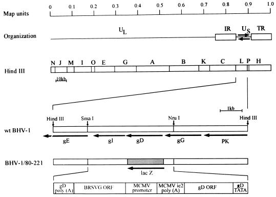
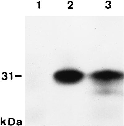
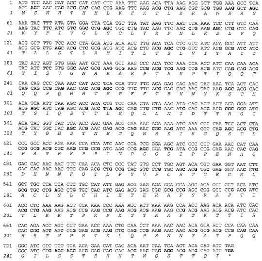
To express the BRSV G glycoprotein by recombinant BHV-1, recombination plasmid pROMeGori, which contains a cDNA fragment encompassing the BRSV G glycoprotein ORF under control of the murine cytomegalovirus (MCMV) early 1 promoter (Fig. 1), was cotransfected with purified BHV-1/80–221 DNA into MDBK cells and the progeny virus was serially diluted and plated on MDBK cells. Since the ORF encoding the essential gD is replaced by a lacZ expression cassette in BHV-1/80–221 (8), only viruses which have acquired the gD ORF by replacement of the lacZ cassette by the insert of the recombination plasmid should be able to replicate on noncomplementing cells (21). Virus from plaques that did not stain blue under a Bluo-Gal-containing agarose overlay were again subjected to titer determination on MDBK cells, and isolates which produced only “white” plaques were plaque purified once more and characterized further (see Fig. 4). Analysis of MDBK cells infected with the resulting recombinant BHV-1/eGori gave no indication of BRSV G glycoprotein expression by indirect immunofluorescence (see Fig. 7), and no stable transcripts were detected within cytoplasmic RNA (data not shown) or whole-cell RNA (see Fig. 5). BRSV G glycoprotein expression was also not detected in transient-expression experiments with plasmid pROMieGori (Fig. 1) in which the BRSV G glycoprotein ORF is under the control of the MCMV immediate-early 1 enhancer/promoter element (data not shown). In vitro translation of mRNA, transcribed in vitro from pSPGori, resulted in synthesis of a polypeptide whose apparent molecular mass of 31 kDa (Fig. 2, lane 2) was in good agreement with the calculated size of 29 kDa (24). This result indicated that mRNA from the BRSV Gori ORF contained within pROMeGori has the potential to direct protein synthesis, and we assumed that the failure to detect BRSV G glycoprotein-specific gene products was due to instability of BRSV G glycoprotein transcripts in the nuclei of BHV-1/eGori-infected cells. Therefore, a modified ORF encoding the BRSV G glycoprotein (Fig. 3) was chemically synthesized, isolated from plasmid pBRSVGsyn, and inserted into pROMe and pSP73. The resulting plasmids were named pROMeGsyn and pSPGsyn, respectively, and RNA transcribed from pSPGsyn directed the synthesis of a 31-kDa protein in a rabbit reticulocyte lysate (Fig. 2, lane 3) that comigrated with the polypeptide synthesized in vitro from the Gori ORF, supporting the conclusion that the 31-kDa protein represents the primary translation product encoded by the BRSV G glycoprotein ORF.
FIG. 1.
Construction of BHV-1 recombinants. The schematic representation of the prototype orientation of the BHV-1 genome is shown above the HindIII restriction fragment map of BHV-1 strain Schönböken (7, 27). The wild-type HindIII L fragment is enlarged, and the location and direction of transcription of genes encoding the putative protein kinase (PK) and glycoproteins G (gG), D (gD), I (gI), and E (gE) are indicated by arrows (15, 25, 40, 45). Relevant restriction enzyme cleavage sites are marked. The respective HindIII fragment of the gD− lacZ+ mutant BHV-1/80–221 is depicted below. The location and direction of transcription of the lacZ cassette (dotted area, not drawn to scale) that replaces the gD ORF is indicated by an arrow. Underneath, a diagram of the integration fragment contained in the recombination vectors is shown. The gD TATA and gD poly(A) segments indicate BHV-1 sequences representing the gD promoter and the gD polyadenylation signal, respectively, which provide homologous regions for recombination. In this study, transcription of the BRSV Gori or Gsyn ORF (BRSVG ORF) was directed by the MCMV ie1 enhancer-promoter element (6) in transient-expression experiments or by the MCMV e1 promoter (2) in recombinant BHV-1 infected cells. Transcription of the BHV-1 gD ORF which is followed by the MCMV ie2 poly(A) signal (28) is under the control of the authentic gD promoter.
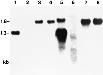
FIG. 4.
Identification of transcripts encompassing the Gsyn ORF. Whole-cell RNA from cells infected with BHV-1/eGsyn (lanes 1, 3, 5, and 7) and BHV-1/eGori (lanes 2, 4, 6, and 8) was prepared at 6 h p.i., and 5-μg samples were transferred to nitrocellulose after 1% agarose gel electrophoresis. The filters were hybridized to 32P-labeled DNA from the BRSV-Gsyn ORF (lanes 1 and 5), the BRSV-Gori ORF (lanes 2 and 6), and the BHV-1 gD ORF (lanes 3, 4, 7, and 8). Bound radioactivity was visualized by autoradiography. Lanes 5 to 8 are longer exposures of lanes 1 to 4. Transcript sizes are indicated in kilobases.
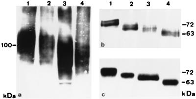
FIG. 7.
Evidence that BHV-1-expressed G glycoprotein contains N- and O-linked sugars. (a and b) Purified BHV-1/eGsyn virions were resuspended in PBS plus 0.5% NP-40 and digested overnight at 37°C with neuraminidase (lanes 2), neuraminidase and O-glycosidase (lanes 3), N-glycosidase F (lanes 4), and without enzyme (lanes 1). Digestion products were analyzed by immunoblotting with G glycoprotein-specific MAb 20 (a) or a polyclonal, BHV-1 gD-specific rabbit antiserum (b). (c) Purified, [35S]methionine-labeled BHV-1/Schö virions were lysed and incubated with gD-specific MAb 21/3/3. Immunoprecipitated proteins were incubated as described above, separated on SDS–10% polyacrylamide gels, and visualized by fluorography. The apparent molecular masses, indicated in kilodaltons on the left, were calculated from the migration of prestained (a and b) or 14C-methylated protein molecular mass standards (c) run on the respective gel.
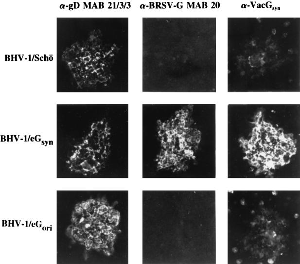
FIG. 5.
BHV-1-encoded BRSV G glycoprotein is expressed on the cell surface. MDBK cell cultures were infected with approximately 100 PFU of BHV-1/Schö, BHV-1/eGsyn, and BHV-1/eGori, fixed after the development of plaques, and used for indirect immunofluorescence with BHV-1 gD-specific MAb 21/3/3, BRSV G glycoprotein-specific MAb 20, and the anti-VacGsyn serum. Bound antibodies were visualized by staining with DTAF-conjugated goat anti-species immunoglobulin G.
FIG. 2.
Cell-free translation of cRNAs encompassing the BRSV Gori or Gsyn ORF. 35S-labeled proteins translated in vitro from RNA transcribed in vitro from plasmids pSPGori (lane 2) or pSPGsyn (lane 3) were size separated in SDS–10% polyacrylamide gels and visualized by fluorography. In lane 1, proteins synthesized from RNA transcribed from pSPGori antisense to the BRSV G glycoprotein ORF were separated. The apparent molecular mass of the translation products, indicated in kilodaltons on the left, was calculated from the migration of 14C-methylated protein molecular mass standards run on the same gel.
FIG. 3.
Comparison of the nucleotide sequences of the BRSV G glycoprotein cDNA ORF (upper sequence) and the Gsyn ORF (lower sequence). Exchanged nucleotides are in boldface type, and the deduced BRSV G glycoprotein amino acid sequence is shown in the single-letter code.
Integration of the BRSV-Gsyn ORF into the genome of BHV-1/80–221.
Recombination plasmid pROMeGsyn was cotransfected with purified BHV-1/80-221 DNA into MDBK cells, and the recombinant virus, BHV-1/eGsyn, was isolated as described above for BHV-1/eGori. To demonstrate integration of the expression cassettes into the genomes of the respective viruses, MDBK cells were infected with BHV-1/eGori, BHV-1/eGsyn, and BHV-1/80–221, phenotypically complemented by propagation on gD-expressing cells (8). Whole-cell DNA was prepared 20 h postinfection (p.i.), cleaved with HindIII, transferred to nitrocellulose filters after size separation in 0.6% agarose gels, and hybridized to Gsyn-, Gori-, and lacZ-specific 32P-labeled DNA fragments. The sizes of the fragments that hybridized to the respective probes were as expected (data not shown). We concluded that BHV-1/eGori and BHV-1/eGsyn were generated by homologous recombination as envisaged.
Transcription of the BRSV Gori and BRSV Gsyn ORFs in recombinant BHV-1-infected cells.
To test for transcription from the recombinant BRSV G genes, MDBK cells were infected with BHV-1/eGsyn (Fig. 4, lanes 1, 3, 5, and 7) and BHV-1/eGori (lanes 2, 4, 6, and 8). Cytoplasmic RNA was isolated at 6 h p.i., size separated by agarose gel electrophoresis, and transferred to nitrocellulose filters. A 32P-labeled DNA probe representing the BRSV Gsyn ORF detected an RNA of 1.3 kb after infection with BHV-1/eGsyn (lane 1) and, after extended exposure, a transcript of 1.8 kb (lane 5) whose synthesis is initiated approximately 500 bp upstream from the MCMV e1 promoter (13). Even after a longer exposure, Gori ORF-specific transcripts of the expected size were not detected (lanes 2 and 6). The faint smear in lane 6 might indicate the presence of fragmented Gori RNA. Hybridization of the filters with 32P-labeled DNA encompassing the BHV-1 gD ORF showed that comparable amounts of gD mRNA were detected in BHV-1/eGsyn-infected cells (lanes 3 and 7) and BHV-1/eGori-infected cells (lanes 4 and 8), demonstrating that the absence of stable transcripts from the BRSV Gori ORF was not due to degradation of the RNA from BHV-1/eGori-infected cells. Substantially the same results were obtained with RNAs isolated 16 h after infection with BHV-1/eGsyn and BHV-1/eGori in the absence or presence of phosphonoacetic acid (data not shown).
To support the assumption that transcripts from the BRSV Gori ORF are unstable in BHV-1/eGori-infected cells, elongation of initiated transcripts in BHV-1/eGori- and BHV-1/eGsyn-infected MDBK cells was analyzed by nuclear runoff transcription assays. 32P-labeled RNA, synthesized in nuclei isolated at 6 h p.i., was hybridized to DNA from the plasmid vector pSP73 or from plasmids pSPGori, pSPGsyn, or pSPgD dotted onto nitrocellulose membranes. Elongation of RNA from both BRSV G ORFs was detected (data not shown). The ratio of bound radioactivity between the respective BRSV G ORFs and the corresponding BHV-1 gD ORFs was 1.7 with RNA from BHV-1/eGori-infected cells and 2.1 with RNA from BHV-1/eGsyn-infected cells, demonstrating that the Gori ORF was transcribed in BHV-1/eGori-infected cells. These results are in accordance with the conclusion that BHV-1-expressed BRSV Gori RNAs are unstable in the nuclei of infected cells.
Identification and characterization of BHV-1-expressed BRSV G glycoprotein.
To test for the expression of the BRSV G glycoprotein in BHV-1/eGsyn-infected cells, BRSV G glycoprotein-specific MAb 20 and a polyclonal antiserum (anti-VacGsyn), raised in rabbits after infection with Gsyn-expressing recombinant vaccinia virus, were used to stain wild-type BHV-1 strain Schönböken (BHV-1/Schö)-, BHV-1/eGsyn-, or BHV-1/eGori-induced plaques by indirect immunofluorescence. The cultures were fixed 2 days after infection and incubated with the BHV-1 gD-specific MAb 21/3/3, MAb 20, or the anti-VacGsyn serum. Antibodies bound on the cell surface were visualized by using DTAF-conjugated anti-species immunoglobulin G and fluorescence microscopy. As shown in Fig. 5, MAb 20 and the anti-VacGsyn serum bound only to cells in plaques generated by BHV-1/eGsyn whereas MAb 21/3/3 reacted with gD on the surface of cells infected with BHV-1/Schö, BHV-1/eGsyn, and BHV-1/eGori. The conclusion that the BRSV Gsyn ORF-encoding protein is expressed on the surface of BHV-1/eGsyn-infected cells was also demonstrated by flow cytometry with living cells and the same antibodies as above (data not shown).
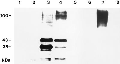
To further characterize the BRSV Gsyn ORF-encoded gene product, proteins from infected cells (Fig. 6, lanes 1 to 4), purified virions (lanes 5 to 7), and the cell culture medium from BHV-1/eGsyn-infected cells (lane 8) were analyzed by immunoblotting because MAb 20 and the anti-VacGsyn serum did not work for immunoprecipitation. MAb 20 did not specifically bind to proteins from cells infected with BHV-1/Schö (Fig. 6, lane 1) and BHV-1/eGori (lane 2) or to proteins from purified BHV-1/Schö and BHV-1/eGori virions (lanes 5 and 6) but reacted strongly with proteins with apparent molecular masses of 38 and 43 kDa from BHV-1/eGsyn-infected cells (lane 3). In addition, several weaker-staining bands ranging in size from 70 to about 100 kDa were detected. To elucidate possible precursor-product relationships among the MAb 20-reactive polypeptides, BHV-1/eGsyn-infected cells were incubated for 2 h with cycloheximide (final concentration, 100 μg per ml) before lysis to inhibit de novo protein synthesis. This treatment resulted in a considerable decrease in the signal at 38 kDa and a slight reduction in the band intensity of the signal at 43 kDa, whereas the bands corresponding to larger proteins increased in size and abundance (Fig. 6, lane 4). Prolongation of the cycloheximide incubation time did not significantly alter this pattern but resulted in a decrease in the intensities of all bands (data not shown). Among proteins from purified virus particles, MAb 20 bound only to polypeptides from BHV-1/eGsyn virions which migrated as a diffuse band with an apparent molecular mass between 70 and 120 kDa (lane 7). No MAb 20-reactive proteins were detected in the medium of BHV-1/eGsyn-infected cells (lane 8). Identical results were obtained with the anti-VacGsyn serum and BRSV G glycoprotein-specific MAb 57 (9). The latter immunoprecipitated the large molecules but not the 38- and 43-kDa proteins from cells infected with BHV-1/eGsyn in the presence of [35S]methionine and [35S]cysteine. From these results, we conclude that the 38- and 43-kDa proteins represent BRSV G glycoprotein precursor molecules which are subsequently modified to the large form, which is also associated with BHV-1/eGsyn virions.
FIG. 6.
Identification of the BHV-1/eGsyn-expressed BRSV G glycoprotein. MDBK cells were infected with BHV-1/Schö (lanes 1 and 5), BHV-1/eGori (lanes 2 and 6), and BHV-1/eGsyn (lanes 3, 4, 7, and 8). Proteins from infected cells, harvested at 10 h p.i. (lanes 1 to 4), from purified virions (lanes 5 to 7), and from cell culture medium (lane 8), were analyzed by immunoblotting with BRSV G glycoprotein-specific MAb 20. Proteins shown in lane 4 were from cells incubated with cycloheximide (100 μg/ml) for 2 h before lysis. Apparent molecular masses are indicated in kilodaltons.
The carbohydrate composition of virus particle-associated BRSV G glycoprotein was analyzed by incubation of purified BHV-1/eGsyn virions with glycosidase F, neuraminidase, and neuraminidase together with O-glycosidase followed by immunoblotting after SDS-PAGE with MAb 20 (Fig. 7a) or a polyclonal rabbit serum raised against purified BHV-1 gD (Fig. 7b) to monitor the reactions. According to the enzymes used, the electrophoretic mobilities of virion-associated gD increased as expected (41), and no differences were detected in comparison to the reaction products of immunoprecipitated virion gD (Fig. 7b and c), indicating the presence of sufficient enzymatic activity for complete deglycosylation within the respective probes. The BRSV G glycoprotein molecules appeared unaffected after incubation with neuraminidase (Fig. 7a, lane 2) and exhibited an increased electrophoretic mobility following digestion with neuraminidase and O-glycosidase (lane 3) or N-glycosidase F (lane 4), which demonstrates the presence of O- and N-linked carbohydrates in the virion-associated BRSV G glycoprotein. However, digestion products obtained after incubation of the virion proteins with neuraminidase and O-glycosidase migrated slower and were more diffuse, as expected for the only N-glycosylated form. Whether this discrepancy is due to additional modifications, the presence of O-glycosidase-resistant O-linked carbohydrates (22), or incomplete removal of O-linked glycans due to specifics of the BRSV G glycoprotein has to be analyzed.
Virus neutralization assays.
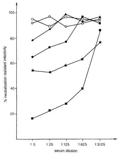
The immunoblot analysis in Fig. 6 indicated that BHV-1/eGsyn virus particles contain the BRSV G glycoprotein and therefore should be susceptible to inactivation by BRSV G glycoprotein-specific antibodies. BHV-1/eGori and BHV-1/eGsyn virions were tested for complement-dependent and -independent neutralization by MAb 57, the anti-VacGsyn serum, and the anti-BRSV serum 2106 (9). With complement, MAb 57, which had no effect on the infectivity of BHV-1/eGori (data not shown), reduced the number of BHV-1/eGsyn-induced plaques to 52% at a final dilution of 1:25. The anti-VacGsyn serum specifically neutralized BHV-1/eGsyn in the presence of complement to 50% neutralization at a dilution of about 1:1,000 (Fig. 8). No activity was detectable without complement (data not shown). The BHV-1/eGsyn-neutralizing activity of anti-BRSV serum 2106 was weak in the absence of complement and more pronounced after the addition of complement. Both polyclonal sera had no significant effect on the infectivity of BHV-1/eGori (Fig. 8) and BHV-1/Schö (data not shown).
FIG. 8.
BHV-1/eGsyn virions are susceptible to neutralization by antibodies against the BRSV G glycoprotein. Approximately 200 PFU of BHV-1/eGori was incubated with 20% fetal calf serum plus complement or serial dilutions of anti-VacGsyn with complement (open squares) or the anti-BRSV hyperimmune serum with complement (open circles), and about 160 PFU of BHV-1/eGsyn was incubated with 20% FCS and complement or with serial dilutions of anti-VacGsyn and complement (solid squares), MAb 57 with complement (stars), anti-BRSV serum 2106 without complement (solid triangles, no complement added to the FCS control), or anti-BRSV serum 2106 with complement (solid circles). After 60 min at 37°C, the virions were plated on MDBK cells and the cultures were overlaid with semisolid medium. Plaques were counted 3 days later. The plaque count of each FCS control was defined as 100% neutralization-resistant infectivity. Serum dilutions are indicated.
DISCUSSION
Analysis of the expression from the authentic BRSV G glycoprotein ORF after infection of cells with BHV-1/eGori indicated that the transcripts are unstable in the nucleus. We assumed that this was due to the nucleotide sequence of the RNA, which might contain signals that do not interfere with the stability of the RNA in the cytoplasm of cells infected with BRSV or recombinant vaccinia virus expressing the BRSV G glycoprotein (24) but result in degradation of the transcripts in the nucleus. Therefore, the complete BRSV G glycoprotein ORF was reconstructed by using synthetic oligonucleotides. The codon usage of the modified ORF was adjusted to that of BHV-1 gD, resulting in an increase of the G+C content from 42 to 62%, and the introduction of putative splice donor signals (30) was avoided. The synthetic ORF was inserted into the genome of BHV-1 under the control of the MCMV e1 promoter (2, 21), and analysis of the gene products in BHV-1/eGsyn-infected cells demonstrated transcription of mRNA, which was translated into proteins recognized by BRSV G glycoprotein-specific MAb 20 and the anti-VacGsyn serum. Although we did not attempt to delineate sequence motifs leading to instability of the Gori transcripts in the nucleus, our results strongly suggest their existence, which might also explain the failure to express the authentic ORF encoding BRSV fusion protein F by BHV-1 (13). Introduction of a series of single restriction enzyme cleavage sites into the Gsyn ORF will be helpful in identifying sequence elements that prevent the generation of mRNA from the genomic BRSV G glycoprotein ORF, which may be beneficial for the development of systems for the expression of RNA virus genes in the nuclei of mammalian cells.
The BRSV G glycoprotein is a type II membrane glycoprotein with a 37-amino-acid N-terminal cytoplasmic domain followed by 29 hydrophobic amino acids representing the transmembrane region. The remaining 191 amino acids are mainly hydrophilic and constitute the extracellular domain (24). Among BHV-1/eGsyn-infected cell proteins, MAb 20, MAb 57, and the anti-VacGsyn serum detected proteins with apparent molecular masses of 38, 43, and 70 to 120 kDa. Inhibition of de novo protein synthesis by cycloheximide resulted in a decrease of the signals generated by the 38- and 43-kDa proteins and a concomitant increase in intensity and size of the large-protein band. Although the exact precursor-product relationship awaits clarification, we assume that the 38- and 43-kDa proteins represent precursor molecules of the mature BRSV G glycoprotein. This assumption is in accordance with results reported by Lerch et al. (24), who demonstrated that in cells infected with a BRSV G glycoprotein-expressing recombinant vaccinia virus, two proteins which migrated with an apparent molecular mass of 43 kDa and as a broad band between 68 and 97 kDa specifically reacted with an anti-BRSV serum in immunoblots. They further provided evidence that the 43-kDa form represents a precursor containing only N-linked carbohydrates whereas the 68- to 97-kDa molecules constitute the mature N- and O-glycosylated BRSV G glycoprotein.
Expression of the BRSV G glycoprotein in BHV-1/eGsyn-infected cells was analyzed only by immunoblotting because the BRSV G glycoprotein-specific MAb 20 and the anti-VacGsyn serum did not work for immunoprecipitation and MAb 57 precipitated only the mature glycoprotein after synthesis in the presence of [35S]methionine and [35S]cysteine (not shown). The observation that antibodies against the BRSV G glycoprotein bind in immunoblots but fail to immunoprecipitate the antigen is, although surprising, not without precedent and has also been reported by Lerch et al. (24). The reason for this apparent paradox, however, remains unclear.
Immunofluorescence and flow cytometry (data not shown) with MAb 20 and the anti-VacGsyn serum demonstrated that BHV-1/eGsyn-infected cells expressed the BRSV G glycoprotein on the cell surface, and we conclude that it is tightly anchored in the cell membrane since no secreted molecules were found in the medium from BHV-1/eGsyn-infected cultures. Thus, our results indicate Golgi processing and post-Golgi transport of the BHV-1/eGsyn-encoded BRSV G glycoprotein to the cell surface, comparable to the situation in BRSV- or recombinant vaccinia virus-infected cells (24).
Since alphaherpesviruses probably do not encode type II glycoproteins, we investigated whether the BRSV G glycoprotein that presumably lacks any herpesvirus-specific targeting signals is incorporated into BHV-1 particles. Western blot analysis with MAb 20 revealed incorporation of the BRSV G glycoprotein into purified virus particles, and this was confirmed by the susceptibility of BHV-1/eGsyn virions to neutralization by the anti-VacGsyn serum, the anti-BRSV immune serum, and MAb 57 in the presence of complement. That these antibodies did not neutralize in the absence of complement was not too surprising, since it is unlikely that the BRSV G glycoprotein exhibits a biological function for the infectivity of BHV-1. In addition, flow cytometry demonstrated that MAb 20 and the anti-VacGsyn serum bound to living cells infected with purified BHV-1/eGsyn virions in the presence of cycloheximide to prevent de novo protein synthesis (13). This further supports the conclusion that the orientation of the BRSV G glycoprotein within the viral envelope is correct.
To our knowledge, this is the first description of a class II membrane glycoprotein becoming incorporated into alphaherpesvirus virions. The presence of the BRSV G glycoprotein in the given background of BHV-1/eGsyn exerted only a slight influence on the entry of virions into target cells in comparison to the situation for wild-type BHV-1 (data not shown). Although no repaired viruses were included in this study, we believe that it is reasonable to assume that BRSV G glycoprotein does not significantly interfere with the envelope structures required for the initial steps of the BHV-1 infection.
This study has practical implications for herpesvirus-based vector vaccine development: (i) it indicates that modification of RNA virus genes may be required for efficient expression, and (ii) it raises the possibility of targeting foreign antigens as class II membrane (glyco)proteins into the envelope of herpesvirus virions, which might be important when booster vaccinations with live recombinant viruses are inefficient due to existing immunity to the vector, a situation observed with recombinant vaccinia viruses (13, 26).
ACKNOWLEDGMENTS
We thank Julie Furze for providing MAb 57, Helmut Stephan and Elke Zorn for photography, J. Naval for corrections, and Bernd Köllner for assistance with the flow cytometry.
Financial support for synthesis and expression of the Gsyn ORF by Intervet International BV is greatly appreciated. G.K. was supported by grant BIOT-CT93-0489 from the Commission of the European Communities.
REFERENCES
- 1.Bello L J, Whitbeck C, Lawrence W C. Bovine herpesvirus 1 as a live vector for expression of foreign genes. Virology. 1992;190:666–673. doi: 10.1016/0042-6822(92)90904-4. [DOI] [PubMed] [Google Scholar]
- 2.Bühler B, Keil G M, Weiland F, Koszinowski U H. Characterization of the murine cytomegalovirus early transcription unit e1 that is induced by immediate-early proteins. J Virol. 1990;64:1907–1919. doi: 10.1128/jvi.64.5.1907-1919.1990. [DOI] [PMC free article] [PubMed] [Google Scholar]
- 3.Chowdhury S. Construction and characterization of an attenuated bovine herpesvirus type 1 (BHV-1) recombinant virus. Vet Microbiol. 1996;52:13–23. doi: 10.1016/0378-1135(96)00043-0. [DOI] [PubMed] [Google Scholar]
- 4.Collins P L. The molecular biology of human respiratory syncytial virus (RSV) of the genus Pneumovirus. In: Kingsburg D W, editor. The paramyxoviruses. New York, N.Y: Plenum Press; 1991. pp. 103–162. [Google Scholar]
- 5.Dormitzer P R, Ho D Y, Mackow E R, Mocarsky E S, Greenberg H B. Neutralizing epitopes on herpes simplex virus-1-expressed rotavirus VP7 are dependent on coexpression of other rotavirus proteins. Virology. 1992;187:18–32. doi: 10.1016/0042-6822(92)90291-v. [DOI] [PubMed] [Google Scholar]
- 6.Dorsch-Häsler K, Keil G M, Weber F, Jasin M, Schaffner W, Koszinowski U H. A long and complex enhancer activates transcription of the gene coding for the highly abundant immediate early mRNA in murine cytomegalovirus. Proc Natl Acad Sci USA. 1985;82:8325–8329. doi: 10.1073/pnas.82.24.8325. [DOI] [PMC free article] [PubMed] [Google Scholar]
- 7.Engels M, Giuilani C, Wild P, Beck T M, Loepfe E, Wyler R. The genome of bovine herpesvirus-1 (BHV-1) exhibiting a neuropathogenic potential compared to known BHV-1 strains by restriction site mapping and cross-hybridization. Virus Res. 1987;6:57–73. doi: 10.1016/0168-1702(86)90057-2. [DOI] [PubMed] [Google Scholar]
- 8.Fehler F, Herrmann J M, Saalmüller A, Mettenleiter T C, Keil G M. Glycoprotein IV of bovine herpesvirus 1-expressing cell line complements and rescues a conditionally lethal viral mutant. J Virol. 1992;66:831–839. doi: 10.1128/jvi.66.2.831-839.1992. [DOI] [PMC free article] [PubMed] [Google Scholar]
- 9.Furze J, Wertz G, Lerch R, Taylor G. Antigenic heterogeneity of the attachment protein of bovine respiratory syncytial virus. J Gen Virol. 1994;75:363–370. doi: 10.1099/0022-1317-75-2-363. [DOI] [PubMed] [Google Scholar]
- 10.Furze J M, Roberts S R, Wertz W G, Taylor G. Antigenically distinct G glycoproteins of BRSV strains share a high degree of genetic homogeneity. Virology. 1997;231:48–58. doi: 10.1006/viro.1997.8490. [DOI] [PubMed] [Google Scholar]
- 11.Greenberg M E, Ziff E B. Stimulation of 3T3 cells induces transcription of the c-fos proto-oncogene. Nature (London) 1984;311:433–438. doi: 10.1038/311433a0. [DOI] [PubMed] [Google Scholar]
- 12.Hanon E, Vanderplasschen A, Lyaku J, Keil G M, Denis M, Pastoret P-P. Inactivated bovine herpesvirus 1 induces apoptotic cell death of mitogen-stimulated bovine peripheral blood mononuclear cells. J Virol. 1996;70:4116–4120. doi: 10.1128/jvi.70.6.4116-4120.1996. [DOI] [PMC free article] [PubMed] [Google Scholar]
- 13.Keil, G. M. Unpublished observation.
- 14.Keil G M, Ebeling-Keil A, Koszinowski U H. Immediate-early genes of murine cytomegalovirus: location, transcripts, and translation products. J Virol. 1987;61:526–533. doi: 10.1128/jvi.61.2.526-533.1987. [DOI] [PMC free article] [PubMed] [Google Scholar]
- 15.Keil G M, Engelhardt T, Karger A, Enz M. Bovine herpesvirus 1 Us open reading frame 4 encodes a glycoproteoglycan. J Virol. 1996;70:3032–3038. doi: 10.1128/jvi.70.5.3032-3038.1996. [DOI] [PMC free article] [PubMed] [Google Scholar]
- 16.Keil G M, Fibi M R, Koszinowski U H. Characterization of the major immediate-early polypeptides encoded by murine cytomegalovirus. J Virol. 1985;54:422–428. doi: 10.1128/jvi.54.2.422-428.1985. [DOI] [PMC free article] [PubMed] [Google Scholar]
- 17.Kit M, Kit S, DiMarchi R D, Little S P, Gale C. Modified-live infectious bovine rhinotracheitis virus vaccine expressing monomer and dimer forms of foot and mouth disease capsid protein epitopes on surface of hybrid virus particles. Arch Virol. 1991;120:1–17. doi: 10.1007/BF01310945. [DOI] [PubMed] [Google Scholar]
- 18.Kit M, Kit S, Little S P, DiMarchi R D, Gale C. Bovine herpesvirus-1 (infectious bovine rhinotracheitis virus)-based viral vector which expresses foot and mouth disease epitopes. Vaccine. 1991;9:564–572. doi: 10.1016/0264-410x(91)90243-y. [DOI] [PubMed] [Google Scholar]
- 19.Kit S, Otsuka H, Kit M. Expression of porcine pseudorabies virus genes by a bovine herpesvirus-1 (infectious bovine rhinotracheitis virus) vector. Arch Virol. 1992;124:1–20. doi: 10.1007/BF01314621. [DOI] [PubMed] [Google Scholar]
- 20.Kühnle, G., and G. M. Keil. Unpublished observation.
- 21.Kühnle G, Collins R A, Scott J E, Keil G M. Bovine interleukins 2 and 4 expressed in recombinant bovine herpesvirus 1 are biologically active secreted glycoproteins. J Gen Virol. 1996;77:2231–2240. doi: 10.1099/0022-1317-77-9-2231. [DOI] [PubMed] [Google Scholar]
- 22.Kurilla M G, Heinemann T, Davenport L C, Kieff E, Hutt-Fletcher L M. A novel Epstein-Barr virus glycoprotein gp150 expressed from the BDLF3 open reading frame. Virology. 1995;209:108–121. doi: 10.1006/viro.1995.1235. [DOI] [PubMed] [Google Scholar]
- 23.Langedijk J P M, Schaaper W M M, Meloen R H, van Oirschot J T. Proposed three-dimensional model for the attachment protein G of respiratory syncytial virus. J Gen Virol. 1996;77:1249–1257. doi: 10.1099/0022-1317-77-6-1249. [DOI] [PubMed] [Google Scholar]
- 24.Lerch R A, Anderson K, Wertz G W. Nucleotide sequence analysis and expression from recombinant vectors demonstrate that the attachment glycoprotein G of bovine respiratory syncytial virus is distinct from that of human respiratory syncytial virus. J Virol. 1990;64:5559–5567. doi: 10.1128/jvi.64.11.5559-5569.1990. [DOI] [PMC free article] [PubMed] [Google Scholar]
- 25.Leung-Tack P, Audonnet J-C, Riviere M. The complete DNA sequence and the genetic organization of the short unique region (Us) of the bovine herpesvirus type 1 (ST strain) Virology. 1994;199:409–421. doi: 10.1006/viro.1994.1139. [DOI] [PubMed] [Google Scholar]
- 26.Mackett M, Smith G L. Vaccinia virus expression vectors. J Gen Virol. 1986;67:2067–2082. doi: 10.1099/0022-1317-67-10-2067. [DOI] [PubMed] [Google Scholar]
- 27.Mayfield J E, Good P J, Vanoort H J, Campbell A R, Reed D E. Cloning and cleavage site mapping of DNA from bovine herpesvirus 1 (Cooper strain) J Virol. 1983;47:259–264. doi: 10.1128/jvi.47.1.259-264.1983. [DOI] [PMC free article] [PubMed] [Google Scholar]
- 28.Messerle M, Keil G M, Koszinowski U H. Structure and expression of murine cytomegalovirus immediate-early gene 2. J Virol. 1991;65:1638–1643. doi: 10.1128/jvi.65.3.1638-1643.1991. [DOI] [PMC free article] [PubMed] [Google Scholar]
- 29.Miethke A, Keil G M, Weiland F, Mettenleiter T C. Unidirectional complementation between glycoprotein B homologues of pseudorabiesvirus and bovine herpesvirus 1 is determined by the carboxy terminal part of the molecule. J Gen Virol. 1995;76:1623–1635. doi: 10.1099/0022-1317-76-7-1623. [DOI] [PubMed] [Google Scholar]
- 30.Mount S M. A catalogue of splice junctions. Nucleic Acids Res. 1982;10:459–472. doi: 10.1093/nar/10.2.459. [DOI] [PMC free article] [PubMed] [Google Scholar]
- 31.Otsuka H, Xuan X. Construction of bovine herpesvirus-1 (BHV-1) recombinants which express pseudorabies virus (PRV) glycoproteins gB, gC, gD and gE. Arch Virol. 1996;141:57–71. doi: 10.1007/BF01718588. [DOI] [PubMed] [Google Scholar]
- 32.Raggo C, Fitzpatrick D R, Babiuk L A, Liang X. Expression of bovine interleukin-1 beta in a bovine herpesvirus-1 vector: in vitro analysis. Virology. 1996;221:78–86. doi: 10.1006/viro.1996.0354. [DOI] [PubMed] [Google Scholar]
- 33.Rijsewijk, F. A. M., and J. T. van Oirschot. Unpublished observation.
- 34.Roizman B, Desrosiers R C, Fleckenstein B, Lopes C, Minson A C, Studdert M J. The family herpesviridae: an update. Arch Virol. 1992;123:425–449. doi: 10.1007/BF01317276. [DOI] [PubMed] [Google Scholar]
- 35.Sambrook J, Fritsch E F, Maniatis T. Molecular cloning: a laboratory manual. 2nd ed. Cold Spring Harbor, N.Y: Cold Spring Harbor Laboratory; 1989. [Google Scholar]
- 36.Satake M, Coligan J E, Elango N, Norrby E, Venkatesan S. Respiratory syncytial virus envelope glycoprotein (G) has a novel structure. Nucleic Acids Res. 1985;13:7795–7812. doi: 10.1093/nar/13.21.7795. [DOI] [PMC free article] [PubMed] [Google Scholar]
- 37.Schmitt J, Keil G M. Identification and characterization of the bovine herpesvirus 1 UL7 gene and gene product which are not essential for virus replication in cell culture. J Virol. 1996;70:1091–1099. doi: 10.1128/jvi.70.2.1091-1099.1996. [DOI] [PMC free article] [PubMed] [Google Scholar]
- 38.Schröder C, Linde G, Fehler F, Keil G M. From essential to beneficial: glycoprotein D loses importance for replication of bovine herpesvirus 1 in cell culture. J Virol. 1997;71:25–33. doi: 10.1128/jvi.71.1.25-33.1997. [DOI] [PMC free article] [PubMed] [Google Scholar]
- 39.Schwyzer M, Ackermann M. Molecular virology of ruminant herpesviruses. Vet Microbiol. 1996;53:17–29. doi: 10.1016/s0378-1135(96)01231-x. [DOI] [PubMed] [Google Scholar]
- 40.Tikoo S, Fitzpatrick D R, Babiuk L A, Zamb T J. Molecular cloning, sequencing, and expression of functional bovine herpesvirus-1 glycoprotein IV in transfected bovine cells. J Virol. 1990;64:5132–5142. doi: 10.1128/jvi.64.10.5132-5142.1990. [DOI] [PMC free article] [PubMed] [Google Scholar]
- 41.van Drunen Littel-vanden Hurk S, Babiuk L A. Synthesis and processing of bovine herpesvirus 1 glycoproteins. J Virol. 1986;59:401–410. doi: 10.1128/jvi.59.2.401-410.1986. [DOI] [PMC free article] [PubMed] [Google Scholar]
- 42.van Engelenburg F A C, Kaashoek M J, Rijsewijk F A M, van den Burg L, Moerman A, Gielkens A L J, van Oirschot J T. A glycoprotein E deletion mutant of bovine herpesvirus 1 is avirulent in calves. J Gen Virol. 1994;75:2311–2318. doi: 10.1099/0022-1317-75-9-2311. [DOI] [PubMed] [Google Scholar]
- 43.van Zijl M, Wensvoort G, de Kluyver E, Hulst M, van der Gulden H, Gielkens A, Berns A, Moormann R. Live attenuated pseudorabies virus expressing envelope glycoprotein E1 of hog cholera virus protects swine against both pseudorabies and hog cholera. J Virol. 1991;65:2761–2765. doi: 10.1128/jvi.65.5.2761-2765.1991. [DOI] [PMC free article] [PubMed] [Google Scholar]
- 44.Wertz G, Collins P L, Huang Y, Gruber C, Levine S, Ball L A. Nucleotide sequence of the G protein gene of human respiratory syncytial virus reveals an unusual type of viral membrane protein. Proc Natl Acad Sci USA. 1985;82:4075–4079. doi: 10.1073/pnas.82.12.4075. [DOI] [PMC free article] [PubMed] [Google Scholar]
- 45.Whitbeck J C, Knapp A C, Enquist L W, Lawrence W C, Bello L J. Synthesis, processing, and oligomerization of bovine herpesvirus 1 gE and gI membrane proteins. J Virol. 1996;70:7878–7884. doi: 10.1128/jvi.70.11.7878-7884.1996. [DOI] [PMC free article] [PubMed] [Google Scholar]


