Abstract
Introduction
Supplementation with dietary neuro-pigments lutein (L) and zeaxanthin (Z) has been shown to improve many aspects of visual and cognitive function in adults. In this study, we tested whether a similar intervention could improve such outcomes in preadolescent children.
Methods
Sixty children (age range 5–12 years) were randomized in a 2:1 ratio in this double-blind, placebo-controlled clinical trial. Subjects were supplemented with gummies containing either a combination of 10 mg lutein and 2 mg zeaxanthin (LZ) or placebo for 180 days. Macular pigment optical density (MPOD) was the primary endpoint. The secondary endpoints included serum levels of L and Z, and brain-derived neurotrophic factor (BDNF), critical flicker fusion (CFF), eye strain and fatigue using visual analogue scales (VAS), Children’s Sleep Habits Questionnaire-Abbreviated (CSHQ-A), and Creyos Health cognitive domains like attention, focus/concentration, episodic memory and learning, visuospatial working memory, and visuospatial processing speed. Safety was assessed throughout the study on the basis of physical examination, vital signs, clinical laboratory tests, and monitoring of adverse events.
Results
The LZ group showed significant increases in MPOD at all visits post-supplementation, with significant increases as early as day 42 compared to placebo. The LZ group showed significant increases in serum lutein levels, reduced eye strain and fatigue, and improved cognitive performance (focus, episodic memory and learning, visuospatial working memory) at days 90 and 180 compared to placebo. Further, the LZ group showed significant increases in processing speed (CFF), attention, visuospatial processing, and serum Z and BDNF levels on day 180 compared to placebo. No safety concerns were observed.
Conclusions
Supplementing LZ resulted in increased MPOD levels, along with increased serum levels of L, Z, and BDNF. These changes were associated with improved visual and cognitive performances and reduction in eye strain and eye fatigue in the children receiving LZ gummies. The investigational product was safe and well tolerated.
Trial Registration
http://ctri.nic.in/ Identifier CTRI/2022/05/042364.
Keywords: BDNF, Blue light, Cognitive performance, Lutein and zeaxanthin, Vision performance, Screen time
Key Summary Points
| Why carry out this study? |
| Supplementing L and Z in young adults improves many aspects of vision and cognitive functions |
| It is not known whether, like in adults and the elderly, an intervention with LZ can actually result in changes in the visual performance and brain function of children |
| What was learned from the study? |
| Supplementing preadolescents with LZ gummies increased MPOD, serum L and Z, and BDNF. These changes improved visual and cognitive performance, and reduced eye strain and fatigue in children |
| The investigational product was safe and well tolerated |
| The sequalae of positive visual and cognitive effects combined with the ease of this intervention suggests that this type of supplementation might be an additional tool that might help optimize central nervous system development, especially in typically undernourished children |
Introduction
A common aphorism in pediatrics observes that children are not simply small adults [1]. This is particularly true when considering the rapid maturation of the central nervous system (CNS). For example, 90% of brain growth occurs before the age of five (doubling in size the first year) [2]. Such rapid change makes adolescents particularly responsive to both environmental and dietary input. Such input, however, can be either positive or negative. For example, the increasing use of digital media and devices early in childhood has likely contributed to increasing rates of myopia [3] and asthenopia (approx. 20%) [4]/digital eye strain [5]. Dietary deficiencies are particularly impactful on a system that is rapidly building and, hence, requires both foundational support and enhanced protection from the increases in metabolism that such building entails. The brain is about 60% fat by volume (the composition being influenced by intake) [6], and the young brain is more vulnerable to oxidative [7] and inflammatory stress [8]. These increased vulnerabilities are likely why so much aging/insult to the CNS occurs in the first two decades. Metabolic stress due to accumulation of metabolites including lactate and free radicals is exacerbated by other susceptibilities like a crystalline lens which is highly transparent in young adults as compared to old and transmits much higher proportions of ultraviolet light (e.g., the young lens transmits UVB up to the age of about 30 years) [9]. Taken together, it creates a system with both extreme potential and vulnerability.
The disproportional impact of development is, of course, why special attention must be paid to both the optimal diet and experience of children. It seems likely that the CNS has evolved mechanisms for optimizing development but such optimization requires proper input. On the environmental side, the system is highly plastic, e.g., the acquisition of languages, visual motor skills, etc. is enhanced. On the dietary side, the brain absorbs a wide array of phytochemicals in regions necessary for cognitive growth. For example, the lipid-soluble antioxidant/anti-inflammatory pigments lutein and zeaxanthin (L and Z) are found in relatively high concentration [10] in areas such as the hippocampus, occipital and frontal cortex (areas critical for information processing) [11]. Interventional studies have shown that supplementing L and Z in young adults results in improvements in many aspects of cognition ranging from cognitive fundamentals (like visual processing speed) to higher-order executive functions (such as verbal fluency and memory) reviewed in Stringham et al., 2019 [12]. Improved cognitive function in adolescents also correlates with higher levels of L and Z [13–17] in CNS tissue (retinal levels are often taken as a biomarker for increased amounts in brain) [10]. What remains is the question of whether, like adults and the elderly, an intervention with LZ can actually result in changes in the vision and brain function of children. That was the purpose of the present study.
L and Z are not synthesized by humans and have to be provided through dietary sources [18]. Epidemiological data indicate that the average intake of L and Z from dietary sources is in the range of 1 to 2 mg/day [19–21] with considerable inter-individual variability in serum concentrations and MPOD [22]. In a US study conducted in 2003–2004 by National Health and Nutrition Examination Survey (NHANES) based on 24 h diet recall estimated an average lutein intake of 311 µg/day among 4–8-year-olds, 335 µg/day for 9–13-year-olds, and 432 µg/day for 14–18-year-olds [23]. However, a similar Canadian study indicated higher (1230 µg/day) lutein intake in children with mean age 5.75 years [24]. The European Food Safety Agency (EFSA) database on children 3–9 years of age indicates that in all the member states, the consumption of fruit and vegetables intake is substantially below the World Health Organization (WHO) recommendations [25]. Overall existing data indicate racial differences in MPOD levels, with Caucasians having significantly lower MPOD levels compared with African Americans and South Asians [26, 27]. To our knowledge, there is no published data on the lutein intake of Indian populations including children.
L and Z at a dietary ratio of 5:1 has been extensively explored in multiple clinical trials at a dose of 10 mg lutein in combination with 2 mg zeaxanthin and has been shown to provide optimal eye-related health benefits while being safe [28–38]. Further, the formulation used in the current study has also been confirmed previously in multiple clinical studies to safely provide visual and cognitive health benefits in healthy subjects in age groups of 18–60 years [39, 40]. Studies show that L and Z supplements are safe for all age groups including infants and children and have been used effectively to increase plasma levels of L and Z with visual benefits and enhanced cognitive performance during childhood [41].
The current study explores the efficacy and safety of L and Z supplementation in the form of gummies in a randomized, double-blind, placebo-controlled study in young children in relation to visual and cognitive benefits.
Methods
Study Design and Procedures
This was a prospective, randomized, double-blind, placebo-controlled, two-arm, parallel, single-center, clinical interventional study in children aged between 5 and 12 years. The study was initiated after obtaining written approval from an institutional ethics committee (Pranav Diabetes Center Ethics Committee, Bengaluru, Karnataka, India). The study was carried out as per the requirements of the Indian Council of Medical Research (ICMR) ethical guidelines, International Council for Harmonization (ICH) Guidance on Good Clinical Practice (E6R2), and the Declaration of Helsinki. The study was registered with the Clinical Trials Registry of India (CTRI/2022/05/042364).
Children who met the eligibility criteria were included in the study after obtaining a voluntary written consent from the subjects and one of their parents. Sixty eligible participants were randomized in a 2:1 ratio to an LZ group and placebo group. A randomization schedule was generated by a non-study-assigned, independent statistician using R statistical software (Version 4.3.1, Auckland, New Zealand). The LZ supplement was a gummy consisting of 10 mg of lutein and 2 mg zeaxanthin isomers (Lutemax Kids). The placebo was an identical gummy but without any active ingredient. Both investigational products were manufactured by OmniActive Health Technologies Ltd, India. After randomization, the subjects were instructed to consume one gummy every morning after breakfast at the same time every day, for 180 consecutive days.
Total study duration was for a maximum of 191 days which included the screening period of 7 days, randomization day, and the supplementation period of 180 days (6 months) followed by an end of study visit at day 180. The study was conducted in five visits over a period of 180 days, i.e., screening/baseline visit (day − 7 to day − 1), randomization visit (day 0), first follow-up visit (day 42 ± 3 days), second follow-up visit (day 90 ± 3 days), and end of study visit (day 180 + 3 days).
Information about gender, age, body weight, height, BMI, medical and medication history, dietary intake including any lutein and zeaxanthin supplements, digital screen exposure time, presence of allergic problems, and drug reactions was obtained during the screening visit.
Inclusion/Exclusion Criteria
Subjects who met all the following inclusion criteria were included in the study; children (male/female) of ≥ 5 and ≤ 12 years of age; had BMI equal to or greater than the 5th percentile and less than the 85th percentile for age, gender, and height; with minimum screen time, i.e., exposure to digital devices, 4 h per day; agreed to maintain their usual dietary habits throughout the trial period; agreed to maintain their usual level of activity throughout the trial period; demonstrated understanding of the study and willingness to participate as evidenced by the participant’s parents or legally authorized representatives by giving voluntary written informed consent; who were willing and able to understand and comply with the requirements of the study, consume the study investigational product as instructed, return for the required treatment period visits, comply with study procedures, and were able to complete the study.
Subjects who met any of the following criteria were excluded from the study: participants < 5 or > 12 years of age; having hypersensitivity or history of allergy to the study product or any of its ingredients; suffering from a metabolic disorder (uncontrolled diabetes, uncontrolled thyroidal condition) and/or from severe chronic disease (cancer, renal failure, HIV, immunodeficiency, hepatic or biliary disorders, uncontrolled cardiac disease) or from a disease found to be inconsistent with the conduct of the study by the investigator; having a current or relevant history of any serious, severe, or unstable physical or psychiatric illness or any medical disorder that would make the subject unlikely to fully complete the study or any condition that presents undue risk from the study product or procedures in the opinion of the investigator; with a recent history (3 months) of serious infections, injuries, and/or surgeries; consuming carotenoid supplements including lutein and zeaxanthin, antioxidant supplements, iron, calcium, and/or other nutritional supplements and/or health food drinks on a regular basis (more than three times a week, in the recommended dosage) in the last 1 month prior to screening visit; have been treated with any investigational drug or investigational device within a period of 3 months prior to study entry; with any other condition which according to the investigator would jeopardize the outcome of the trial; those who, in the judgment of the investigator, were unlikely to comply with the requirements of this protocol.
Safety and Efficacy Parameters
Safety parameters included monitoring of adverse events, physical examination, and vital signs that was conducted by the investigator at all the visits. Blood samples for safety assessment (hematology, fasting glucose, total bilirubin, aspartate aminotransferase (AST), alanine aminotransferase (ALT), alkaline phosphatase (ALP), blood urea nitrogen (BUN), creatinine, and total cholesterol) were collected after an overnight fasting at screening visit and day 180.
The primary efficacy parameter for this study was macular pigment optical density (MPOD). The macular densitometer, supplied by Vekaria Healthcare LLP, is the gold standard device for measuring MPOD using heterochromatic flicker photometry technology (originally developed by Macular Metrics, LLC) [42]. This method has been shown to be reliable when used with children [43]. The instrument measures MPOD by viewing a small circular stimulus at center and periphery alternating between a test wavelength absorbed by the macular pigment (MP) (460 nm) and a reference wavelength (570 nm) which is not absorbed. The standard protocol originally outlined by Wooten et al. [42] and refined for children by McCorkle et al. [43] was used. MPOD assessment was done at all visits except randomization visit (day 0).
Secondary parameters consisted of visual processing speed using (Flicker Fusion Apparatus, Anand Agencies, Pune, India) critical flicker fusion (CFF), cognitive performance assessed by Creyos Health (CH) (formerly Cambridge Brain Sciences) (Version 20220219040902) [attention (Feature Match test), focus/concentration (Double Trouble test), episodic memory and learning (Paired Associates test), visuospatial working memory (Monkey Ladder test) and visuospatial processing speed (Polygons test)], eye strain and eye fatigue using visual analogue scales (VAS), concentration of serum lutein and serum zeaxanthin, serum brain-derived neurotrophic factor (BDNF) levels, and sleep quality using Children’s Sleep Habits Questionnaire-Abbreviated (CSHQ-A).
Visual Processing Speed by Critical Flicker Fusion
CFF thresholds are determined as the rate of flicker (using square wave alternation) at which a rapidly flickering light fuses. Since the retina keeps responding to flicker past the fusion point, it is generally regarded as being determined as post-receptoral. Data on children have shown it correlates with high-order cognitive processes [44]. CFF frequency was measured with a standard CFF apparatus developed by Anand Agencies (Pune, India). Subjects were oriented to the device and explained the procedure before turning off the room lights. They were asked to sit in a chair and view the target through an eyepiece. CFF thresholds were determined on the basis of the average of three ascending trials and three descending trials. CFF thresholds were assessed at all visits except randomization visit (day 0).
Contrast Sensitivity
Contrast sensitivity (CS) was measured using Chart 2020® software (Johannesburg, South Africa) using sine gratings at 1.5, 3, 6, 12, and 18 cpd. The software uses datacolor’s spyder calibration device to calibrate the monitor to a consistent luminance of 85 cd/m2. The CS assessment was done at all visits except randomization visit (day 0).
Cognitive Performance Testing Using Creyos Health
Cognitive performance was measured via a computer-based assessment tool from Creyos Health. Processing speed, sustained attention, and focus/concentration memory and learning were assessed through the software’s research platform. The Feature Match test was used for attention, Double trouble test for focus/concentration, Paired Associates test for episodic memory and learning, Monkey Ladder test for visuospatial working memory, and the Polygons test for visuospatial processing. The cognitive assessment was done at all visits except randomization visit (day 0).
Feature Match test: Feature Match is a game of “spot the difference” between two similar sets of shapes which are not quite as similar as they first appear and measures ability of the brain to perceive and process complex visual stimuli.
Double Trouble test: Double Trouble test measures a key component of concentration, specifically the ability of the brain to concentrate on relevant information to make an appropriate response, even when distracting information or interference is present. Double Trouble test is a modified Stroop effect, referring to the increased difficulty in naming the print color of a word when the text of the word refers to a different color.
Paired Associates test: Assesses episodic memory and measures ability to remember and recall specific events, paired with the context in which they occurred. The test asks subjects to remember specific objects they previously saw, along with the location where they were seen.
Monkey Ladder test: Monkey ladder test evaluates visuospatial working memory which is the ability to hold information in memory and update it on the basis of changing circumstances. Monkey Ladder requires storing numbers and their locations, then translating that memory into a series of movements in space.
Polygons test: Polygon test measures visuospatial processing and assesses the ability to effectively interpret visual information, such as complex visual stimuli and relationships between objects, by picking out subtle differences between shapes.
Eye Strain and Eye Fatigue Assessment Using VAS
Visual analogue scale (VAS) is a psychometric measurement that documents severity of symptoms in the form of statistically measurable and reproducible score in a subject. Eye strain and eye fatigue were determined using a VAS that ranged from 0 to 10 (0 = no fatigue/strain to 10 = maximal fatigue/straining). These assessments were done at all site visits except on randomization visit (day 0). The change/reduction in the aforementioned parameters with respect to baseline screening visit/baseline visit (day − 7 to day − 1) was assessed.
Sleep Quality Assessment Using Children’s Sleep Habits Questionnaire-Abbreviated (CSHQ-A)
The CSHQ-A was used to assess sleep behavior. It is the most commonly used tool that is conducted through parent-rated questionnaire to assess the frequency of behaviors associated with common pediatric sleep difficulties. The 22 items of the CSHQ-A were rated on a five-point scale. These questions were given to the parents/caregivers to evaluate sleep patterns, disturbances, or behaviors (bedtime, waking during the night, morning wake up, etc.) at each site visit except the randomization visit (day 0).
Analysis of Serum Lutein and Zeaxanthin Levels
Serum levels of lutein and zeaxanthin were analyzed by Anthem Biosciences Limited, Bengaluru using high-performance liquid chromatography (HPLC). All serum samples were analyzed using the same standard curve. Quality control samples were distributed through each batch of study samples assayed as per standard operating procedure of the bioanalytical facility. Study samples were stored in a deep freezer. The sample was then allowed to thaw at room temperature followed by vortexing. A 10-μL aliquot of internal standard working solution (about 10 g/mL) was added to radioimmunoassay (RIA) vials. RIA vials were filled with 200 μL of human serum and 200 μL of ethanol and vortexed for 1 min. All samples were vortexed for 10 min with 0.7 mL of n-hexane. The samples were centrifuged for 15 min at 4 °C at 4500 revolution per minute. The organic layer in the supernatant separated, and the extraction was done twice. The organic layer in the supernatant was separated and evaporated to dryness and reconstituted with 200 μL of mobile phase. The reconstituted samples were then transferred into pre-labelled autosampler vials and submitted to the HPLC apparatus at 10 °C for analysis. All sample processing was done in monochromatic light. Serum lutein and zeaxanthin analysis was done at screening/baseline visit, day 90, and day 180.
Analysis of Serum BDNF Levels
Quantitative analysis of BDNF in serum was analyzed using an ELISA kit. Samples were stored at − 20 °C until the analysis was done. Serum BDNF analysis was done at screening/baseline visit, day 90, and day 180.
Statistical Analysis
Determination of Sample Size
The study was designed to have a power of 80% in order to detect a difference in the LZ arm over placebo of approximately 0.044 in terms of mean change in the primary endpoint, standard deviation of 0.06, two-sided level of significance as 5%, with a total 60 subjects. As demonstrated in numerous studies, when subjects consume a placebo, neither their serum nor retinal levels of lutein change appreciably [45, 46]. As MPOD was our primary outcome measure, and our primary interest was strictly in relation to change in serum or retinal concentrations of lutein, a smaller placebo (control) group (n = 20) was employed and compared to 40 subjects in the LZ group.
Evaluation of Efficacy Endpoints
Statistical analyses were performed using R Statistical Software (Version 4.3.1, Auckland, New Zealand) after all subjects had completed the study and the database was locked. Demographic measurements were summarized descriptively. Summary statistics was provided for all collected parameters, including mean, standard error (SE), median, minimum, and maximum for the continuous variables. The categorical variables were presented with frequency and percentages.
Primary endpoint evaluation: Mean change in MPOD was evaluated from baseline to each follow-up visit (day 42 ± 3 days and 90 ± 3 days) and end of the study visit (day 180 + 3 days).
Secondary endpoint evaluation: Mean change in CFF, cognitive performance by CH online assessments, CSHQ-A, and VAS was evaluated from baseline to each follow-up visit (day 42 ± 3 days and 90 ± 3 days) and end of the study visit (day 180 + 3 days). Mean changes in serum lutein, zeaxanthin, and BDNF levels were analyzed from baseline to visit 4 (90 ± 3 days) and end of the study visit (day 180 + 3 days).
Statistical analyses were conducted using mixed model for repeated measures (MMRM) with change as the dependent variable, treatment, visit, and treatment by visit interaction as the fixed effects, and baseline score as a covariate. Baseline P values were based on two-sample t test, and within-group analysis was conducted using paired t test. Non-parametric tests such Wilcoxon signed rank and Wilcoxon rank sum tests were used for between-group and within-group comparisons.
Safety Analysis
Safety analyses were performed using hematology, biochemistry, the incidence of adverse events, physical examination, and vital sign measurements for all the randomized subjects who received at least one dose of the study supplement. Descriptive statistics [n (number of subjects), mean, standard deviation, median, minimum, and maximum] for continuous safety variables and frequency and percentage for categorical safety variables such as adverse events were summarized by treatment.
Results
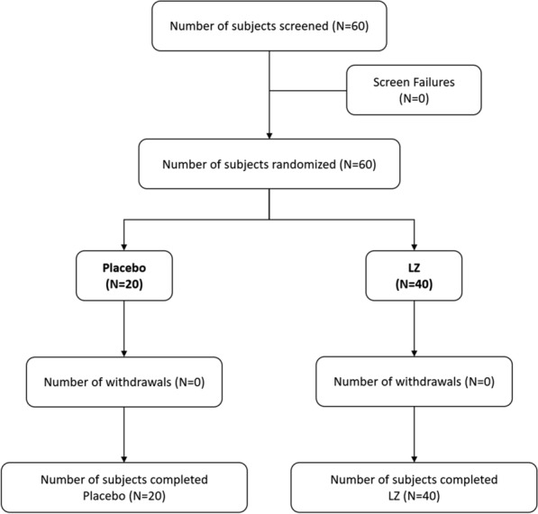
A total of 60 subjects were screened and enrolled into the study. All 60 subjects completed the study as per protocol and were included in the efficacy and safety analysis (Fig. 1).
Fig. 1.

Consolidated Standards of Reporting Trials (CONSORT) diagram
The demographic information of the study subjects is presented in Table 1. Mean (± SD) age of participants was 10.30 ± 2.03 years in the placebo group and 9.73 ± 2.18 years in the LZ group. Gender distribution was 9 boys and 11 girls in the placebo group and 22 boys and 18 girls in the LZ group. Mean (± SD) BMI was 16.80 ± 0.87 kg/m2 for the placebo group and 16.75 ± 1.18 kg/m2 for the LZ group.
Table 1.
Demographics of the study participants at baseline
| Parameters | Placebo | LZ |
|---|---|---|
| N | 20 | 40 |
| Age (years), mean ± SD | 10.30 ± 2.03 | 9.73 ± 2.18 |
| Male, n (%) | 9 (45) | 22 (55) |
| Female, n (%) | 11 (55) | 18 (45) |
| BMI (kg/m2), mean ± SD | 16.80 ± 0.87 | 16.75 ± 1.18 |
Percentages are based on the number of subjects in the specified treatment
N number of subjects in the specified treatment, n number of subjects in the specified category, SD standard deviation
Efficacy
Efficacy analyses were performed on the data from all 60 subjects. The mean change (± SE) values were used to compare LZ against placebo.
Primary Efficacy Analyses
Macular Pigment Optical Density
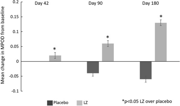
MPOD mean (± SE) change is summarized in Fig. 2. A significant increase (p < 0.05) was observed in MPOD for the LZ group (approx. 25% increase) compared to placebo from baseline to day 42 (0.02 ± 0.01 for LZ vs. 0.00 ± 0.00 for placebo), day 90 (0.06 ± 0.01 for LZ vs. − 0.04 ± 0.01 for placebo), and day 180 (0.13 ± 0.01 for LZ vs. − 0.06 ± 0.01 for placebo).
Fig. 2.

Summary of mean change in macular pigment optical density (MPOD) from baseline
Secondary Efficacy Analyses
Visual Processing Speed Measured by Critical Flicker Fusion
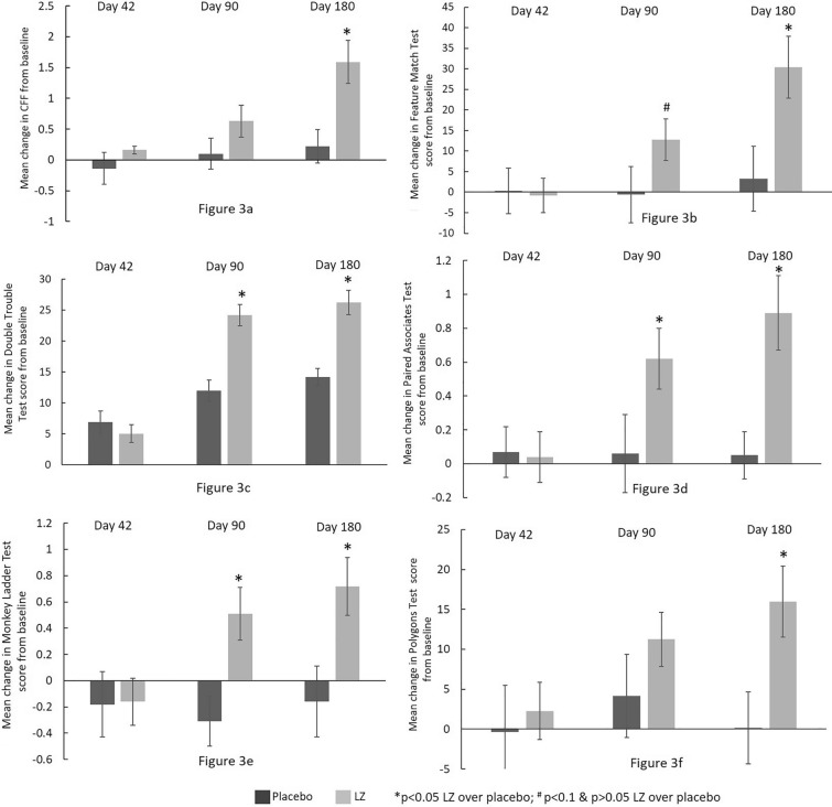
The LZ group showed significant (albeit modest, approx. 7%) increase in processing speed as measured by CFF at day 180 (1.59 ± 0.35 for LZ vs. 0.22 ± 0.27 for placebo) as compared to placebo (Fig. 3a).
Fig. 3.
Summary of mean change from baseline. a Critical flicker fusion (CFF) scores; b Creyos Health attention—Feature Match test scores; c Creyos Health focus/concentration—Double Trouble test score; d episodic memory and learning—Paired Associates test scores; e visuospatial working memory—Monkey Ladder test scores; and f visuospatial processing speed—Polygons test scores
Cognitive Performance: Creyos Health Assessments
Feature Match test: The LZ group showed significant increases (p < 0.05) in attention measured by Feature Match test at day 180 (30.40 ± 7.49 for LZ vs. 3.30 ± 7.90 for placebo) and an increasing trend (p = 0.0623) at day 90 (12.75 ± 5.02 for LZ vs. − 0.60 ± 6.81 for placebo) as compared to placebo (Fig. 3b).
Double Trouble test: The LZ group showed significant increases (p < 0.05) in focus/concentration measured by Double Trouble test at days 90 (24.23 ± 1.72 for LZ vs. 11.95 ± 1.71 for placebo) and 180 (26.26 ± 1.94 for LZ vs. 14.20 ± 1.39 for placebo) as compared to placebo (Fig. 3c).
Paired Associates test: The LZ group showed significant increases (p < 0.05) in episodic memory and learning measured by Paired Associates test at days 90 (0.62 ± 0.18 for LZ vs. 0.06 ± 0.23 for placebo) and 180 (0.89 ± 0.22 for LZ vs. 0.05 ± 0.14 for placebo) as compared to placebo (Fig. 3d).
Monkey Ladder test: The LZ group showed significant increases (p < 0.05) in visuospatial working memory measured by Monkey Ladder test at days 90 (0.51 ± 0.20 for LZ group vs. − 0.31 ± 0.19 for placebo) and 180 (0.72 ± 0.22 for LZ vs. − 0.16 ± 0.27 for placebo) as compared to placebo (Fig. 3e).
Polygons test: The LZ group showed significant increases (p < 0.05) in visuospatial processing measured by Polygons test at day 180 (15.97 ± 4.47 for LZ vs. 0.15 ± 4.49 for placebo) as compared to placebo (Fig. 3f).
Visual Analogue Scales (VAS)
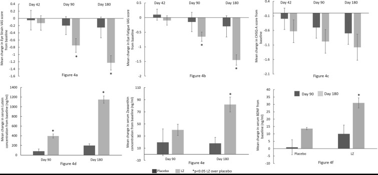
Eye strain: The LZ group showed significant decreases (p < 0.05) in eye strain measured by VAS at days 90 (− 0.75 ± 0.19 for LZ vs. − 0.20 ± 0.25 for placebo) and 180 (− 1.23 ± 0.20 for LZ vs. − 0.25 ± 0.29 for placebo) as compared to placebo (Fig. 4a).
Fig. 4.
Summary of mean change in a visual analogue scales (VAS)—eye strain, b VAS—eye fatigue, c Children’s Sleep Habits Questionnaire-Abbreviated (CSHQ-A) scores, d serum lutein level (in ng/ml), e serum zeaxanthin level (ng/ml), and f serum BDNF level (g/ml)
Eye fatigue: The LZ group showed significant decreases (p < 0.05) in eye fatigue measured by VAS at days 90 (− 0.65 ± 0.16 for LZ vs. − 0.15 ± 0.33 for placebo) and 180 (− 1.45 ± 0.18 for LZ vs. − 0.30 ± 0.36 for placebo) as compared to placebo (Fig. 4b).
Children’s Sleep Habits Questionnaire-Abbreviated (CSHQ-A):
The LZ group did not show any significant difference in sleep at any visit as compared to placebo (Fig. 4c).
Serum Lutein Concentration
The LZ group showed significant increases (p < 0.05) in serum lutein levels at days 90 (394.4 ± 41.80 ng/mL for LZ vs. 80.96 ± 44.08 ng/mL for placebo) and 180 (1149 ± 74.55 ng/mL for LZ vs. 198.6 ± 34.40 ng/mL for placebo) as compared to placebo (Fig. 4d).
Serum Zeaxanthin Concentration
The LZ group showed significant increases (p < 0.05) in serum zeaxanthin levels at day 180 (82.12 ± 12.37 ng/mL for LZ vs. 18.11 ± 9.74 ng/mL for placebo) as compared to placebo (Fig. 4e).
Serum BDNF Concentration
Although the placebo group had significantly higher levels of serum BDNF vs. the LZ group at baseline, by day 180, BDNF levels were significantly increased (p < 0.05) with LZ supplementation (31.03 ± 3.47 ng/mL for LZ vs. 13.61 ± 5.88 ng/mL for placebo). There were no differences in BDNF levels at day 90 (Fig. 4f).
Contrast Sensitivity Measured Using Chart 2020®
No significant differences were observed between the groups on contrast sensitivity measures at any visits (data not shown).
Safety
During the study, a total of eight adverse events were reported by 6 (10%) subjects, five adverse events were reported by 4 (10%) subjects from the LZ group, and three adverse events were reported by 2 (10%) subjects from the placebo group. Subjects in the LZ group reported adverse events of fever (n = 3), viral fever (n = 1), and toothache (n = 1) and subjects in the placebo group reported adverse events of fever (n = 2) and viral fever (n = 1). All the adverse events reported by the subjects were mild in severity, and the causality of the adverse events was diagnosed by the study investigator as not related to the investigational product. The outcomes of all the adverse events were noted as resolved before the end of the study.
None of the subjects reported any serious adverse events or were withdrawn from the study because of an adverse event or a serious adverse event. No clinically significant changes in the vital signs and laboratory parameters were found throughout the study duration in any of the groups.
Discussion
A general evaluation of the dietary habits of younger children led to an unfortunate but inescapable conclusion: they are often poorly nourished. This is certainly true of children in the USA [47] but it is also the case worldwide (e.g., in Indian samples) [48]. Johnson [49] showed that American adults had a relatively low dietary intake of L and Z, about 1–2 mg/day but children had about half those levels. In this study, we supplemented a sample of Indian children with 12 mg L and Z/day in gummy form for 6 months. This supplementation resulted in an increase in serum L and Z in the treated group over the test period by a factor of over 12 (from a mean baseline of 96.9 to 1246 ng/mL). MPOD increased by about 25% in the LZ group and showed significant increases over 6 weeks and 3 and 6 months, whereas MPOD levels were slightly lower than their baseline levels for the placebo at these visits which could be attributed to the lower intake of these carotenoids from the diet. Further, these changes were accompanied by a significant increase in BDNF, a key molecule for promoting plasticity specific to neural processing speed, learning, and memory (Stringham et al. [12] found a similar effect on this neurotrophic factor when supplementing young adults with L and Z). All of these increases were not seen in the placebos.
The treated group also improved on a number of secondary endpoints such as improvement in visual processing speed (CFF) and in cognitive categories such as attention and visuospatial processing speed at day 180. We also observed significant improvement in other cognitive parameters such as focus and concentration, episodic memory and learning, and visuospatial working memory associated with reduction in eye strain and eye fatigue on days 90 and 180 post-supplementation.
Although the exact mechanisms of lutein’s neuroprotective and promoting effects are not fully known, ex vivo data suggest the pigments modulate functional properties of synaptic membranes [50], influence brain morphology and enhance neural response [51], and improve blood flow in the brain [37]. Carotenoids are believed to influence the differentiation of pluripotent neural stem cells [52] and the production of connexin proteins promoting intercellular communication [53, 54].
BDNF is a neuroprotective factor that supports development of new neurons and their differentiation, maturation, and survival in the nervous system [55]. Pro-inflammatory cytokines such as interleukin-6 and tumor necrosis factor attenuate neuroplasticity via reduction of BDNF and negatively affect brain function [56]. It is believed that macular carotenoids act as anti-inflammatory agents and protect synaptic vesicle protein which further helps to induce expression of BDNF levels [57]. Although the BDNF level of the placebo group was higher than the treatment group at baseline, the BDNF levels of the treated group were 24% higher than the controls by the end of the study. An earlier study involving L and Z supplementation in adults also increased levels of serum BDNF levels. The mechanisms behind this change in BDNF as a result of L and Z are yet to be fully understood.
Photooxidation is a light-induced chemical reaction responsible for vision also produces free radicals and reactive oxygen species in the eyes and they in turn damage ocular tissues [58, 59]. Children’s retinas are at high risk from short-wavelength blue light with a peak absorption at 460 nm as a result of the lack of crystallin oxidation that absorbs UV light otherwise reaching the retina [60, 61]. L and Z screen foveal cones from short-wave light and exhibit antioxidant properties that can quench singlet oxygen and free radicals [62]. Therefore, higher macular density reduces ocular tissue damage. Excessive blue light exposure resulting from extended exposure to digital devices and LED screens also causes eye strain and fatigue [63]. Our results showed a significant effect on eye strain and fatigue in children taking L and Z compared to placebo. This is consistent with a previous study of L and Z isomer supplementation on visual function, performance, and sleep quality in individuals using computer devices [40].
L and Z are safe to consume and they are part of normal foods and beverages. There were no adverse events observed due to the investigational product in any of the children participating in this study throughout its entire duration. Hence, L and Z are safe to be consumed by children as well.
The fact that the researchers did not quantify the amount of lutein and zeaxanthin that the participants consumed through their diet was a limitation of the study. On the other hand, both the participants and their parents were instructed to maintain their regular diet without making any adjustments and to accurately recollect their diet for a period of 3 days before every visit.
Conclusions
The levels of L and Z in retina and brain during the early stage of life play an important developmental role in visual and cognitive health. Humans likely evolved mechanisms for accumulating L and Z in order to protect and promote optimal nervous system development. Protection, historically, likely involved reducing actinic (exposure to ultraviolet rays from sunlight and UV lamps), oxidative, and inflammatory stress. One novel finding of the present study is that L and Z might also attenuate some modern stressors such as digital eye strain in children. An American Optometric Association survey reported that 83% of children between the ages of 10 and 17 use electronic devices for at least 3 h each day [64]. Our study also demonstrated the high bioavailability of L and Z when administered in the kid-friendly form of gummies. The sequalae of positive visual and cognitive effects combined with the ease of this intervention suggests that this type of supplementation might be an additional tool that might help optimize CNS development, especially in typically undernourished children. It also emphasizes the importance of optimal nutrition in the developing eye and brain.
Acknowledgements
The authors thank participants and their families for their involvement in the trial. We also thank our recruitment and follow-up teams.
Medical Writing and Editorial Assistance.
The authors did not use any medical writing and editorial assistance for this article.
Authorship.
All named authors meet the International Committee of Medical Journal Editors (ICMJE) criteria for authorship for this article, take responsibility for the integrity of the work as a whole, and have given their approval for this version to be published.
Author Contributions
Rajesh Parekh, Billy R. Hammond Jr, and Divya Chandradhara contributed to the study conception and design. Study product was developed by OmniActive Health Technologies Limited. Study conduct, subject recruitment and data collection were performed at respective study center by Rajesh Parekh. Study was monitored by Divya Chandradhara in a blinded fashion. Statistical analysis and study report were prepared by Divya Chandradhara. Overall study data interpretation was done by Rajesh Parekh, Billy R. Hammond Jr, and Divya Chandradhara. The first draft of the manuscript was written by Rajesh Parekh, Billy R. Hammond Jr, and Divya Chandradhara. All authors commented on previous versions of the manuscript. All authors read and approved the final manuscript.
Funding
This study and the journal’s Rapid Service and Open Access Fee was supported by OmniActive Health Techonologies Limited (Mumbai, India).
Data Availability
The datasets generated during and/or analyzed during the current study are available from the corresponding author on reasonable request.
Declaration
Conflict of Interest
Rajesh Parekh is affiliated to the clinical study site, Sanjeevani Netralaya, where the study was conducted. Billy R. Hammond Jr is a professor at the University of Georgia, Athens and supported as a consultant for the sponsor in study and manuscript development. Divya Chandradhara is an employee of Bioagile Therapeutics Pvt. Ltd., the Contract Research Organization contracted to conduct the presented study.
Ethical Approval
Institutional ethics approval was obtained from Pranav Diabetes Center Ethics Committee, Bengaluru, Karnataka, India. This study was conducted in compliance with the ethical principles originating in or derived from the Declaration of Helsinki and in compliance with the International Conference on Harmonization (ICH), Good Clinical Practice (GCP) Guidelines, as well as in strict compliance with the “The New Drugs and Clinical Trial Rules- 2019”, the Ministry of Health and the Government of India at all stages of the trial for adherence to protocol and compliance with ethical and regulatory guidelines. Voluntary consent was obtained in written from all the study participants before commencing any study related activities. The EC was duly apprised of the progress and updates of the trial at regular intervals as per prescribed guidelines.
References
- 1.Klassen TP, Hartling L, Craig JC, Offringa M. Children are not just small adults: the urgent need for high-quality trial evidence in children. PLoS Med. 2008;5:e172. doi: 10.1371/journal.pmed.0050172. [DOI] [PMC free article] [PubMed] [Google Scholar]
- 2.Steiner P. Brain fuel utilization in the developing brain. Ann Nutr Metab. 2020;75:8–18. doi: 10.1159/000508054. [DOI] [PubMed] [Google Scholar]
- 3.Yang Z, Wang X, Zhang S, Ye H, Chen Y, Xia Y. Pediatric myopia progression during the COVID-19 pandemic home quarantine and the risk factors: a systematic review and meta-analysis. Front Public Health. 2022;10:835449. doi: 10.3389/fpubh.2022.835449. [DOI] [PMC free article] [PubMed] [Google Scholar]
- 4.Vilela MA, Pellanda LC, Fassa AG, Castagno VD. Prevalence of asthenopia in children: a systematic review with meta-analysis. J Pediatr (Rio J) 2015;91:320–325. doi: 10.1016/j.jped.2014.10.008. [DOI] [PubMed] [Google Scholar]
- 5.Mohan A, Sen P, Shah C, Jain E, Jain S. Prevalence and risk factor assessment of digital eye strain among children using online e-learning during the COVID-19 pandemic: digital eye strain among kids (DESK study-1) Indian J Ophthalmol. 2021;69:140. doi: 10.4103/ijo.IJO_2535_20. [DOI] [PMC free article] [PubMed] [Google Scholar]
- 6.Innis SM. Dietary omega 3 fatty acids and the developing brain. Brain Res. 2008;1237:35–43. doi: 10.1016/j.brainres.2008.08.078. [DOI] [PubMed] [Google Scholar]
- 7.Ikonomidou C, Kaindl AM. Neuronal death and oxidative stress in the developing brain. Antioxid Redox Signal. 2011;14:1535–1550. doi: 10.1089/ars.2010.3581. [DOI] [PubMed] [Google Scholar]
- 8.Andersen SL. Neuroinflammation, early-life adversity, and brain development. Harv Rev Psychiatry. 2022;30:24–39. doi: 10.1097/HRP.0000000000000325. [DOI] [PMC free article] [PubMed] [Google Scholar]
- 9.Hammond BR, Jr, Renzi-Hammond L. Individual variation in the transmission of UVB radiation in the young adult eye. PLoS ONE. 2018;13:e0199940. doi: 10.1371/journal.pone.0199940. [DOI] [PMC free article] [PubMed] [Google Scholar]
- 10.Vishwanathan R, Neuringer M, Snodderly DM, Schalch W, Johnson EJ. Macular lutein and zeaxanthin are related to brain lutein and zeaxanthin in primates. Nutr Neurosci. 2013;16:21–29. doi: 10.1179/1476830512Y.0000000024. [DOI] [PMC free article] [PubMed] [Google Scholar]
- 11.Lieblein-Boff JC, Johnson EJ, Kennedy AD, Lai C-S, Kuchan MJ. Exploratory metabolomic analyses reveal compounds correlated with lutein concentration in frontal cortex, hippocampus, and occipital cortex of human infant brain. PLoS ONE. 2015;10:e0136904. doi: 10.1371/journal.pone.0136904. [DOI] [PMC free article] [PubMed] [Google Scholar]
- 12.Stringham JM, Johnson EJ, Hammond BR. Lutein across the lifespan: from childhood cognitive performance to the aging eye and brain. Curr Dev Nutr. 2019;3:nzz066. [DOI] [PMC free article] [PubMed]
- 13.Barnett SM, Khan NA, Walk AM, et al. Macular pigment optical density is positively associated with academic performance among preadolescent children. Nutr Neurosci. 2018;21:632–640. doi: 10.1080/1028415X.2017.1329976. [DOI] [PMC free article] [PubMed] [Google Scholar]
- 14.Hassevoort KM, Khazoum SE, Walker JA, et al. Macular carotenoids, aerobic fitness, and central adiposity are associated differentially with hippocampal-dependent relational memory in preadolescent children. J Pediatr. 2017;183:108–114. doi: 10.1016/j.jpeds.2017.01.016. [DOI] [PubMed] [Google Scholar]
- 15.Walk AM, Khan NA, Barnett SM, et al. From neuro-pigments to neural efficiency: the relationship between retinal carotenoids and behavioral and neuroelectric indices of cognitive control in childhood. Int J Psychophysiol. 2017;118:1–8. doi: 10.1016/j.ijpsycho.2017.05.005. [DOI] [PMC free article] [PubMed] [Google Scholar]
- 16.Walk AM, Edwards CG, Baumgartner NW, et al. The role of retinal carotenoids and age on neuroelectric indices of attentional control among early to middle-aged adults. Frontiers in aging neuroscience. 2017;9;183. [DOI] [PMC free article] [PubMed]
- 17.Saint SE, Renzi-Hammond LM, Khan NA, Hillman CH, Frick JE, Hammond BR., Jr The macular carotenoids are associated with cognitive function in preadolescent children. Nutrients. 2018;10:193. doi: 10.3390/nu10020193. [DOI] [PMC free article] [PubMed] [Google Scholar]
- 18.Renzi LM, Hammond BR., Jr The relation between the macular carotenoids, lutein and zeaxanthin, and temporal vision. Ophthalmic Physiol Opt. 2010;30:351–357. doi: 10.1111/j.1475-1313.2010.00720.x. [DOI] [PubMed] [Google Scholar]
- 19.Institute of Medicine (US) Panel on dietary antioxidants and related compounds. β-Carotene and other carotenoids. Dietary reference intakes for vitamin C, vitamin E, selenium, and carotenoids. Washington (DC): National Academies Press; 2000. https://www.ncbi.nlm.nih.gov/books/NBK225483/, 10.17226/9810. [PubMed]
- 20.EFSA Panel on Food Additives and Nutrient Sources added to Food (ANS). Scientific opinion on the re-evaluation of lutein (E 161b) as a food additive. EFSA Journal. 10.2903/j.efsa.2010.1678. Accessed 2024 Jan 7.
- 21.Shao A, Hathcock JN. Risk assessment for the carotenoids lutein and lycopene. Regul Toxicol Pharmacol. 2006;45:289–298. doi: 10.1016/j.yrtph.2006.05.007. [DOI] [PubMed] [Google Scholar]
- 22.Norkus EP, Norkus KL, Dharmarajan TS, Schierle J, Schalch W. Serum lutein response is greater from free lutein than from esterified lutein during 4 weeks of supplementation in healthy adults. J Am Coll Nutr. 2010;29:575–585. doi: 10.1080/07315724.2010.10719896. [DOI] [PubMed] [Google Scholar]
- 23.Johnson EJ, Maras JE, Rasmussen HM, Tucker KL. Intake of lutein and zeaxanthin differ with age, sex, and ethnicity. J Am Diet Assoc. 2010;110:1357–1362. doi: 10.1016/j.jada.2010.06.009. [DOI] [PubMed] [Google Scholar]
- 24.Mulder KA, Innis SM, Rasmussen BF, Wu BT, Richardson KJ, Hasman D. Plasma lutein concentrations are related to dietary intake, but unrelated to dietary saturated fat or cognition in young children. J Nutr Sci. 2014;3:e11. doi: 10.1017/jns.2014.10. [DOI] [PMC free article] [PubMed] [Google Scholar]
- 25.European Commission. Fruit and vegetables intake in European countries: knowledge for policy. https://knowledge4policy.ec.europa.eu/health-promotion-knowledge-gateway/fruit-vegetables-5_en. Accessed 2024 Jan 7.
- 26.Wolf-Schnurrbusch UE, Röösli N, Weyermann E, Heldner MR, Höhne K, Wolf S. Ethnic differences in macular pigment density and distribution. Invest Ophthalmol Vis Sci. 2007;48:3783–3787. doi: 10.1167/iovs.06-1218. [DOI] [PubMed] [Google Scholar]
- 27.Huntjens B, Asaria TS, Dhanani S, Konstantakopoulou E, Ctori I. Macular pigment spatial profiles in South Asian and white subjects. Invest Ophthalmol Vis Sci. 2014;55:1440–1446. doi: 10.1167/iovs.13-13204. [DOI] [PubMed] [Google Scholar]
- 28.Chew EY, Clemons TE, SanGiovanni JP, et al. Secondary analyses of the effects of lutein/zeaxanthin on age-related macular degeneration progression: AREDS2 report No. 3. JAMA Ophthalmol. 2014;132:142–9. [DOI] [PMC free article] [PubMed]
- 29.Liu R, Wang T, Zhang B, et al. Lutein and zeaxanthin supplementation and association with visual function in age-related macular degeneration. Invest Ophthalmol Vis Sci. 2015;56:252–258. doi: 10.1167/iovs.14-15553. [DOI] [PubMed] [Google Scholar]
- 30.Dawczynski J, Jentsch S, Schweitzer D, Hammer M, Lang GE, Strobel J. Long term effects of lutein, zeaxanthin and omega-3-LCPUFAs supplementation on optical density of macular pigment in AMD patients: the LUTEGA study. Graefes Arch Clin Exp Ophthalmol. 2013;251:2711–2723. doi: 10.1007/s00417-013-2376-6. [DOI] [PubMed] [Google Scholar]
- 31.Chew EY, Clemons TE, Keenan TD, Agron E, Malley CE, Domalpally A. The results of the 10 year follow-on study of the age-related eye disease study 2 (AREDS2) Invest Ophthalmol Vis Sci. 2021;62:1215–1215. [Google Scholar]
- 32.Stringham JM, Hammond BR. Macular pigment and visual performance under glare conditions. Optom Vis Sci. 2008;85:82–88. doi: 10.1097/OPX.0b013e318162266e. [DOI] [PubMed] [Google Scholar]
- 33.Hammond BR, Fletcher LM, Roos F, Wittwer J, Schalch W. A double-blind, placebo-controlled study on the effects of lutein and zeaxanthin on photostress recovery, glare disability, and chromatic contrast. Invest Ophthalmol Vis Sci. 2014;55:8583–8589. doi: 10.1167/iovs.14-15573. [DOI] [PubMed] [Google Scholar]
- 34.Stringham JM, O’Brien KJ, Stringham NT. Macular carotenoid supplementation improves disability glare performance and dynamics of photostress recovery. Eye and Vis. 2016;3:1–8. doi: 10.1186/s40662-016-0060-8. [DOI] [PMC free article] [PubMed] [Google Scholar]
- 35.Johnson EJ, McDonald K, Caldarella SM, Chung H, Troen AM, Snodderly DM. Cognitive findings of an exploratory trial of docosahexaenoic acid and lutein supplementation in older women. Nutr Neurosci. 2008;11:75–83. doi: 10.1179/147683008X301450. [DOI] [PubMed] [Google Scholar]
- 36.Hammond BR, Jr, Miller LS, Bello MO, Lindbergh CA, Mewborn C, Renzi-Hammond LM. Effects of lutein/zeaxanthin supplementation on the cognitive function of community dwelling older adults: a randomized, double-masked, placebo-controlled trial. Front Aging Neurosci. 2017;9:254. doi: 10.3389/fnagi.2017.00254. [DOI] [PMC free article] [PubMed] [Google Scholar]
- 37.Lindbergh CA, Renzi-Hammond LM, Hammond BR, et al. Lutein and zeaxanthin influence brain function in older adults: a randomized controlled trial. J Int Neuropsychol Soc. 2018;24:77–90. doi: 10.1017/S1355617717000534. [DOI] [PubMed] [Google Scholar]
- 38.Renzi-Hammond LM, Bovier ER, Fletcher LM, et al. Effects of a lutein and zeaxanthin intervention on cognitive function: a randomized, double-masked, placebo-controlled trial of younger healthy adults. Nutrients. 2017;9:1246. doi: 10.3390/nu9111246. [DOI] [PMC free article] [PubMed] [Google Scholar]
- 39.Culver MF, Bowman J, Juturu V. Lutein and zeaxanthin isomers effect on sleep quality: a randomized placebo-controlled trial. Biomed J Sci Tech Res. 2018;9:7018–7024. [Google Scholar]
- 40.Juturu V, Bowman JP, Stringham JM. Lutein and zeaxanthin isomers (L/Zi) supplementation improves visual function, performance and sleep quality in individuals using computer devices (CDU)–a double blind randomized placebo controlled study. Biomed J. 2018;2:7. [Google Scholar]
- 41.Ranard KM, Jeon S, Mohn ES, Griffiths JC, Johnson EJ, Erdman JW. Dietary guidance for lutein: consideration for intake recommendations is scientifically supported. Eur J Nutr. 2017;56:37–42. doi: 10.1007/s00394-017-1580-2. [DOI] [PMC free article] [PubMed] [Google Scholar]
- 42.Wooten BR, Hammond BR, Land RI, Snodderly DM. A practical method for measuring macular pigment optical density. Invest Ophthalmol Vis Sci. 1999;40:2481–2489. [PubMed] [Google Scholar]
- 43.McCorkle SM, Raine LB, Hammond BR, Jr, Renzi-Hammond L, Hillman CH, Khan NA. Reliability of heterochromatic flicker photometry in measuring macular pigment optical density among preadolescent children. Foods. 2015;4:594–604. doi: 10.3390/foods4040594. [DOI] [PMC free article] [PubMed] [Google Scholar]
- 44.Saint SE, Hammond BR, Jr, Khan NA, Hillman CH, Renzi-Hammond LM. Temporal vision is related to cognitive function in preadolescent children. Appl Neuropsychol Child. 2021;10:319–326. doi: 10.1080/21622965.2019.1699096. [DOI] [PubMed] [Google Scholar]
- 45.Landrum JT, Bone RA, Joa H, Kilburn MD, Moore LL, Sprague KE. A one year study of the macular pigment: the effect of 140 days of a lutein supplement. Exp Eye Res. 1997;65:57–62. [DOI] [PubMed]
- 46.Bovier ER, Hammond BR. A randomized placebo-controlled study on the effects of lutein and zeaxanthin on visual processing speed in young healthy subjects. Arch Biochem Biophys. 2015;572:54–57. doi: 10.1016/j.abb.2014.11.012. [DOI] [PubMed] [Google Scholar]
- 47.Liu J, Micha R, Li Y, Mozaffarian D. Trends in food sources and diet quality among US children and adults, 2003–2018. JAMA Netw Open. 2021;4:e215262. doi: 10.1001/jamanetworkopen.2021.5262. [DOI] [PMC free article] [PubMed] [Google Scholar]
- 48.Joe W, Rajpal S, Kim R, et al. Association between anthropometric-based and food-based nutritional failure among children in India, 2015. Matern Child Nutr. 2019;15:e12830. doi: 10.1111/mcn.12830. [DOI] [PMC free article] [PubMed] [Google Scholar]
- 49.Johnson EJ. Role of lutein and zeaxanthin in visual and cognitive function throughout the lifespan. Nutr Rev. 2014;72:605–612. doi: 10.1111/nure.12133. [DOI] [PubMed] [Google Scholar]
- 50.Tan JS, Wang JJ, Flood V, Rochtchina E, Smith W, Mitchell P. Dietary antioxidants and the long-term incidence of age-related macular degeneration: the Blue Mountains Eye Study. Ophthalmology. 2008;115:334–341. doi: 10.1016/j.ophtha.2007.03.083. [DOI] [PubMed] [Google Scholar]
- 51.Zuniga KE, Bishop NJ, Turner AS. Dietary lutein and zeaxanthin are associated with working memory in an older population. Public Health Nutr. 2021;24:1708–1715. doi: 10.1017/S1368980019005020. [DOI] [PMC free article] [PubMed] [Google Scholar]
- 52.Kuchan M, Wang F, Geng Y, Feng B, Lai C. Lutein stimulates the differentiation of human stem cells to neural progenitor cells in vitro. Advances and Controversies in Clinical Nutrition, Washington, DC Abstract. 2013;
- 53.Stahl W, Nicolai S, Briviba K, et al. Biological activities of natural and synthetic carotenoids: induction of gap junctional communication and singlet oxygen quenching. Carcinogenesis. 1997;18:89–92. doi: 10.1093/carcin/18.1.89. [DOI] [PubMed] [Google Scholar]
- 54.Bertram JS. Carotenoids and gene regulation. Nutr Rev. 1999;57:182–191. doi: 10.1111/j.1753-4887.1999.tb06941.x. [DOI] [PubMed] [Google Scholar]
- 55.Bathina S, Das UN. Brain-derived neurotrophic factor and its clinical implications. Arch Med Sci. 2015;6:1164–78. [DOI] [PMC free article] [PubMed]
- 56.García-Bueno B, Caso JR, Leza JC. Stress as a neuroinflammatory condition in brain: damaging and protective mechanisms. Neurosci Biobehav Rev. 2008;32:1136–1151. doi: 10.1016/j.neubiorev.2008.04.001. [DOI] [PubMed] [Google Scholar]
- 57.Stringham NT, Holmes PV, Stringham JM. Lutein supplementation increases serum brain-derived neurotrophic factor (BDNF) in humans. FASEB J. 2016;30:689–693. doi: 10.1096/fasebj.30.1_supplement.689.3. [DOI] [Google Scholar]
- 58.Lion Y, Delmelle M, Van de Vorst A. New method of detecting singlet oxygen production. Nature. 1976;263:442–443. doi: 10.1038/263442a0. [DOI] [PubMed] [Google Scholar]
- 59.Straight R, Spikes J. Photosensitized oxidation of biomolecules. Singlet O2. 1985;4:143.
- 60.Dillon J. New trends in photobiology: the photophysics and photobiology of the eye. J Photochem Photobiol B. 1991;10:23–40. doi: 10.1016/1011-1344(91)80209-Z. [DOI] [PubMed] [Google Scholar]
- 61.Dillon J, Atherton SJ. Time resolved spectroscopic studies on the intact human lens. Photochem Photobiol. 1990;51:465–468. doi: 10.1111/j.1751-1097.1990.tb01738.x. [DOI] [PubMed] [Google Scholar]
- 62.Böhm F, Edge R, Truscott TG. Interactions of dietary carotenoids with singlet oxygen (1O2) and free radicals: potential effects for human health. Acta Biochim Pol. 2012;59:27–30. [PubMed]
- 63.Wilkins AJ, Wilkinson P. A tint to reduce eye-strain from fluorescent lighting? Preliminary observations. Ophthalmic Physiol Opt. 1991;11:172–175. doi: 10.1111/j.1475-1313.1991.tb00217.x. [DOI] [PubMed] [Google Scholar]
- 64.American Optometric Association. Survey reveals parents drastically underestimate the time kids spend on electronic devices. Cision PR Newswire. 2014.
Associated Data
This section collects any data citations, data availability statements, or supplementary materials included in this article.
Data Availability Statement
The datasets generated during and/or analyzed during the current study are available from the corresponding author on reasonable request.


