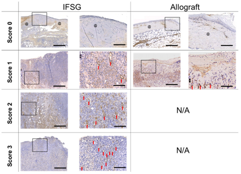
Figure 4.
Day 7 alpha-smooth muscle actin staining. Representative images of slides stained with α-SMA that consist of wound bed samples collected prior to skin graft application on day 7. Sections are shown that align with the pathologist scoring listed in Table 2. No allograft-treated wounds scored a 2 or 3 and, hence, N/A is indicative of no wounds with those scores. Myofibroblasts and new blood vessels can be identified in the images by the positive brown staining. The left image of the biopsy punch has scale bars = 500 µm. The image on the right is the magnified black box region from the left image with scale bars = 125 µm. Residual pieces of product = @; new blood vessels = red arrow. IFSG = intact fish skin graft.

