Abstract
Endoscopic Retrograde Cholangiopancreatography (ERCP) is the primary therapeutic procedure for pancreaticobiliary disorders, and studies highlighted the impact of papilla anatomy on its efficacy and safety. Our objective was to quantify the influence of papilla morphology on ERCP outcomes. We systematically searched three medical databases in September 2022, focusing on studies detailing the cannulation process or the rate of adverse events in the context of papilla morphology. The Haraldsson classification served as the primary system for papilla morphology, and a pooled event rate with a 95% confidence interval was calculated as the effect size measure. Out of 17 eligible studies, 14 were included in the quantitative synthesis. In studies using the Haraldsson classification, the rate of difficult cannulation was the lowest in type I papilla (26%), while the highest one was observed in the case of type IV papilla (41%). For post-ERCP pancreatitis, the event rate was the highest in type II papilla (11%) and the lowest in type I and III papilla (6–6%). No significant difference was observed in the cannulation failure and post-ERCP bleeding event rates between the papilla types. In conclusion, certain papilla morphologies are associated with a higher rate of difficult cannulation and post-ERCP pancreatitis.
Subject terms: Pancreatic disease, Therapeutic endoscopy, Gastroenterology
Introduction
Endoscopic Retrograde Cholangiopancreatography (ERCP) is the most used therapeutic procedure for pancreaticobiliary disorders. However, how to best achieve safe and effective bile duct cannulation is still debated. Despite notable developments in the past decades, the failure rate is still 5–20% in experienced hands1. Moreover, the incidence of the procedure’s adverse events is high; post-ERCP pancreatitis (PEP) has an incidence rate of 9.7%, with a mortality rate of 0.7%2.
Endoscopists performing ERCP recognize the differences in the macroscopic appearance of the major papilla. This has led to a conception that certain appearances of the papilla are more challenging to cannulate and, therefore, more prone to adverse events. Despite the essential role of bile duct cannulation in procedural safety and success, research on this topic is still limited.
A Scandinavian research group published the first inter- and intraobserver-validated classification of the major papilla’s endoscopic appearance in 20173. In the same year, they also published a multicentric prospective cohort study, indicating that the anatomy of the major papilla affects both the difficulty of the bile duct cannulation and the procedural adverse events4. Further, their results suggest that the morphology of the papilla should be considered in the training of fellow endoscopists4. Other identified studies support their results5,6.
Recently, several articles have been published assessing the influence of papilla morphology on ERCP outcomes, with contradicting results. Therefore, we aimed to systematically review and quantify the magnitude of its effect and investigate its importance and relevance in the endoscopic practice.
Methods
A systematic review and meta-analysis were conducted following the Preferred Reporting Items for Systematic Review and Meta-Analysis (PRISMA) Statement (see Supplementary Table 12) and the recommendations of the Cochrane Handbook7,8. The review protocol was registered in advance on PROSPERO with the registration number CRD42022360894.
Systematic search
Three databases: MEDLINE (via PubMed), Embase, and Cochrane Central Register of Controlled Trials (CENTRAL), were systematically searched from inception until the 29th of September 2022. We did not apply any filters or restrictions to our search. The main parts of the search query included terms in connection with ERCP and papilla morphology. For the detailed search strategy, see Table S1. Additionally, we systematically searched for relevant articles by reviewing the included articles’ bibliographic references and citation lists.
Eligibility criteria
The condition-context-population (CoCoPop) framework was used to identify eligible studies9. The conditions were (Co): difficult cannulation, cannulation attempts, cannulation time, cannulation failure, post-ERCP pancreatitis, and other post-ERCP adverse events (bleeding, perforation, infection) in the context of the different papilla morphologies (Co). Studies with adult patients (> 18) undergoing ERCP with a native papilla (Pop) were selected.
Randomized controlled trials, case–control, cross-sectional, and cohort studies were eligible for inclusion. Both full-text articles and conference abstracts with sufficient data were considered eligible. Regarding the definition of difficult cannulation, cannulation failure, and post-ERCP adverse events, the definitions provided in the included studies were used.
Morphology of the papilla
Primarily, for the classification of the morphology of the papilla, as the first validated intra- and interobserver classification, the Haraldsson system was used4. They classified the papilla into four types: regular (type 1), small (type 2), protruding or pendulous (type 3), and creased or ridged (type 4)3.
Secondarily, a comparison between the Haraldsson and the other identified classification systems was attempted with the following method: two endoscopists (PJH, EB) assessed the description of the morphology and the imagery of the studies. They chose the identical papilla types to Haraldsson’s. In case of any disagreement, a third reviewer was included in the decision process (ET). After the comparison, additional analyses were conducted.
Study selection and data extraction
After the systematic search, the yielded articles were imported into a reference management program (EndNote X7.4, Clarivate Analytics, Philadelphia, PA, USA) to remove the duplicates automatically and manually. After removing duplicates, two independent authors (ET, EBG) screened the remaining publications first by title and abstract and then by full text. We used Rayyan for the selection process10. Cohen’s kappa coefficient (κ) was calculated on both levels of selection to measure inter-reviewer reliability11.
Two investigators extracted data independently (ET, EBG) and manually populated it into a purpose-designed Excel 2016 sheet (Office 365, Microsoft, Redmond, WA, USA). Data were collected on the first author, year of publication, digital object identifier, period of data collection, study location, number of centers, study design, the mean or median age of the patients (with standard deviation or interquartile range), the total number of patients, the number of women, the number of patients with each papilla morphology, and data regarding the primary and secondary outcomes in the context of the different papilla types. For statistical analysis, raw data were extracted into two-by-four tables (condition yes/no; papilla morphologies).
Statistical analysis
The statistical analysis was performed by a biostatistician (DSV) with R (R Core Team 2022, v4.2.2)12. Forest plots were used to display the results of the meta-analytical calculations. The minimum study number to perform the meta-analytical calculation were three. Event rates with a 95% confidence interval (CI) were used for the effect size measure. As we anticipated considerable between-study heterogeneity, a random-effects model was used to pool effect sizes. For assessing the small study publication bias, funnel plots were used with a visual inspection. Additional sensitivity analyses were conducted using the leave-one-out method, with a minimum study number of four (see additional details in the supplementary material).
See supplementary material for additional details on the statistical analyses.
Risk of bias assessment
Two investigators (ET, EBG) independently assessed the risk of bias for each outcome using the Joanna Briggs Institute Critical Appraisal tool for studies reporting prevalence13.
Quality of evidence
Certainty of evidence was assessed following the Grading of Recommendations Assessment, Development, and Evaluation (GRADE) recommendation14. Two independent investigators (ET, EBG) evaluated all criteria for all outcomes. Disagreements were resolved by the senior review author (BE).
Results
Search and selection
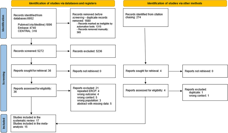
The details of the study selection process are summarised in the PRISMA flow chart shown in Fig. 1.
Figure 1.
PRISMA 2020 flowchart representing the study selection process.
A total of 6,952 studies were identified through database searching. Finally, our narrative synthesis comprised 17 studies4–6,15–28. Of those, 14 could be included in the quantitative synthesis4–6,15–17,19,21–27.
Basic characteristics of included studies
The main characteristics of the included studies are summarised in Table 1. Eligible studies were reported between 2016 and 2022. Of the 17 studies, 15 were cohort studies, eight had prospective (5, 6, 19–22, 26, 27), and seven had retrospective designs. There was also one case–control24 and one cross-sectional study19. 13 of the studies were full-text articles4–6,15–17,19,21,22,24,26–28, and four of them were conference abstracts18,21,23,25. Seven studies used the Haraldsson classification4–6,19,22,24,25, with seven additional ones using comparable classifications15–17,21,23,26,27. Three studies used classification systems that were not comparable to the Haralddson classification. The number of study participants ranged from 72 to 11,090.
Table 1.
Basic characteristics of included studies.
| Author | Year | Country | Centers | Study type | Study period | Age (*:mean; #:median) | Sex (female %) | Number of patients | Classification | Outcomes |
|---|---|---|---|---|---|---|---|---|---|---|
| Balan et al.15 | 2020 | Romania | 1 | Prospective cohort | January 2018 to August 2018 | NA | NA | 322 |
Regular: 52% Canard type I 11%: Canard type II: 19% Canard type III: 10% Canard type IV: 8% |
Difficult cannulation Cannulation time Cannulation attempts Post-ERCP pancreatitis Post-ERCP bleeding Post-ERCP infection |
| Canena et al.16 | 2021 | Portugal | 3 | Prospective cohort | May 2018 to October 2020 | *69.6 | 56.8% | 361 |
Viana type I: 13% Viana type IIa: 35% Viana type IIb: 30% Viana type IIc:10% Viana type IIIa: 4% Viana type IIIb: 4% Viana type IV: 4% |
Cannulation failure Cannulation time Post-ERCP pancreatitis Post-ERCP bleeding Post-ERCP perforation |
| Chen et al.5 | 2020 | Taiwan | 1 | Prospective cohort | October 2017 to October 2018 | *64 (SD: 16.5) | 47.5% | 286 |
Haraldsson type I: 41% Haraldsson type II: 9% Haraldsson type III: 22% Haraldsson type IV: 28% |
Cannulation failure Cannulation time Post-ERCP pancreatitis Post-ERCP bleeding Post-ERCP perforation Post-ERCP cholangitis |
| Fernandes et al.18 | 2018 | Portugal | 3 | Prospective cohort | August 2017 to January 2018 | #79 | 59.4% | 106 |
Leés type I: 50% Leés type II: 32% Leés type III: 12% Leés type IV: 6% |
Cannulation time |
| Gutierrez- De Aranguren et al.19 | 2021 | Peru | 1 | Retrospective cross-sectional | July 2019 to April 2021 | *55 (SD:2 0) | 66.5% | 188 |
Haraldsson type I: 32% Haraldsson type II: 25% Haraldsson type III: 27% Haraldsson type IV: 16% |
Difficult cannulation |
| Haraldsson et al.4 | 2019 | Nordic countries | 9 | Prospective cohort | NA | 66 (SD: 16) | 52% | 1377 |
Haraldsson type I: 56% Haraldsson type II: 13% Haraldsson type III: 23% Haraldsson type IV: 8% |
Difficult cannulation Cannulation time Post-ERCP pancreatitis |
| Liu et al.20 | 2021 | China | 1 | Retrospective cohort | January 2008 to December 2017 | NA | NA | 11 090 |
Normal: 44% Thick and long: 11%: Peridiverticular: 27% Intradiverticular: 5% Ectopic: 1% Edematous 10%: Ulcerative: 2% |
Difficult cannulation |
| Mohamed et al.6 | 2021 | Canada | 1 | Retrospective cohort | September 2018 to January 2020 | NA | 51.8% | 637 |
Haraldsson type I: 62% Haraldsson type II: 5% Haraldsson type IIIa: 9% Haraldsson type IIIb: 9% Haraldsson type IV: 3% Type D: 12% |
Cannulation failure Cannulation time Cannulation attempts Post-ERCP pancreatitis Post-ERCP bleeding Post-ERCP infection Post-ERCP cholangitis or sepsis |
| Nakeeb et al.17 | 2016 | Egypt | 1 | Prospective cohort | August 2012 to September 2014 | *58.4 (SD: 14.7) | 44.4% | 996 |
Normal: 60% Atrophic: 3% Pregnant: 7% Tumor: 7% Redundant: 8% Juxtadivertcular: 8% Small: 6% Long: 1% |
Post-ERCP pancreattis |
| Onilla et al.21 | 2021 | Philippines | 1 | Retrospective cohort | January 2017 to December 2019 | NA | NA | 347 |
Regular protrusion: 57% Small protrusion: 31% Large protrusion: 12% Annular pattern: 72% Unstructured pattern: 11% Longitudinal pattern 11%: Isolated pattern: 1% Gyrus pattern: 5% |
Difficult cannulation Cannulation failure |
| Quiroga-Purizaca et al.22 | 2022 | Peru | 1 | Propective cohort | NA | *51.5 ( CI 48.8–54.1) | 68.4% | 138 |
Haraldsson type I: 59% Haraldsson type II: 8% Haraldsson type III: 29% Haraldsson type IV: 4% |
Difficult cannulation Cannulation time Cannulation attempts Post-ERCP pancreatitis Post-ERCP bleeding Post-ERCP perforation |
| Sadeghi et al.23 | 2019 | Iran | 1 | Prospective cohort | September 2017 to March 2018 |
*62.3 (SD: 15.5) |
51.4% | 72 |
Small: 33%: Bulging: 28% Long: 39% |
Cannulation success |
| Saito et al.24 | 2022 | Japan | 3 | Retrospective case–control | April 2012 to February 2020 | *74.9 | 47.5% | 1406 |
Haraldsson type I: 45% Haraldsson type II: 44% Haraldsson type III: 7% Haraldsson type IV: 4% |
Difficult cannulation |
| Thongsuwan et al.25 | 2021 | Thailand | 1 | Retrospective cohort | January 2013 to May 2017 | NA | 50.4% | 558 |
Haraldsson type I: 66% Haraldsson type II: 16% Haraldsson type III: 12% Haraldsson type IV: 6% |
Difficult cannulation Cannulation failure Post-ERCP pancreatitis, Post-ERCP bleeding Post-ERCP infection |
| Watanabe et al.26 | 2019 | Japan | 1 | Retrospective cohort | September 2013 to June 2017 | #70 | 36% | 589 |
Regular protrusion: 12% Small protrusion: 78% Large protrusion: 10% Annular pattern: 67% Unstructured pattern: 7% Longitudinal pattern: 7% Isolated pattern: 1% Gyrus pattern:16% Unclassified pattern: 2% |
Difficult cannulation Cannulation failure Cannulation attempts |
| Zhang et al.27 | 2016 | China | 1 | Retrospective cohort | February 2012 to March 2015 | *75 (SD: 2.2) | 42.7% | 82 |
bulging: 44% normal: 22% small: 16% unusual location: 18% |
Cannulation failure Cannulation time |
| Zheng et al.28 | 2020 | China | 1 | Retrospective cohort | January 2016 to December 2019 | NA | 46.1% | 2385 |
others:18% villous: 74% granular: 8% |
Post-ERCP pancreatitis |
Quantitative synthesis
Difficult cannulation
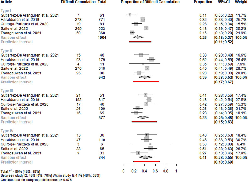
Nine studies were identified regarding the event rate of difficult cannulation4,15,19–22,24–26, of which eight were included in the quantitative synthesis4,15,19,21,22,24–26. In the case of studies using the classification proposed by Haraldsson, in type I papilla, the rate of difficult cannulation was lower (26%; CI 18–37) compared to the other papilla types (type III: 35%; CI 25–48; type II: 39%; CI 28–52; type IV: 41%; CI 28–55). The difference was statistically no significant; however, the p-value referred for a higher tendency for difficult cannulation in certain papilla types (p: 0.075). The heterogeneity was high (total I2: 89%; CI 48–98). Sensitivity analyses did not reveal outlier studies or relevant changes in the estimate (see Figs. 2 and S1).
Figure 2.
Forest plot representing the pooled event rate of difficult cannulation in the different papilla types in studies using the Haraldsson classification, showing a lower tendency for difficult cannulation in type I papilla compared to the other papilla types.
A similar but statistically significant result with no outlier study was observed, including all the studies with different classifications (p: 0.019; total I2: 87%; CI 55–96) (see Figures S2-3).
Cannulation failure
Eight studies detailed the event rate of cannulation failure, all using Haraldsson’s or classifications comparable to it5,6,16,21,23,25,26,28. In the analysis, including studies only using the Haraldsson classification, no statistically significant difference was observed in the rate of failed cannulation between the different papilla types (p: 0.262, total I2: 61%; CI 0–97) (see Fig. 3).
Figure 3.
Forest plot representing the pooled event rate of cannulation failure in the different papilla types in studies using the Haraldsson classification, showing no statistically significant difference in the event rates between the papilla types.
In the case of including all eight studies, the difference was statistically significant (p: 0.047, I2: 64%; CI 0–91). The rate of cannulation failure was the highest in the case of type II papilla (8%, CI 4–14) and the lowest in type I (3%; CI 2–6) (see Figure S4). Sensitivity analyses did not reveal outlier studies or relevant changes in the estimate (see Figure S5).
Post-ERCP pancreatitis
Nine of the identified studies reported the event rate of PEP in the different papilla types4–6,15–17,22,25,28, of which eight articles were included in the quantitative synthesis4–6,15–17,22,25. In the case of studies using the Haraldsson classification, in type II papilla, the rate of post-ERCP pancreatitis was higher (11%; CI 8–15) compared to the other papilla types (type IV: 7%; CI 4–12; type I: 6%; CI 5–8; type III: 6%; CI 4–8). The result was statistically significant (p: 0.0441). Total homogeneity was observed (total I2: 0.044) (see Fig. 4).
Figure 4.
Forest plot representing the pooled event rate of post-ERCP pancreatitis in the different papilla types in studies using the Haraldsson classification, showing a statistically significantly higher rate of post-ERCP pancreatitis in type II papilla, compared to the other papilla types.
A similar tendency was observed in the case of including all eight studies; however, the difference between the papilla types was not statistically significant (p: 0.103) (see Figure S6). Sensitivity analyses did not reveal outlier studies or relevant changes in the estimate (see Figures S7-8).
Post-ERCP bleeding
Six eligible studies reported information about a bleeding episode after an ERCP procedure, all using the Haraldsson classification or classifications comparable to it5,6,15,16,22,25. In the analyses with only studies using the Haraldsson classification and with all classification systems, no statistically significant difference was observed in the event rate of the post-ERCP bleeding between the papilla types (p: 0.8585 and p: 0.8078, respectively) (see Figs. 5 and S9). Sensitivity analyses did not reveal outlier studies or relevant changes in the estimate (see Figures S10-11).
Figure 5.
Forest plot representing the pooled event rate of post-ERCP bleeding in the different papilla types in studies using the Haraldsson classification, showing no statistically significant difference in the event rates between the papilla types.
Qualitative synthesis
Cannulation time
Eight studies investigated cannulation time in the context of papilla morphology4–6,15,16,18,22,27, and four used the Haraldsson classification4–6,22. The time for cannulation was the lowest in type I papilla, without exception. Two-two studies reported the highest cannulation time in type II4,5 and type IV papilla6,22.
Cannulation attempts
Four studies investigated the number of cannulation attempts in the context of papilla morphology6,15,22,26, from which two used the Haraldsson classification6,22. In both cases, the cannulation attempts were the highest in type IV and the lowest in type I and III papillae.
Post-ERCP perforation
Three studies investigated the perforation rate after an ERCP procedure, all using the Haraldsson classification5,16,22. The meta-analytical calculation was impossible due to the number of zero events.
Post-ERCP infection
Four studies reported the proportion of patients with an infection after ERCP5,6,15,25; of those, three studies used the Haraldsson classification5,6,25. Chen et al. reported the highest event rate of cholangitis in type I (2.5%) and no event in type II and III papillae5. Mohammed et al. found the highest event rate of cholangitis and/or sepsis in type II (3.2%) and no event in type III and IV papillae, meanwhile in the study by Thongsuwan et al., the event rate of infection was the highest in type III (10.5%) and the lowest in type I papilla (6%)6,25.
Risk of bias and publication bias assessment
Most of the included studies carried a low risk of bias. Among the eight studies detailing difficult cannulation, two (25%) had high, and six (75%) had low risk of bias. The results of the risk of bias assessments are shown in Figures S12-19. Publication bias could not be observed in the conducted analyses. The results of the assessments are shown in Figures S20-27.
Quality of evidence
Since we included only cohort studies, the certainty of evidence ranged between very low and low for each outcome. Detailed results of the GRADE assessment can be found in Tables S4-11.
Discussion
Our systematic review and meta-analysis assessed the impact of papilla morphology on ERCP and its outcomes. We found that in studies using the Haraldsson classification, compared to the other papilla types, the event rate of difficult cannulation was lower in type I papilla. Type II papilla was associated with a twofold increase in the event rate of PEP compared to the other papilla types. There was no difference in the cannulation failure and post-ERCP bleeding event rates between the different papilla types.
Since its introduction, there have been debates regarding ERCP’s safety and success rate. Several factors seem to influence cannulation difficulties, such as age and age-related factors, including duodenal distortion; procedure-related aspects, such as duodenal positioning or certain etiologies, for example, malignant biliary obstruction. The morphology of the papilla is also assumed to be related to multiple perspectives of the procedure29.
First, papilla morphology should be considered in the training of fellow endoscopists. In the studies selected for inclusion, there are contradicting data regarding how the endoscopist’s expertise influences cannulation difficulty. Mohamed et al. found no relationship between the rate of difficult cannulation and the endoscopist’s expertise (7). In contrast, in the study by Haraldsson et al., the rate of difficult cannulation was the highest in type II papilla, where the number of trainees starting the cannulation process was the highest (5). Other studies also suggest that the operator’s experience may decrease the rate of difficult cannulation and cannulation failure (34, 35). Further data in the literature suggest that the rate of PEP and other adverse events also decreases with the endoscopist’s experience (36).
Secondly, papilla morphology also influences the rate of PEP, the procedure’s most common adverse event2. We found the highest rate of PEP in type II papilla, which is consistent with the result of the individual studies. However, the definite explanation for this pattern is still uncertain. According to Chen et al. hypothesis, it could be due to the fact that endoscopic papilla balloon dilatation (EPBD) was used more often in this papilla type in their cohort5. The same trend could be observed in the study by Mohamed et al.6. Further data in the literature suggest that EPBD with small-caliber balloons (diameter: 8–10 mm) increases the rate of PEP30.
Lastly, all the included studies observed differences in rescue techniques’ use in different papilla morphologies. It could be one of the explanations for the non-significant difference in cannulation failure between the different papilla types. We hypothesize that the morphology of the papilla should be considered when choosing a rescue cannulation technique since it decreases the difference in the tendency for cannulation failure or difficult cannulation between the papilla types. Studies suggest that a pre-cut sphincterotomy or needle-knife fistulotomy (NKF) may be used in normal papillae. Trans-pancreatic sphincterotomy could be the recommended rescue technique in small papillae. In protruding/pendulous or creased/ridged papillae, also NKF could be the preferred method31,32.
Several classification systems were identified; the Haraldsson was the most widely used and well-recognized one. Despite being the first validated classification system developed by expert endoscopists and, therefore, the basis of our analysis, it has one major limitation: it ignores the presence of a periampullary diverticulum. A modified version of the classification was proposed by Mohamed et al. in 2021, introducing an additional papilla type (type D) for papillae involved with a periampullary diverticulum6. In addition, a meta-analysis by Mui et al. found that the presence of PAD may increase the risk of cannulation failure and may also be associated with a higher risk for post-ERCP adverse events33. These results suggest that this modified version of the classification should be used.
Strengths
Despite the topic’s importance, to our knowledge, this is the first meta-analysis focusing on papilla morphology and its relation to the most relevant endpoints of the ERCP cannulation process and the rate of adverse events. A rigorous methodology was applied, with a comprehensive search key. No publication bias or outlier study was detected in any conducted analyses, and most studies carried a low risk of bias. Moreover, the number of included patients was above 20,000.
Limitations
Regardless of all the strengths, this study also had some limitations: (1) In certain analyses, considerable statistical heterogeneity was observed. Its explanation could be the clinical heterogeneity across studies, such as the difference in the applied definitions in connection with the endoscopic procedure. Most studies used the definition of the European Society of Gastrointestinal Endoscopy for difficult cannulation; however, Thongsuwan et al. used its simplified version. (2) Some of the included cohort studies were retrospective analyses. (3) The certainty of the evidence was low or very low. (4) Abstracts were also eligible for inclusion; however, all were high-quality, containing all the necessary data.
Implication for practice
Based on our results, during training of fellow endoscopists, papilla morphology should be determined, and trainees should start their learning with type I (“regular”) papillae. Using a unified classification system for papilla morphology is recommended to promote transparency in clinical practice.
Implication for research
Large sample cohorts are needed to validate the Mohammed version of the classification and assess the presence of a periampullary diverticulum. Besides the event rate, future research should also focus on the severity of PEP in the different papilla types. Furthermore, developing a recommendation system for advanced cannulation techniques in the context of papilla morphologies should be considered.
Conclusion
In conclusion, other types are associated with a higher rate of difficult cannulation compared to the regular papilla type. The small papilla is associated with a higher rate of post-ERCP pancreatitis.
Supplementary Information
Author contributions
TE: conceptualization, project administration, methodology, formal analysis, writing—original draft; EBG: formal analysis, visualization, writing—review & editing; AR: conceptualization, writing—review & editing; DSV: formal analysis, data curation, writing—review & editing; SzV: conceptualization, writing—review & editing; PJH: conceptualization, writing—review & editing; KH: conceptualization, writing—review & editing; PH: conceptualization, writing—review & editing; BE: conceptualization; supervision; writing—original draft. All authors certify that they have participated sufficiently in the work to take public responsibility for the content, including participation in the manuscript’s concept, design, analysis, writing, or revision.
Funding
Open access funding provided by Semmelweis University.
Data availability
All data is provided within the manuscript or supplementary information files.
Competing interests
The authors declare no competing interests.
Footnotes
Publisher's note
Springer Nature remains neutral with regard to jurisdictional claims in published maps and institutional affiliations.
Change history
5/20/2024
The original online version of this Article was revised: In the original version of this Article, the Supplementary Information file, which was included with the initial submission, was omitted. The correct Supplementary Information file now accompanies the original Article.
Supplementary Information
The online version contains supplementary material available at 10.1038/s41598-024-57758-9.
References
- 1.Tse F, Yuan Y, Moayyedi P, Leontiadis GI. Guidewire-assisted cannulation of the common bile duct for the prevention of post-endoscopic retrograde cholangiopancreatography (ERCP) pancreatitis. Cochrane Database Syst. Rev. 2012;12:Cd009662. doi: 10.1002/14651858.CD009662.pub2. [DOI] [PMC free article] [PubMed] [Google Scholar]
- 2.Kochar B, et al. Incidence, severity, and mortality of post-ERCP pancreatitis: A systematic review by using randomized, controlled trials. Gastrointest Endosc. 2015;81:143–149.e149. doi: 10.1016/j.gie.2014.06.045. [DOI] [PubMed] [Google Scholar]
- 3.Haraldsson E, et al. Endoscopic classification of the papilla of Vater. Results of an inter- and intraobserver agreement study. United Eur. Gastroenterol. J. 2017;5:504–510. doi: 10.1177/2050640616674837. [DOI] [PMC free article] [PubMed] [Google Scholar]
- 4.Haraldsson E, et al. Macroscopic appearance of the major duodenal papilla influences bile duct cannulation: a prospective multicenter study by the Scandinavian Association for Digestive Endoscopy Study Group for ERCP. Gastrointest. Endosc. 2019;90:957–963. doi: 10.1016/j.gie.2019.07.014. [DOI] [PubMed] [Google Scholar]
- 5.Chen PH, et al. Duodenal major papilla morphology can affect biliary cannulation and complications during ERCP, an observational study. BMC Gastroenterol. 2020 doi: 10.1186/s12876-020-01455-0. [DOI] [PMC free article] [PubMed] [Google Scholar]
- 6.Mohamed R, et al. Morphology of the major papilla predicts ERCP procedural outcomes and adverse events. Surg. Endosc. 2021;35:6455–6465. doi: 10.1007/s00464-020-08136-9. [DOI] [PMC free article] [PubMed] [Google Scholar]
- 7.Cumpston M, et al. Updated guidance for trusted systematic reviews: A new edition of the cochrane handbook for systematic reviews of interventions. Cochrane Database Syst. Rev. 2019;10:Ed000142. doi: 10.1002/14651858.Ed000142. [DOI] [PMC free article] [PubMed] [Google Scholar]
- 8.Page MJ, et al. The PRISMA 2020 statement: An updated guideline for reporting systematic reviews. Bmj. 2021;372:n71. doi: 10.1136/bmj.n71. [DOI] [PMC free article] [PubMed] [Google Scholar]
- 9.Munn Z, Stern C, Aromataris E, Lockwood C, Jordan Z. What kind of systematic review should I conduct? A proposed typology and guidance for systematic reviewers in the medical and health sciences. BMC Med. Res. Methodol. 2018;18:5. doi: 10.1186/s12874-017-0468-4. [DOI] [PMC free article] [PubMed] [Google Scholar]
- 10.Ouzzani M, Hammady H, Fedorowicz Z, Elmagarmid A. Rayyan—A web and mobile app for systematic reviews. Syst. Rev. 2016;5:210. doi: 10.1186/s13643-016-0384-4. [DOI] [PMC free article] [PubMed] [Google Scholar]
- 11.McHugh ML. Interrater reliability: the kappa statistic. Biochem Med (Zagreb) 2012;22:276–282. doi: 10.11613/BM.2012.031. [DOI] [PMC free article] [PubMed] [Google Scholar]
- 12.R Core Team. R: A Language and Environment for Statistical Computing (Vienna, 2022).
- 13.Munn Z, Moola S, Lisy K, Riitano D, Tufanaru C. Methodological guidance for systematic reviews of observational epidemiological studies reporting prevalence and incidence data. Int. J. Evid. Based Healthc. 2015;13(3):147–153. doi: 10.1097/XEB.0000000000000054. [DOI] [PubMed] [Google Scholar]
- 14.GRADEpro GDT: GRADEpro Guideline Development Tool [Software]. (McMaster University and Evidence Prime).
- 15.Balan GG, et al. Anatomy of major duodenal papilla influences ERCP outcomes and complication rates: A single center prospective study. J. Clin. Med. 2020 doi: 10.3390/jcm9061637. [DOI] [PMC free article] [PubMed] [Google Scholar]
- 16.Canena J, et al. Influence of a novel classification of the papilla of Vater on the outcome of needle-knife fistulotomy for biliary cannulation. BMC Gastroenterol. 2021 doi: 10.1186/s12876-021-01735-3. [DOI] [PMC free article] [PubMed] [Google Scholar]
- 17.El Nakeeb A, et al. Post-endoscopic retrograde cholangiopancreatography pancreatitis: Risk factors and predictors of severity. World J. Gastrointest. Endosc. 2016;8:709–715. doi: 10.4253/wjge.v8.i19.709. [DOI] [PMC free article] [PubMed] [Google Scholar]
- 18.Fernandes J, et al. Does the morphology of the major papilla influence biliary cannulation?-A multicenter prospective study. United Eur. Gastroenterol. J. 2018;6:A200–A201. doi: 10.1177/2050640618792819. [DOI] [Google Scholar]
- 19.Gutierrez-De Aranguren C, et al. Association between the type of major duodenal papilla and difficult biliary cannulation in a private tertiary center. Rev. Gastroenterol. Peru. 2021;41:66–71. doi: 10.47892/rgp.2021.413.1255. [DOI] [PubMed] [Google Scholar]
- 20.Liu Y, et al. Causes and countermeasures of difficult selective biliary cannulation: A large sample size retrospective study. Surg. Laparosc. Endosc. Percutaneous Tech. 2021;31:533–538. doi: 10.1097/SLE.0000000000000924. [DOI] [PubMed] [Google Scholar]
- 21.Onilla J, et al. Duodenal papilla morphology and ERCP cannulation difficulties, failure and complications: A cross-sectional study. J. Gastroenterol. Hepatol. 2021;36:218. doi: 10.1111/jgh.15607. [DOI] [Google Scholar]
- 22.Quiroga-Purizaca WG, Paucar-Aguilar DR, Barrientos-Pérez JA, Vargas-Blacido DA. Morphological characteristics of the duodenal papilla and its association with complications post-endoscopic retrograde cholangiopancreatography (ERCP) in a Peruvian hospital. Rev. Colomb. Gastroenterol. 2022;37:296–301. doi: 10.22516/25007440.859. [DOI] [Google Scholar]
- 23.Sadeghi A, et al. Characteristics of major duodenal papill a in efficacy and safety of needle-knife fistulotomy. United Eur. Gastroenterol. J. 2019;7:860. doi: 10.1177/205064061985467. [DOI] [Google Scholar]
- 24.Saito H, et al. Factors predicting difficult biliary cannulation during endoscopic retrograde cholangiopancreatography for common bile duct stones. Clin. Endosc. 2022;55:263–269. doi: 10.5946/ce.2021.153. [DOI] [PMC free article] [PubMed] [Google Scholar]
- 25.Thongsuwan C, et al. Influence of major duodenal papilla morphology on the biliary cannulation and post-ERCP complications. Gastrointest. Endosc. 2021;93:152–153. doi: 10.1016/j.gie.2021.03.311. [DOI] [Google Scholar]
- 26.Watanabe M, et al. Transpapillary biliary cannulation is difficult in cases with large oral protrusion of the duodenal papilla. Dig. Dis. Sci. 2019;64:2291–2299. doi: 10.1007/s10620-019-05510-z. [DOI] [PubMed] [Google Scholar]
- 27.Zhang QS, Han B, Xu JH, Gao P, Shen YC. Needle-knife papillotomy and fistulotomy improved the treatment outcome of patients with difficult biliary cannulation. Surg. Endosc. 2016;30:5506–5512. doi: 10.1007/s00464-016-4914-x. [DOI] [PubMed] [Google Scholar]
- 28.Zheng R, et al. Development and validation of a risk prediction model and scoring system for post-endoscopic retrograde cholangiopancreatography pancreatitis. Ann. Transl. Med. 2020 doi: 10.21037/ATM-20-5769. [DOI] [PMC free article] [PubMed] [Google Scholar]
- 29.Berry R, Han JY, Tabibian JH. Difficult biliary cannulation: Historical perspective, practical updates, and guide for the endoscopist. World J. Gastrointest. Endosc. 2019;11:5–21. doi: 10.4253/wjge.v11.i1.5. [DOI] [PMC free article] [PubMed] [Google Scholar]
- 30.Fujisawa T, et al. Is endoscopic papillary balloon dilatation really a risk factor for post-ERCP pancreatitis? World J. Gastroenterol. 2016;22:5909–5916. doi: 10.3748/wjg.v22.i26.5909. [DOI] [PMC free article] [PubMed] [Google Scholar]
- 31.Halttunen J, et al. Difficult cannulation as defined by a prospective study of the Scandinavian Association for Digestive Endoscopy (SADE) in 907 ERCPs. Scand. J. Gastroenterol. 2014;49:752–758. doi: 10.3109/00365521.2014.894120. [DOI] [PubMed] [Google Scholar]
- 32.Löhr JM, et al. How to cannulate? A survey of the Scandinavian association for digestive endoscopy (SADE) in 141 endoscopists. Scand. J. Gastroenterol. 2012;47:861–869. doi: 10.3109/00365521.2012.672588. [DOI] [PubMed] [Google Scholar]
- 33.Mu P, et al. Does periampullary diverticulum affect ERCP cannulation and post-procedure complications? An up-to-date meta-analysis. Turk. J. Gastroenterol. 2020;31:193–204. doi: 10.5152/tjg.2020.19058. [DOI] [PMC free article] [PubMed] [Google Scholar]
Associated Data
This section collects any data citations, data availability statements, or supplementary materials included in this article.
Supplementary Materials
Data Availability Statement
All data is provided within the manuscript or supplementary information files.


