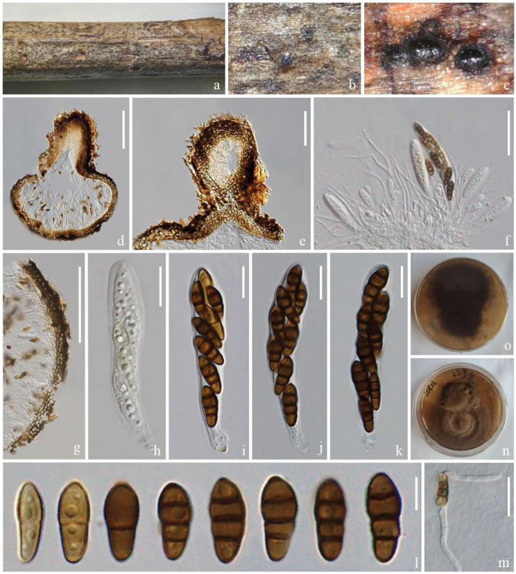
Figure 4.
Nigrogranalincangensis (ZHKU 23–0104, holotype) a–c appearance of ascomata on the host surface d vertical section of an ascoma e vertical section of ostiole f hamathecium and asci g section of peridium h–k asci l ascospores m a germinated ascospore n, o colonies on PDA (n-front and o-reverse views). Scale bars: 100 µm (d); 50 µm (e); 30 µm (f); 200 µm (g); 10 µm (h–k); 5 µm (l); 20 µm (m).

