Abstract
Garnet-type Li6.5La3Zr1.5Ta0.5O12 (LLZO) is considered a promising solid electrolyte, but the surface degradation in air hinders its application for all-solid-state battery. Recent studies have mainly focused on the final products of the LLZO surface reactions due to lacking of powerful in situ characterization methods. Here, we use ambient pressure X-ray spectroscopies to in situ investigate the dynamical evolution of LLZO surface in different gas environments. The newly developed ambient pressure mapping of resonant Auger spectroscopy clearly distinguishes the lithium containing species, including LiOH, Li2O, Li2CO3 and lattice oxygen. The reaction of CO2 with LLZO to form Li2CO3 is found to be a thermodynamically favored self-limiting reaction. On the contrary, the reaction of H2O with LLZO lags behind that of CO2, but intensifies at high pressure. More interestingly, the results provide direct spectroscopic evidence for the existence of Li+/H+ exchange and reveal the importance of the initial layer formed on clean electrolyte surface in determining their air stability. This work demonstrates that the newly developed in situ technologies pave a new way to investigate the oxygen evolution and surface degradation mechanism in energy materials.
Subject terms: Characterization and analytical techniques, Batteries, Batteries
Li6.5La3Zr1.5Ta0.5O12 (LLZO) is a promising solid electrolyte but suffers from severe surface degradation in air. Here, authors use mapping of resonant Auger spectroscopy and various ambient pressure X-ray spectroscopies to elucidate the dynamical evolution of CO2 and H2O on clean LLZO surfaces.
Introduction
The increasing desire for higher capacity and better safety energy storage technology is fostering the revolution of lithium-ion batteries (LIBs)1–3. Among all possible anode materials, Li metal is considered as the ultimate choice to boost the energy density in LIBs because of its high theoretical capacity (3869 mA h g−1) and low redox potential1,4. However, the growth of Li dendrites and the severe volume changes limit the applications of Li metal batteries5–7. Non-flammable solid-state electrolytes (SSEs) with high shear modulus and Li+ transference number have potential to solve these problems8,9. Among various types of SSEs, garnet-based solid electrolyte Li7La3Zr2O12 is highly promising due to its high ionic conductivity (up to 1 mS cm−1 at 25 °C), high electrochemical stability towards Li metal, the feasibility of mass production and possible storage in air10,11. Nevertheless, previous findings have pointed out that a lithiophobic Li2CO3 layer forms on the surface of LLZO as a result of exposure to CO2 and H2O in air12,13. This thin Li2CO3 layer, moreover, have shown to surprisingly governs the interface property, leading to poor interfacial contact and high interfacial impedance between LLZO and electrode materials14.
Different pathways for the surface reaction have been proposed to describe the actual reaction between LLZO and air. The density functional theory (DFT) calculations indicate that, thermodynamically, CO2 can react directly with LLZO to produce Li2CO313. However, the reaction seems kinetically slow as only a negligible amount of Li2CO3 was observed experimentally on the surface of LLZO after long-term exposure to anhydrous air15. In another highly probable pathway, H2O first reacts with LLZO to form LiOH through Li+/H+ exchange, then a substantial part of LiOH transforms into Li2CO3 after exposure to CO216. Spontaneous Li+/H+ ion exchange is believed not to change the cubic garnet structure of LLZO, but blocks Li+ transmission channel16,17. Therefore, a core−shell structure is proposed, comprising a garnet core surrounded by a proton-rich garnet shell and a LiOH/Li2CO3 outer layer. Up to now, this core-shell structure has only been studied by destructive characterization techniques such as argon ion sputter etching18, and the reaction mechanism of Li2CO3 formation has not been fully understood. Thus, it is of great significance to develop in situ techniques with depth-profiling capability to comprehensively probe the initial reactions at gas/solid interface, which can help us understand the different thermodynamic and kinetic processes for CO2 and H2O on LLZO surface, and eventually provide guidance for avoiding the formation of Li2CO3 and building an excellent interface.
Synchrotron-based core-level X-ray spectroscopies, including X-ray photoelectron spectroscopy (XPS) and X-ray absorption spectrum (XAS), have been widely used to monitor the gas/solid interface in catalysis and environmental science19–21. Recent developed in situ spectroscopy techniques can be utilized to investigate the reaction between gas and the solid surface with elemental and chemical sensitivities. Additionally, for soft-X-rays, XAS in total-electronic-yield (TEY) mode can support a detection depth of ~10 nm, while signals in auger-electron-yield (AEY) mode usually come from a depth of ~3 nm22. Thus, simultaneous detection of XAS in both TEY and AEY mode along with XPS allows us to obtain an in-depth analysis of the chemical evolution on the surface and sub-surface. However, due to the poor air stability and very similar spectroscopic fingerprints, lithium containing species such as Li2O, Li2O2, LiOH, Li2CO3 and LLZO are very difficult to identify, thus very limited studies have combined all these surface methods in ambient pressure for energy materials characterization until now.
In this paper, the initial surface chemistry and evolution mechanism of LLZO in H2O and CO2 were in situ investigated by ambient pressure X-ray photoelectron and absorption spectroscopy. In particular, the newly developed ambient pressure mapping of resonant Auger spectroscopy (AP-mRAS) method clearly identifies lattice oxygen (LLZO) and various surface oxygen species such as LiOH, Li2O and Li2CO3, and the in situ depth-profiling technology can deduce the flow direction of lithium. We find that CO2 reacts directly with LLZO thermodynamically, but the reaction is limited by surface active sites and a lack of oxygen supply. The reaction of H2O with LLZO needs a relatively higher pressure than CO2 through Li+/H+ exchange. However, the reaction is more intense and continuous, forming LiOH. Comparing LLZO with Li1.5Al0.5Ge1.5P3O12 (LAGP), we found that the characteristics of the interface layer initially formed on the surface and the stability of the material structure may be the decisive factors for its resistance to air degradation.
Results
Clean LLZO surface obtained by low temperature treatment
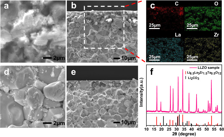
The LLZO pellet suffers severe surface degradation after exposure to air. Figure 1a, b shows the surface and cross-section morphology of the LLZO pellet after exposure to air for 2 months. Large amounts of amorphous structures are visible around the voids of LLZO pellet, assignable to Li2CO3 which is evidenced by the presence of C and O in energy-dispersive X-ray spectroscopy (EDS) analysis in Fig. 1c23,24. After polishing by sandpaper and wiping by alcohol, the LLZO particles can be clearly observed on the surface with a small amount of residual debris Fig. 1d, e. The results also indicate that completely clean LLZO surface cannot be obtained only by physical polishing. The X-ray diffraction (XRD) pattern of the aged LLZO pellet is displayed in Fig. 1f. The diffraction peaks match well with the cubic garnet phase with no other impurities, which means that the phase of the bulk LLZO is barely changed during the surface chemical evolution in air, and the Li2CO3 contamination may have an amorphous structure.
Fig. 1. Surface degradation of LLZO after exposure to air.
a Surface and b cross-section scanning electron microscope (SEM) images of aged LLZO pellet. c EDS elemental mappings of the aged sample. d Surface and e cross-section SEM images of polished LLZO pellet. f XRD patterns of the aged LLZO pellet.
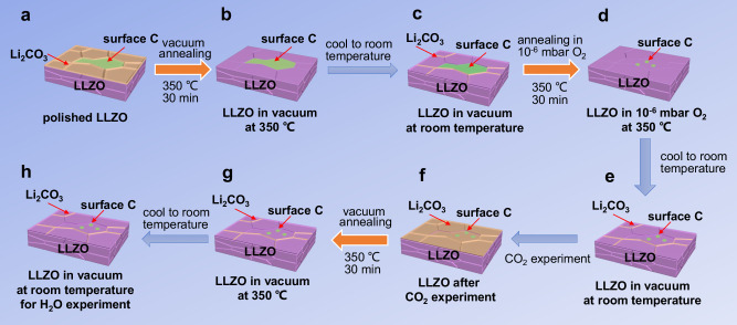
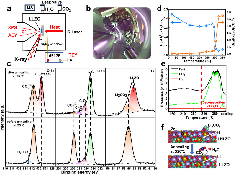
The schematic diagram of the entire in situ experimental processes in this work are shown in Fig. 2. A clean LLZO surface is necessary for monitoring of the degradation mechanism. To achieve a clean LLZO surface, the polished LLZO pellet was annealed at 350 °C in vacuum for 30 min and then annealed in 1 × 10⁻⁶ mbar O2 at 350 °C for 30 min, as shown in Fig. 2a–e. During the vacuum annealing process, in the range of 30 to 350 °C under ultra-high vacuum (UHV) condition, XPS, mass spectrum and XAS in AEY and TEY mode were carried out to characterize the surface chemical evolution. The schematic and the photo of the instrument are shown in Fig. 3a, b. XPS and XAS in AEY mode were conducted by the Scienta Hipp3 analyzer, while the XAS in TEY mode was measured by a pico-ammeter25,26.
Fig. 2. Schematic diagram of the entire in situ experimental processes in this work.
a, b Polished LLZO pellet was vacuum annealing at 350 °C for 30 min to remove surface Li2CO3, but surface contaminated carbon species cannot be removed. c During the cooling process, a small amount of Li2CO3 will be generated on the surface of LLZO. d,e Most of the surface contaminated carbon species can be removed by annealing in 1 × 10⁻⁶ mbar O2 at 350 °C for 30 min. f–h The clean LLZO surface then be used to investigate the reaction with CO2 and H2O.
Fig. 3. Clean LLZO surface obtained by low temperature vacuum annealing.
a Schematic and b real instrument for the operando ambient pressure experiment. c XPS spectra of the LLZO pellet before and after vacuum annealing at room temperature. d The influence of temperature rise on the ratios of O(lattice)/O(CO32-) and C(CO32-)/C(C-C). e In situ mass spectrum results in the temperature range of 160 – 370 °C. CO2 and H2O are simultaneously observed to be the decomposition products at around 300 °C. f The schematic illustration of reaction of xLi2CO3 + Li6.5-2xH2xLa3Zr1.5Ta0.5O12 → Li6.5La3Zr1.5Ta0.5O12 + xH2O + xCO2 at the LLZO surface during the vacuum annealing process.
The polished LLZO pellet was mounted onto a sample holder under ambient air (which took about 15 min) and were pumped into the instrument. Thus, a strong signal of Li2CO3 and weak signals of La and Zr could be observed in XPS spectra as shown in Fig. 3c and Supplementary Fig. 1. The peak at 284.6 eV in C 1 s spectrum can be assigned to the C-C (sp2) bond of the surface contamination27. The O K edge AEY and TEY spectra in Supplementary Fig. 2 are consistent with our XPS results that the surface of LLZO is mainly Li2CO3 before annealing. A small signal of LLZO is present at ~531.9 eV in the AEY spectrum, which is slightly weaker than in the TEY spectrum with a larger investigation depth of ~10 nm, indicating that the thickness of the contamination layer is several nanometers.
In order to determine whether the surface Li2CO3 can be completely removed by vacuum annealing, the O 1 s and C 1 s XPS spectra were measured at 350 °C. Only the peak of LLZO at around 529 eV labeled as O(lattice) can be detected in the O 1 s XPS spectrum as shown in Supplementary Fig. 3a. The results indicate that a clean LLZO surface without other O containing species can be achieved by low-temperature vacuum annealing28. However, after cooling down the LLZO pellet to room temperature, the peak of Li2CO3 appears again, as evident from Fig. 3c, indicating the clean LLZO surface may be very sensitive to residual CO229 and can form Li2CO3 on the surface even at a pressure below 1 × 10⁻⁸ mbar. Li2CO3 is difficult to be observed in the AEY and TEY spectra of annealed LLZO at RT in Supplementary Fig. 2, indicating that very little Li2CO3 is generated on the surface during the cooling process. Thus, a clean surface of LLZO with very small amount of Li2CO3 is obtained by vacuum annealing.
Further, we systematically investigated the influence of temperature rise on the ratios of O(lattice)/O(CO32-) and C(CO32-)/C(C-C), delineated in Fig. 3d. The decomposition temperature of the surface Li2CO3 can be identified to be around 300 °C, which is much lower than the reported reaction temperature (over 620 °C for the reaction Li2CO3 → Li2O + CO2)30,31. No signal of Li2O was detected in the O 1 s XPS and O K edge mRAS data of annealed LLZO as shown in Supplementary Fig. 4. To probe the decomposition reaction at low-temperature, the decomposition gaseous products were detected by in situ mass spectrum, and the results are exhibited in Fig. 3e. Both CO2 and H2O were simultaneously observed as the decomposition products around 300 °C. Thus, the actual reaction at the LLZO surface during the vacuum annealing process could be: xLi2CO3 + Li6.5-2xH2xLa3Zr1.5Ta0.5O12 → Li6.5La3Zr1.5Ta0.5O12 + xH2O + xCO2 as depicted in Fig. 3f18,32. However, surface contaminated carbon species cannot be removed by vacuum annealing, leading to the relatively weak signal of carbonate in C 1 s as seen in Fig. 3c. In order to prevent the strong signal of contaminated C-C sp2 from affecting the observation of the evolution of carbonate, we then annealed the sample in 1 × 10⁻⁶ mbar O2 at 350 °C for 30 min. After annealing, the signals of other elements remained basically unchanged, and the signal of contaminated carbon species significantly decreased.
Clear identification of lithium containing species using AP-mRAS and XAS
Due to the poor air stability and very similar spectroscopic fingerprints, lithium containing species are very difficult to identify by surface sensitive characterization methods33,34. Here, using the newly developed AP-mRAS method, we can carefully identify lattice and surface oxygen in lithium containing species before studying the reaction mechanism. The mechanism and characteristics of mRAS compared with mapping of resonant inelastic X-ray scattering (mRIXS) are shown in Supplementary Fig. 5, the incident photon energy is scanned across the absorption edge, and the emitted Auger electrons at each resonant energy are further resolved in kinetic energy (KE).
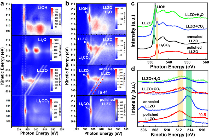
Li metal was in situ scraped using a wobble-stick with sharp blade as shown in Supplementary Fig. 4. By utilizing near ambient pressure technology, we can obtain mRAS of pure Li2O, LiOH and Li2CO3 for comparison. Figure 4a compares annealed LLZO with Li2O, LiOH and Li2CO3, which shows completely different characteristics. The results support that the surface of annealed LLZO is clean. The AP-mRAS spectra of LLZO surface at different states are shown in Fig. 4b: LLZO surface after physical polishing mainly contains Li2CO3 on the surface, which displays a vertical symmetrical feature at photon energy (h) of 533.7 eV. In the kinetic energy (KE) direction, the strongest point of Li2CO3 is located at around 512.2 eV and the intensity extends to both high and low KE directions. After surface treatment, clean LLZO surface shows an oblique spot at photon energy around 531.9 eV. The inclination of the spot depends on the d orbital property that hybridized with the O 2p orbital. Furthermore, clean LLZO surface shows a localized feature in KE direction with a much smaller photon energy which is quite different from Li2O, which confirms the surface contamination is fully wiped out. Two parallel structures with the same binding energy are Ta 4 f peaks, which come from the doped Ta in LLZO. For the Ap-mRAS of LLZO after the introduction of 1 mbar CO2, the coexistence of LLZO and Li2CO3 features are observed, which proves that only a small amount of Li2CO3 (<3 nm) is produced on the surface. Compared with the signal of Li2CO3, the signal of LiOH is much stronger after 0.5 mbar H2O is introduced. LiOH also displays a vertical symmetrical feature at photon energy of 532.8 eV and its intensity extends along the KE direction. Li2CO3 and LiOH show very close KE values, which is 1.5 eV lower than that of LLZO as shown in Fig. 4d. These results reveal that the inclination, localization and the KE position of the intensity center in the two-dimensional spectrum can be used to accurately identify lattice oxygen (LLZO) and surface oxygen in lithium containing species such as LiOH, Li2O and Li2CO3. Combined with in situ mass spectrometry, AP-mRAS is a potential method to study anionic redox behavior in various cathode and catalytic materials. After the identification of lithium containing species, we can use the in situ ambient pressure technologies to study the specific reaction kinetics and thermodynamic process, which will be detailed in the following.
Fig. 4. The mRAS and XAS spectra of pure LiOH, Li2O, Li2CO3 and LLZO surface at different states.
a The color-coded mRAS spectra comparison of clean LLZO with pure LiOH, Li2O, Li2CO3. b The color-coded mRAS spectra comparison of LLZO surface at different states, corresponding to the process a, e, f, h in flowchart Fig. 2. The pressure of CO2 and H2O are 1 mbar and 0.5 mbar separately. c Distribution of LLZO surface at different states in photon energy direction. d Distribution of LLZO surface at different states in kinetic energy direction. LLZO (blue line) is extracted from h = 531.9 eV, Li2CO3 (red line) is extracted from h = 533.7 eV and LiOH (green line) is extracted from h = 532.8 eV.
The detailed reaction process of clean LLZO surface with CO2
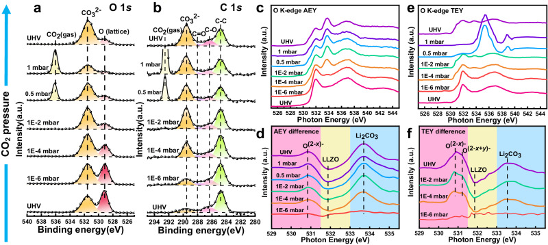
The advanced synchrotron radiation ambient pressure technology helped us to slow down the rapid reactions of CO2/H2O on clean LLZO surface, so that we can clearly observe the thermodynamic and kinetic reaction processes. The kinetics of the reaction of clean LLZO with CO2 was investigated by in situ ambient pressure APXPS and XAS measurements to achieve a depth-profiling analysis of the surface reaction. Figure 5a, b shows the O 1 s and C 1 s APXPS spectra of the LLZO at increasing pressure of CO2. During the reaction, the reaction product is almost pure Li2CO3 which is illustrated in Supplementary Fig. 6. The peak intensity of CO32- at around 531.5 eV enhances with the increase of CO2 pressures from 1 × 10⁻⁶ – 1 × 10⁻² mbar, implying that CO2 can rapidly react with LLZO to form Li2CO3 even at low CO2 pressure. However, the peak intensity of CO32- remains almost unchanged upon increasing the pressure of CO2 from 1×10⁻² to 1 mbar. The results reveal that the reaction of CO2 on the LLZO surface may be restricted by surface active sites or oxygen supply from the sub-layer.
Fig. 5. The evolution of clean LLZO surface with the introduction of CO2 studied by APXPS and APXAS.
a The variation of O 1 s and b C 1 s APXPS spectra at increasing CO2 pressure from UHV to 1 mbar. c,d AEY spectra and difference images at different CO2 pressures. A new feature at 530.8 eV appears, which can be assigned to the high valence oxygen labeled as O(2-x)-. e,f TEY spectra and difference images at different CO2 pressures. Except the O(2-x)-, a new peak at 531.2 eV is observed above 1 × 10⁻⁴ mbar, attributed to the O(2-x+y)-.
The O K edge AEY and AP-mRAS spectra of the LLZO pellet at increasing CO2 pressures are displayed in Fig. 5c and Supplementary Fig. 7. The variation in the intensity of the Li2CO3 peak is consistent with that of XPS; the peak increases from 1 × 10⁻⁶ to 1 × 10⁻² mbar and then stabilizes from 1 × 10⁻² to 1 mbar. We normalized the AEY data at different CO2 pressures to achieve a clearer internal reaction image and then subtracted the UHV data (Ipressures-IUHV). The difference results are presented in Fig. 5d, where the spectrum at 1 × 10⁻⁶ mbar CO2 is close to the horizontal line, meaning that the reaction is insignificant at this pressure. When the CO2 pressure is escalated to 1 × 10⁻⁴ mbar, the signal of Li2CO3 intensifies significantly while the peak intensity of LLZO dramatically decreases, indicating the formation of Li2CO3 on the LLZO surface. Noteworthy, a new feature at ~530.8 eV appears, which can be assigned to high valence O(2-x)- caused by the Li extraction35–37. The O(2-x)- formation is induced when the lithium in the sub-layer are pulled to the surface to form Li2CO3 because there is not enough lithium around the oxygen atom on the surface. Our TEY results with a higher detective depth of ~10 nm are shown in Fig. 5e. Due to the interference of signal from CO2, effective TEY signals from the LLZO surface could not be obtained above 0.5 mbar. Besides the 530.8 eV signal, a new peak at 531.2 eV is observed in difference spectra in Fig. 5f above 1 × 10⁻⁴ mbar, which can be assigned to the O(2-x+y)-. The observation of high valence O(2-x)- and O(2-x+y)- at different depths suggests the existence of Li gradient in the sub-layer of LLZO after exposure to CO2, confirming our hypothesis that Li from the sub-layer is pulled to the surface to form Li2CO3, while oxygen remain in their original positions.
The variations of Li 1 s, Zr 3d, and La 4d APXPS spectra at increasing CO2 pressure are shown in Supplementary Fig. 8. The Li 1 s peak shifts to higher binding energy as the CO2 pressure rises, indicating the formation of Li2CO3 on the surface. No noticeable spectral line shape changes are seen in Zr 3d and La 4d spectra. Thus, the possible reaction between CO2 and clean LLZO surface can be described as following: the CO2 molecules may easily adsorb on particular oxygen sites on the surface of LLZO at very low CO2 pressure. Subsequently, the lithium in the sub-layer are pulled to the surface to form Li2CO3 through the reaction: Li6.5La3Zr1.5Ta0.5O12 + xCO2 → Li6.5−2xLa3Zr1.5Ta0.5O12-x + xLi2CO3. In contrast, LLZO is not a good oxygen ionic conductor at room temperature, the oxygen in the sub-layer have difficulty migrating to the surface. When the above definite oxygen sites are fully occupied, CO2 cannot react with LLZO further, resulting in the Li2CO3 layer being only 1–3 nm thick. Consequently, the CO2 reaction pathway is considered kinetically slow which is coincided with the negligible amount of Li2CO3 formed on garnet pellets after exposure to dry air18,38,39.
The detailed reaction process of clean LLZO surface with H2O
After the CO2 experiment, the LLZO pellet was treated using the same annealing method as shown in Fig. 2f–h. Interestingly, the Li2CO3 could not be removed completely by vacuum annealing as the signal of Li2CO3 was still observable in the O 1 s XPS spectrum at 350 °C in Supplementary Fig. 9a. The results confirm the following decomposition reaction of Li2CO3 at 350 °C: xLi2CO3 + Li6.5-2xH2xLa3Zr1.5Ta0.5O12 → Li6.5La3Zr1.5Ta0.5O12 + xH2O + xCO2. Thus, the decomposition reaction of Li2CO3 would require a higher temperature if the hydrogens in the sub-layer LLZO are fully removed.
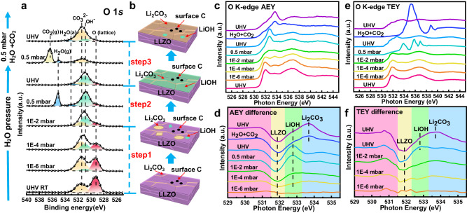
Furthermore, the reaction of H2O with the LLZO pellet was studied by APXPS and APXAS. The O 1 s XPS spectra and the product evolution diagram at increasing H2O pressure are displayed in Fig. 6a, b. Surprisingly, the peak intensity of CO32- at 531.5 eV enhances with increasing H2O pressure from UHV to 1 × 10⁻⁴ mbar, probably due to the gas path can only be cleaned to 1 × 10⁻⁷ mbar and there is a small amount of residual CO2. The results also indicate that the reaction of Li6.5La3Zr1.5Ta0.5O12 + xCO2 → Li6.5−2xLa3Zr1.5Ta0.5O12-x + xLi2CO3 at clean LLZO surface may be a thermodynamically favorable route compared to the reaction of LLZO with H2O, which is consistent with a just published article by Grey’s group40. As the pressure of H2O increases to 0.5 mbar, the signal of OH- at 530.9 eV41 almost covers the peak of CO32-, revealing that H2O mostly reacts with LLZO at 0.5 mbar and the reaction products are much more than that of CO2. Moreover, we compared the variation of O(CO32-)/O(lattice) and O(OH-)/O(lattice) during the introduction of CO2 and of H2O, respectively, and the corresponding results can be viewed in Supplementary Fig. 10. The reaction with CO2 shows a deceleration process, while that of H2O accelerates as the pressure increases. These results corroborate that the reaction of CO2 is a thermodynamically favorable route compared with that of H2O at low pressure. Next, we introduced a mixture gas of 0.5 mbar H2O and 0.5 mbar CO2, most of the surface LiOH is converted into Li2CO3 as shown in Fig. 6a and Supplementary Table 1.
Fig. 6. The evolution of LLZO surface with the introduction of H2O studied by APXPS and APXAS.
a The variation of O 1 s APXPS spectra at increasing H2O pressure from UHV to 0.5 mbar, followed by the introduction of a mixture gas of 0.5 mbar H2O and 0.5 mbar CO2. The high binding energy peaks appear at high pressure are the gas peaks of H2O and CO2. The peak at around 533.3 eV is the adsorbed H2O on LLZO surface. b Evolution diagram of LLZO surface reaction products. c,d AEY spectra and difference images at different H2O and CO2 pressures. No O(2-x)- feature is found in the difference spectra at 530.8 eV. e,f TEY spectra and difference images at different H2O and CO2 pressures. The slight fluctuations below 532 eV in the difference spectra may come from the signal noise.
The O K edge AEY and AP-mRAS spectra, shown in Fig. 6c and Supplementary Fig. 11, give a detailed analysis of the surface reaction, and the obtained results are consistent with the APXPS. The intensity of Li2CO3 enhances as the pressure of H2O increases from UHV to 1 × 10⁻⁴ mbar. However, the signal of LiOH at 532.8 eV nearly covers the signal of LLZO over 0.5 mbar, indicating that the reaction of H2O is more severe than CO2. Since no effective TEY spectra could be collected at the 0.5 mbar H2O pressure in Fig. 6e, we collected the spectrum when returning from 0.5 mbar to UHV instead. Even though the peak of LiOH is significantly intense, the LLZO signal can still be detected in the TEY spectrum at UHV, indicating that the thickness of LiOH layer at 0.5 mbar H2O is slightly <10 nm, much thicker than the Li2CO3 layer formed at the pressure of 1 mbar CO2. Notably, signals of O(2-x)- and O(2-x+y)- at around 530.8 eV were absent in all of the AEY and TEY difference spectra shown in Fig. 6d, f, signifying no valence change of oxygen in LLZO sub-layer after exposure to H2O. These results establish the existence of Li+/H+ exchange, i.e., the H+ fill the vacancies after the Li+ are pulled to the surface. After introducing a mixture gas of 0.5 mbar H2O and 0.5 mbar CO2, the peak of Li2CO3 in both AEY and TEY spectra is unambiguously observed, indicating the existence of the surface reaction: 2LiOH + CO2 → Li2CO3 + H2O.
Comparison of LLZO with LAGP
In order to find the possible determining factor for the air stability of electrolyte materials, we used the same in situ methods to study LAGP, which is an air stable solid electrolyte. The LAGP ceramic pellet was removed from its sealed packaging and placed directly into the test chamber from air without polishing. The signals of P, Ge and O can still be seen on the surface, indicating that LAGP has much better air stability than LLZO. Vacuum annealing at 350 °C hardly changes the composition of the surface. Then, we annealed the sample in 1 × 10⁻⁶ mbar O2 at 350 °C for 30 min. As shown in the Supplementary Fig. 12, it can be seen that the surface C of the sample at 350 °C significantly decreases, while the signals of O and P are greatly enhanced. However, when the sample was cooled to room temperature, the signal of C increased again while the signal of O and P decreased. No such carbonization phenomenon is observed on the surface of LLZO.
We conducted APXPS experiments on clean LAGP and the results are shown in Supplementary Fig. 13. LAGP cannot react with CO2 and H2O even at a high pressure of 0.5 mbar. By comparing two electrolyte materials, we infer that the stability of LAGP may come from the hydrophobic carbon layer generated on the surface and the stability of PO4 structure42. Due to the stability of surface O, it is difficult for LAGP to directly react with CO2 to generate Li2CO3. On the contrary, LLZO surface can react directly with CO2 to form a Li2CO3, which is a hydrophilic layer, may cause the occurrence of subsequent severe reactions. The results indicate that the hydrophilicity and hydrophobicity of the initial layer formed on the clean electrolyte surface, as well as the stability of the surface O structure, may play an important role in determining the air stability of solid electrolyte materials.
Discussion
The human factor in peak fitting has always been a problem in XPS data processing, especially in the overlapped O 1 s XPS data. Thus, the mRAS and AEY method are developed to assist XPS in species identification. The mRAS and AEY method can be implemented at any synchrotron radiation XPS end-station without requiring additional hardware. The mRAS and AEY has a detection depth slightly higher than XPS which not only can assist XPS in species identification, but also provide deep analysis abilities. In addition, its identification of species is also clearer and more intuitive because additional dimensions can result in more fingerprint features just like mRIXS as shown in Supplementary Fig. 5. Due to the dimensional improvement, mRIXS in O K edge has played an important role in the study of oxygen redox in cathode materials. The detection efficiency of mRAS is much higher than that of mRIXS, and the detection is also not affected by the gas and electrochemical environment. Thus, the mRAS method combined with near ambient pressure depth-profiling characterization methods possess the substantial potential and, therefore, should be widely popularized to study the air stability and the thermodynamics and kinetics process of gas/solid interface in energy materials.
In conclusion, in situ ambient pressure depth-profiling techniques were initiatively used to elucidate the dynamical evolution of LLZO pellet with CO2 and H2O. Low-temperature vacuum annealing helped us to obtain a clean LLZO surface through the reaction: xLi2CO3 + Li6.5-2xH2xLa3Zr1.5Ta0.5O12 → Li6.5La3Zr1.5Ta0.5O12 + xH2O + xCO2, where the H travel from LHLZO on the surface of LLZO. The APXPS, AP-mRAS and APXAS results experimentally prove that the reaction of CO2 with LLZO to form Li2CO3 is a thermodynamically favored path. However, the CO2 reaction is restricted by the hindered oxygen supply from the sub-layer, affording only 1–3 nm thick Li2CO3 layer. The driving force of the reaction leads to the formation of a lithium gradient in the sub-layer of LLZO. Moreover, Li+/H+ exchange was directly observed as no lithium gradient appeared during the reaction. This exchange is more intense when H2O reaches a higher pressure. Our results give a precise mechanism of the initial reaction of LLZO with CO2 and H2O and reveal the initial layer may play an important role in determining the air stability of solid electrolyte materials.
Methods
Materials and characterizations
The starting materials of Li2CO3 (Alfa Aesar, 99.9%), La2O3 (Alfa Aesar, 99.9%), ZrO2 (Alfa Aesar, 99.5%) and Ta2O5 (Aladdin, 99.5%) were mixed in stoichiometric amounts with 15 mol% Li2CO3 in excess. The mixture was ball-milled in 2-propanol for 12 h with agate balls in an agate vial, and then dried and heated in air at 1150 °C for 12 h. Then the ball-milling was repeated once, and the powder was sieved with a mesh number of 600 to obtain fine particles. The pellets were made by hot-pressing of the as-prepared LLZO powder in a flowing argon atmosphere at a temperature of 1050 °C under a constant pressure of 50 MPa for 1 h. The size of pellet for the operando experiment was 0.8 mm in thickness and 12 mm in diameter23. 0.3 mm thick LAGP ceramic pellet was purchased from Hefei Kejing Material Technology Co., Ltd. The X-ray powder diffraction (XRD) was performed using a Bruker D8 advance with Cu Kα radiation (λ = 1.54178 Å). Sintered and densified LLZO pellet was carried out from the glove box, then dry polished in air progressively using polishing paper with grit number from 800 to 1500, and the surface was wiped by alcohol. After polishing treatment, the polished and aged pellets were quickly transferred to the test chamber for the surface and cross section scanning electron microscopy (SEM) images, using a JEOL JSM-7800F field emission microscope.
Operando ambient pressure experiment
In situ annealing, ambient pressure mapping of resonant Auger electronic spectroscopy (AP-mRAS), ambient pressure X-ray photoelectron spectroscopy (APXPS) and X-ray absorption spectroscopy (APXAS) were carried out at BL02B at the Shanghai Synchrotron Radiation Facility (SSRF). The pellets were brought to the SSRF through an aluminum plastic bag sealed in a glove box. Before transferred to UHV chamber, the LLZO pellet was polished in air by sandpapers with grit number from 800 – 1500 to achieve parallel faces. The polishing thickness was sufficient to ensure that the surface contamination layer is completely polished off and then the surface was wiped by alcohol. The polished pellet was mounted onto a sample holder under ambient air (which took about 15 min) and were pumped into the instrument. LAGP and Li metal were pumped into the chamber without polishing process. Li metal was in situ scraped using a wobble-stick with sharp blade surface. For the vacuum annealing process, an infrared laser heater (912 nm wavelength, PREVAC) was used for heating from backside of the sample holder. The temperature was monitored by the K type thermocouple attached onto the LLZO pellet. The temperature was increased at the rate of 3 °C/min from room temperature to 370 °C and then kept at 350 °C for 30 min before spectroscopic measurements. The mass spectrum was collected by using MKS e-Vision 2 residual gas analyzer (RGA) installed on the analysis chamber. After conducting high-temperature measurements, LLZO pellet was naturally cooled to room temperature at a base pressure ~1 × 10⁻⁸ mbar for subsequent in situ characterization. After vacuum annealing, the LLZO pellet was annealed in 1×10⁻⁶ mbar O2 at 350 °C to reduce surface contaminated carbon.
The beamline 02B was a bending magnet beamline providing photons with energy range from 40 – 2000 eV. The photon flux was about 1011 photons/s and the energy resolving power was up to 13000. The C 1 s, O 1 s, Li 1 s, Zr 3d and La 3d XPS spectra were all collected at the photon energy of 650 eV with a step size of 0.1 eV using the Hipp-3 electron energy analyzer (Scienta Omicron). The photon energy was calibrated by a gold foil on the sample holder and the binding energy was calibrated by the C 1 s peak on LLZO at 284.6 eV. The APXAS data of O K-edge were collected simultaneously by using Auger electron yield (AEY) mode with investigation depth ~3 nm and total electron yield (TEY) mode with the penetration depth ~10 nm. The photon energy step was set at 0.2 eV for O K-edge mRAS and TEY experiments and the collection time of each mRAS mapping was about 12 min. The window of kinetic energy for O K-edge mRAS was set to 512 ±7 eV. All the spectra have been normalized to the beam flux measured by the upstream gold mesh. The stainless-steel tubes for CO2 (99.999%) and water vapor were baked in vacuum conditions and then flushed several times using high-purity CO2 and water vapor, respectively, before introducing the gases into the chamber. High-purity CO2 and water vapor were introduced into the reaction chamber by controlling two independent all-metal leak valves (VACGEN) as shown in Fig. 3a. The gas pressure was read by a capacitance film vacuum gauge (PFEIFFER CMR 363) attached on the chamber. All spectra were collected after the pressure had stabilized for 15 min. For the experiments of a mixture of H2O + CO2, the water vapor of 0.5 mbar was firstly maintained, then CO2 was introduced until the total pressure stabilized at 1 mbar.
Supplementary information
Source data
Acknowledgements
This work is supported by the National Key R&D Program of China (Grant No. 2019YFA0405601 X.S.L.), the National Natural Science Foundation of China (Grant Nos. 11905283 N.Z.) and the National Key R&D Program of China (Grant No. 2022YFA1503801 Z.L., No. 2022YFE0198500 H.Z.). The authors thank BL02B of Shanghai Synchtrotron Radiation Facility supported by National Science Foundation of China under contract No. 11227902 Z.L.. The authors thank prof Yong Han for disscussion.
Author contributions
N.Z., G.X.R., X.B.L. and H.Z. conducted the experiments. L.L.L, J.Z. and L.J.Z. participated in discussions and established the possible mechanism model. N.Z., G.X.R., H.Z. and Z.L. are responsible for the in situ ambient pressure depth-profiling X-ray spectroscopies and analysis. N.Z., Z.W., P.F.Y., H.Z. and X.S.L. designed the project, analyzed the results, and wrote the manuscript.
Peer review
Peer review information
Nature Communications thanks Lei Cheng, Hirotoshi Yamada and the other, anonymous, reviewers for their contribution to the peer review of this work. A peer review file is available.
Data availability
The data supporting the findings of this study are available within the article and its Supplementary Information. Additional data are available from the corresponding authors on request. Source data are provided with this paper.
Competing interests
The authors declare no competing interests.
Footnotes
Publisher’s note Springer Nature remains neutral with regard to jurisdictional claims in published maps and institutional affiliations.
These authors contributed equally: Nian Zhang, Guoxi Ren, Lili Li.
Contributor Information
Hui Zhang, Email: huizhang@mail.sim.ac.cn.
Zhi Liu, Email: liuzhi@shanghaitech.edu.cn.
Xiaosong Liu, Email: xsliu19@ustc.edu.cn.
Supplementary information
The online version contains supplementary material available at 10.1038/s41467-024-47071-4.
References
- 1.Cheng XB, Zhang R, Zhao CZ, Zhang Q. Toward safe lithium metal anode in rechargeable batteries: a review. Chem. Rev. 2017;117:10403–10473. doi: 10.1021/acs.chemrev.7b00115. [DOI] [PubMed] [Google Scholar]
- 2.Shen X, Liu H, Cheng X-B, Yan C, Huang J-Q. Beyond lithium ion batteries: higher energy density battery systems based on lithium metal anodes. Energy Storage Mater. 2018;12:161–175. doi: 10.1016/j.ensm.2017.12.002. [DOI] [Google Scholar]
- 3.Goodenough JB, Park KS. The Li-ion rechargeable battery: a perspective. J. Am. Chem. Soc. 2013;135:1167–1176. doi: 10.1021/ja3091438. [DOI] [PubMed] [Google Scholar]
- 4.Zhang Y, et al. Towards better Li metal anodes: challenges and strategies. Mater. Today. 2020;33:56–74. doi: 10.1016/j.mattod.2019.09.018. [DOI] [Google Scholar]
- 5.Harry KJ, Hallinan DT, Parkinson DY, MacDowell AA, Balsara NP. Detection of subsurface structures underneath dendrites formed on cycled lithium metal electrodes. Nat. Mater. 2014;13:69–73. doi: 10.1038/nmat3793. [DOI] [PubMed] [Google Scholar]
- 6.Lu D, et al. Failure mechanism for fast-charged lithium metal batteries with liquid electrolytes. Adv. Energy Mater. 2015;5:1400993. doi: 10.1002/aenm.201400993. [DOI] [Google Scholar]
- 7.Kinzer B, et al. Operando analysis of the molten Li| LLZO interface: understanding how the physical properties of Li affect the critical current density. Matter. 2021;4:1947–1961. doi: 10.1016/j.matt.2021.04.016. [DOI] [Google Scholar]
- 8.Janek, J. & Zeier, W. G. A solid future for battery development. Nat. Energy1, 1–4 (2016).
- 9.Tsai CL, et al. Li7La3Zr2O12 interface modification for Li dendrite prevention. ACS Appl Mater. Interfaces. 2016;8:10617–10626. doi: 10.1021/acsami.6b00831. [DOI] [PubMed] [Google Scholar]
- 10.Awaka J, Kijima N, Hayakawa H, Akimoto J. Synthesis and structure analysis of tetragonal Li7La3Zr2O12 with the garnet-related type structure. J. Solid State Chem. 2009;182:2046–2052. doi: 10.1016/j.jssc.2009.05.020. [DOI] [Google Scholar]
- 11.Murugan R, Thangadurai V, Weppner W. Fast lithium ion conduction in garnet-type Li7La3Zr2O12. Angew. Chem. Int Ed. Engl. 2007;46:7778–7781. doi: 10.1002/anie.200701144. [DOI] [PubMed] [Google Scholar]
- 12.Cheng L, et al. The origin of high electrolyte-electrode interfacial resistances in lithium cells containing garnet type solid electrolytes. Phys. Chem. Chem. Phys. 2014;16:18294–18300. doi: 10.1039/C4CP02921F. [DOI] [PubMed] [Google Scholar]
- 13.Cheng L, et al. Interrelationships among grain size, surface composition, air stability, and interfacial resistance of Al-substituted Li7La3Zr2O12 Solid Electrolytes. ACS Appl. Mater. Interfaces. 2015;7:17649–17655. doi: 10.1021/acsami.5b02528. [DOI] [PubMed] [Google Scholar]
- 14.Huo H, et al. Li2CO3: A critical issue for developing solid garnet batteries. ACS Energy Lett. 2019;5:252–262. doi: 10.1021/acsenergylett.9b02401. [DOI] [Google Scholar]
- 15.Sharafi A, et al. Surface chemistry mechanism of ultra-low interfacial resistance in the solid-state electrolyte Li7La3Zr2O12. Chem. Mater. 2017;29:7961–7968. doi: 10.1021/acs.chemmater.7b03002. [DOI] [Google Scholar]
- 16.Ma C, et al. Excellent stability of a lithium-ion-conducting solid electrolyte upon reversible Li+ /H+ exchange in aqueous solutions. Angew. Chem. Int. Ed. Engl. 2015;54:129–133. doi: 10.1002/anie.201408124. [DOI] [PubMed] [Google Scholar]
- 17.Liu X, et al. Elucidating the mobility of H+ and Li+ ions in (Li6.25−xHxAl0.25)La3Zr2O12 via correlative neutron and electron spectroscopy. Energy Environ. Sci. 2019;12:945–951. doi: 10.1039/C8EE02981D. [DOI] [Google Scholar]
- 18.Sharafi A, et al. Impact of air exposure and surface chemistry on Li–Li7La3Zr2O12 interfacial resistance. J. Mater. Chem. A. 2017;5:13475–13487. doi: 10.1039/C7TA03162A. [DOI] [Google Scholar]
- 19.Han Y, Zhang H, Yu Y, Liu Z. In situ characterization of catalysis and electrocatalysis using APXPS. ACS Catal. 2021;11:1464–1484. doi: 10.1021/acscatal.0c04251. [DOI] [Google Scholar]
- 20.Starr D, Liu Z, Hävecker M, Knop-Gericke A, Bluhm H. Investigation of solid/vapor interfaces using ambient pressure X-ray photoelectron spectroscopy. Chem. Soc. Rev. 2013;42:5833–5857. doi: 10.1039/c3cs60057b. [DOI] [PubMed] [Google Scholar]
- 21.Zhang N, et al. Mechanism study on the interfacial stability of a lithium garnet-type oxide electrolyte against cathode materials. ACS Appl. Energy Mater. 2018;1:5968–5976. doi: 10.1021/acsaem.8b01035. [DOI] [Google Scholar]
- 22.Zhang H, et al. Ambient pressure mapping of resonant auger spectroscopy at BL02B01 at the shanghai synchrotron radiation facility. Rev. Sci. Instrum. 2020;91:123108. doi: 10.1063/5.0020469. [DOI] [PubMed] [Google Scholar]
- 23.Fu J, et al. In situ formation of a bifunctional interlayer enabled by a conversion reaction to initiatively prevent lithium dendrites in a garnet solid electrolyte. Energy Environ. Sci. 2019;12:1404–1412. doi: 10.1039/C8EE03390K. [DOI] [Google Scholar]
- 24.Wu JF, et al. In situ formed shields enabling Li2CO3-free solid electrolytes: a new route to uncover the intrinsic lithiophilicity of garnet electrolytes for dendrite-free Li-metal batteries. Acs Appl. Mater. Interfaces. 2019;11:898–905. doi: 10.1021/acsami.8b18356. [DOI] [PubMed] [Google Scholar]
- 25.Cai J, et al. An APXPS endstation for gas–solid and liquid–solid interface studies at SSRF. Nucl. Sci. Tech. 2019;30:81. doi: 10.1007/s41365-019-0608-0. [DOI] [Google Scholar]
- 26.Ren G, et al. Photon-in/photon-out endstation for studies of energy materials at beamline 02B02 of shanghai synchrotron radiation facility. Chin. Phys. B. 2020;29:016101. doi: 10.1088/1674-1056/ab5d04. [DOI] [Google Scholar]
- 27.Zhang H, et al. Comprehensive electronic structure characterization of pristine and nitrogen/phosphorus doped carbon nanocages. Carbon. 2016;103:480–487. doi: 10.1016/j.carbon.2016.03.042. [DOI] [Google Scholar]
- 28.Zhu, Y. et al. Dopant‐dependent stability of garnet solid electrolyte interfaces with lithium metal. Adv. Energy Mater.9, 1803440 (2019).
- 29.Cai J, et al. Stoichiometric irreversibility of aged garnet electrolytes. Mater. Today Energy. 2021;20:100669. doi: 10.1016/j.mtener.2021.100669. [DOI] [Google Scholar]
- 30.Ren Y, et al. Effects of Li source on microstructure and ionic conductivity of Al-contained Li6. 75La3Zr1.75Ta0.25O12 ceramics. J. Eur. Ceram. Soc. 2015;35:561–572. doi: 10.1016/j.jeurceramsoc.2014.09.007. [DOI] [Google Scholar]
- 31.Timoshevskii A, Ktalkherman M, Emel’Kin V, Pozdnyakov B, Zamyatin A. High-temperature decomposition of lithium carbonate at atmospheric pressure. High. Temp. 2008;46:414–421. doi: 10.1134/S0018151X0803019X. [DOI] [Google Scholar]
- 32.Larraz G, Orera A, Sanjuán ML. Cubic phases of garnet-type Li7La3Zr2O12: the role of hydration. J. Mater. Chem. A. 2013;1:11419. doi: 10.1039/c3ta11996c. [DOI] [Google Scholar]
- 33.Paul PP, et al. A review of existing and emerging methods for lithium detection and characterization in Li‐ion and Li‐metal batteries. Adv. Energy Mater. 2021;11:2100372. doi: 10.1002/aenm.202100372. [DOI] [Google Scholar]
- 34.Cheng D, et al. Unveiling the stable nature of the solid electrolyte interphase between lithium metal and LiPON via cryogenic electron microscopy. Joule. 2020;4:2484–2500. doi: 10.1016/j.joule.2020.08.013. [DOI] [Google Scholar]
- 35.Roychoudhury S, et al. Deciphering the oxygen absorption pre‐edge: a caveat on its application for probing oxygen redox reactions in batteries. Energy Environ. Mater. 2020;4:246–254. doi: 10.1002/eem2.12119. [DOI] [Google Scholar]
- 36.Zhang J-N, et al. Trace doping of multiple elements enables stable battery cycling of LiCoO2 at 4.6 V. Nat. Energy. 2019;4:594–603. doi: 10.1038/s41560-019-0409-z. [DOI] [Google Scholar]
- 37.Qiao R, Chuang YD, Yan S, Yang W. Soft x-ray irradiation effects of Li2O2, Li2CO3 and Li2O revealed by absorption spectroscopy. PLoS One. 2012;7:e49182. doi: 10.1371/journal.pone.0049182. [DOI] [PMC free article] [PubMed] [Google Scholar]
- 38.Xia W, et al. Reaction mechanisms of lithium garnet pellets in ambient air: the effect of humidity and CO2. J. Am. Ceram. Soc. 2017;100:2832–2839. doi: 10.1111/jace.14865. [DOI] [Google Scholar]
- 39.Yamada H, Ito T, Kammampata SP, Thangadurai V. Toward understanding the reactivity of garnet-type solid electrolytes with H2O/CO2 in a glovebox using X-ray photoelectron spectroscopy and electrochemical ethods. ACS Appl. Mater. Interfaces. 2020;12:36119–36127. doi: 10.1021/acsami.0c09135. [DOI] [PubMed] [Google Scholar]
- 40.Vema S, et al. Understanding the surface regeneration and reactivity of garnet solid-state electrolytes. ACS Energy Lett. 2023;8:3476–3484. doi: 10.1021/acsenergylett.3c01042. [DOI] [PMC free article] [PubMed] [Google Scholar]
- 41.Wood KN, Teeter G. XPS on Li-battery-related compounds: analysis of inorganic SEI phases and a methodology for charge correction. ACS Appl. Energy Mater. 2018;1:4493–4504. doi: 10.1021/acsaem.8b00406. [DOI] [Google Scholar]
- 42.Yang, H., Zhang, Q., Chen, M., Yang, Y. & Zhao, J. Unveiling the origin of air stability in polyanion and layered‐oxide cathode materials for sodium‐ion batteries and their practical application considerations. Adv. Funct. Mater.34, 2308257 (2024).
Associated Data
This section collects any data citations, data availability statements, or supplementary materials included in this article.
Supplementary Materials
Data Availability Statement
The data supporting the findings of this study are available within the article and its Supplementary Information. Additional data are available from the corresponding authors on request. Source data are provided with this paper.


