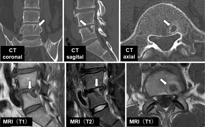
A 45-year-old man visited the emergency department with a 2-day history of lasting lower back pain that was aggravated by movement and alleviated by rest. A physical examination uncovered tenderness over the L5 spinous process. His blood laboratory test values were normal. Computed tomography (CT) demonstrated a small, radiolucent nodular lesion, suggesting an irregular bony defect in the L5 vertebra. Magnetic resonance imaging (MRI) demonstrated a focal herniation of the intervertebral disc through the end plate into the L5 vertebral body (Picture, arrows). He was diagnosed with a Schmorl's node and successfully managed with conservative treatment. A Schmorl's node is an intravertebral disc herniation referring to protrusions of the intervertebral disc cartilage through the vertebral body endplate and into the neighboring vertebra (1,2). Although there was no radiological evidence of a new onset, the acute lower back pain seen in this patient was most likely caused by a Schmorl's node based on the location of the pain. We herein report the CT and MRI features of the Schmorl's node, one cause of acute back pain. The specific imaging findings shown here may be helpful for emergency physicians.
Picture.
The authors state that they have no Conflict of Interest (COI).
References
- 1.Grivé E, Rovira A, Capellades J, Rivas A, Pedraza S. Radiologic findings in two cases of acute Schmörl's nodes. AJNR Am J Neuroradiol 20: 1717-1721, 1999. [PMC free article] [PubMed] [Google Scholar]
- 2.Mattei TA, Rehman AA. Schmorl's nodes: current pathophysiological, diagnostic, and therapeutic paradigms. Neurosurg Rev 37: 39-46, 2014. [DOI] [PubMed] [Google Scholar]


