Abstract
The Objective of the study was to assess the ossicular status in chronic otitis media (COM)—mucosal and squamosal type and statistically evaluate the extent of ossicular destruction intraoperatively in COM patients. The findings of this study could help us to predict preoperatively the probability of having ossicular chain destruction in COM ears and thus patients could therefore be properly consented about these potential issues before surgery. The study was carried out in ENT department of tertiary health care hospital, between January 2019 to January 2020. All patients of all age groups and both genders, diagnosed with COM Mucosal and Squamosal Type with complaints of ear discharge and hearing loss with good cochlear reserve and requiring surgery were included in the study, after taking informed written consent in vernacular language. All the patients included in the study were evaluated with detailed history, clinical examination including otomicroscopy, tuning fork tests and pure tone audiometry. The patients were then posted for ear surgery and the middle ear status and ossicular chain status were assessed using a microscope intraoperatively. Out of 98 patients, 45(45.9%) had mucosal and 53 (54.08%) had squamosal disease. Ossicular chain was eroded in 69 cases (70.5%). 23 out of 45 (51.1%) mucosal cases and 46 out of 53 squamosal cases (86.7%) reported ossicular erosion. Most frequently involved was long process of incus > stapes > malleus. From our study, we concluded that there is a significant relationship between type of disease pathology in middle ear and ossicular erosion being higher in Squamosal type of COM, with malleus being the most resistant and incus being the most susceptible ossicle.
Keywords: Mucosal, Squamosal, Tympanic membrane, Ear ossicles
Introduction
Chronic Otitis Media has been broadly classified as Mucosal disease (safe) and Squamosal (unsafe/dangerous) disease. Central perforation of tympanic membrane exposes the mucosa of middle ear and Eustachian tube orifice, their presence is denoted by the term Mucosal disease. Marginal and attic defects of tympanic membrane expose the anatomical structures of the attic, antrum and mastoid cell system and are referred to as Squamosal disease. Chronic Otitis Media (COM) is a leading cause of conductive hearing impairment secondary to damage of ear drum and middle ear ossicle induced by chronic inflammation present in tympanic cavity [1]. Although, ossicular chain erosion can occur in both types of COM, propensity for ossicular destruction is much greater in case of Squamosal COM due to presence of cholesteatoma and/or granulations [2–4].
The affected ossicles typically show areas of hyperaemia with proliferation of capillaries and prominent granulation tissue. Bone resorption or destruction occurs by osteoclast activity. High Resolution Computed Tomography (CT) of Temporal Bone also provides good information regarding status ofossicles. But they may not give accurate anatomical detail in all cases. Knowing the ossicular chain status is important. The status can be typically confirmed only intraoperatively under microscope or endoscope. It allows the surgeon to plan the need for an ossiculoplasty and type of ossiculoplasty for the particular patient. The patient can also be properly counseled about the likely outcome of the surgery.
Objective
To assess the status of Ossicular Chain in Chronic Otitis Media – Mucosal and Squamosal type.
Methods
Study type: Prospective Observational
Sample size: Sample size is 98 as per calculation
It was conducted in ENT department at a Tertiary health care hospital.
All the data were entered on Excel sheet® and analyzed. All the quantitative data were summarized as in the form of Mean ± SD. The difference between mean value of all groups was analyzed using ANOVA test in Open EPI software. All the qualitative data were summarized in the form of number and percentage. Data presented in the form of charts wherever applicable. The levels of significance and α error were kept 95% and 5% respectively, for all statistical analysis. p value < 0.05 was considered as Significant (S) and > 0.05 as Nonsignificant (NS).
Inclusion Criteria
All patients giving consent for participating in the study belonging to both genders and all age group with chronic otitis media (Mucosal and Squamosal type) with complaints of ear discharge and hearing loss having good cochlear reserve and requiring surgery and willing to come for regular follow up and follow the medical advice.
Exclusion Criteria
Patients with revision cases, presenting with clinical features suggestive of complications and not willing for surgery and not giving consent for the study.
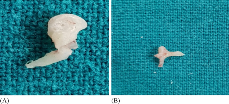
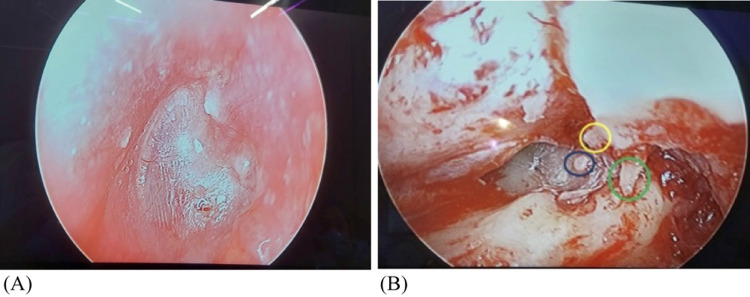
Patients of all age group with chronic otitis media (Mucosal and Squamosal type) were included in the study after taking informed written consent in vernacular language. All the patients included in the study were evaluated with detailed history, clinical examination including otomicroscopy, tuning fork test and pure tone audiometry. The patients were then posted for ear surgery and using otomicroscopy the status of ossicles was analyzed intraoperatively (Figs. 1, 2).
Fig. 1.
Eroded Malleus (a) and Incus (b)
Fig. 2.
Intraoperative status of ossicular chain (A) in Posterosuperior quadrant Cholesteatoma (B) Yellow circle—Eroded lenticular process of incus, blue circle—Intact stapes, green circle- short process of incus)
Results
Total 98 patients were enrolled for this study. All of these patients and divided into Mucosal and Squamosal type of Chronic Otitis Media based on the history and clinical findings. The number of cases of Mucosal COM were 45(45.9%) and Squamosal COM were 53(54.08%). Most patients 37(37.75%) belong to age group 11–20 years, followed by age group of 21–30 years in 24(24.48%) patients, there was equal gender distribution of males and females (49) each. The unilateral pathology was more common (67 patients) than bilateral disease (31 patients). The most common complaint in the present study was ear discharge in 95.9% patients followed by decreased hearing in 67.3%.
Ossicular chain erosion was correlated among patients with symptoms, type of COM, condition of middle ear mucosa, ear discharge, duration of symptoms, hearing loss in terms of air conduction threshold level and air bone gap, otoscopic findings. All the patients in the study were evaluated for ossicular chain intraoperatively.
Regarding the chief complaint of ear discharge, it was seen that most of the patients complained of ear discharge for less than 5 years (23/45 patients with mucosal type, and 37/53 patients with squamosal type). Patients in whom duration of discharge was present for 11–15 years was less (8/45 with mucosal type and 6/53 with squamosal type) (Table 1).
Table 1.
Correlation ofsymptoms withossicular chain erosion
| Symptoms | Mucosal n = 45 | Squamosal n = 53 | ||
|---|---|---|---|---|
| Intact | Erosion | Intact | Erosion | |
| Ear discharge | 21(46.6%) | 23(51.1%) | 04(7.5%) | 46(86.7%) |
| Decreased hearing | 13(28.8%) | 16(35.5%) | 05(9.4%) | 32(60.3%) |
| Ear ache | 19(42.2%) | 17(37.3%) | 02(3.7%) | 22(41.5%) |
| Others | 04(8.8%) | 03(6.6%) | 01(1.8%) | 03(5.6%) |
Decreased hearing was the next complaint being present in 66 out of 98 patients. Most of the patient in both mucosal type and Squamosal group, had the complaint for less than 5 years. In Mucosal COM, 29 patients out of 45 had complaint of decreased hearing; out of which 14 patients had complaint from 0.1- 5 years, 8 patients had complaint from 6–10 years and 7 had the complaint from 11–15 years. In Squamosal COM, 37 patients out of 53 had complaint of decreased hearing out of whom 24 patients had the complaint from 0.1–5 years, 8 had it from 6–10 years and 5 had from 11–15 years.
In Mucosal COM, 36 patients out of 45 had complaint of ear ache; out of which 18 patients had the complaint from 0.1 to 5 years, 10 had complaint from 6 to 10 years and 8 had complaint from 11 to 15 years. While in Squamosal COM, 24 out of 53 patients had complaint of ear ache; out of whom 14 patients had the complaint from 0.1 to 5 years, 5 patients had complaint from 6 to 10 years and 5 had complaint from 11 to 15 years (Table 2).
Table 2.
Correlation of hearing loss (air conduction threshold level) with ossicular chain status
| Hearing loss (dB) Air conduction |
Mucosal n = 45 |
Squamosal n = 53 |
||
|---|---|---|---|---|
| Intact | Erosion | Intact | Erosion | |
| 20–40 (mild) | 08(17.7%) | 02(4.4%) | 01(1.8%) | 14(26.4%) |
| 41–70 (moderate) | 14(31.1%) | 18(40%) | 03(5.6%) | 28(52.8%) |
| 71–95 (severe) | 01(2.2%) | 02(4.4%) | 00 | 07(13.2%) |
| > 96 (profound) | 00 | 00 | 00 | 00 |
| p value | < 0.05 | < 0.05 | ||
In this study, in Mucosal COM, ossicular erosion causes moderate hearing loss (air conduction threshold) in 18(40%) cases followed by severe hearing loss in 02(4.4%) cases and mild hearing loss in 02(4.4%) cases.
In Squamosal COM, presence of ossicular erosion resulted in moderate hearing loss (air conduction threshold) in 28(52.8%) followed by mild hearing loss in 14(26.4%) and severe hearing loss in 7(13.2%) (Table 3).
Table 3.
Correlation of middle ear mucosa status with ossicular chain status
| M E mucosa | Mucosal n = 45 |
Squamosal n = 53 |
||
|---|---|---|---|---|
| Intact | Erosion | Intact | Erosion | |
| Healthy | 16(35.5%) | 08(17.78%) | 00 | 01(1.88%) |
| Congested | 02(4.44%) | 01(2.22%) | 00 | 00 |
| Edematous | 05(11.1%) | 05(11.1%) | 01(1.88%) | 05(9.43%) |
| Granulations | 02(4.44%) | 12(26.6%) | 04(7.54%) | 48(90.56%) |
In patients with Mucosal COM, the ossicular chain was found to be eroded when there was presence of granulation in middle ear. The chain was found eroded in 12 patients out of 14 who had granulation. When the middle ear mucosa was found healthy, the ossicular chain was intact in 16 out of 20 patients.
The ossicular chain was eroded in 48 patients out of 52 who had granulation in the middle ear in Squamosal COM. Intact ossicular chain was seen in 4 patients with granulations in middle ear and 1 patient with edematous mucosa. These features are not exclusive of one another (Table 4).
Table 4.
Correlation of otoscopic finding with ossicular chain erosion (Intra-operative)
| Otoscopic finding | Malleus | Incus | Stapes |
|---|---|---|---|
| Tympanosclerosis | 0 | 3 | 0 |
| Small perforation | 0 | 3 | 0 |
| Moderate perforation | 1 | 6 | 0 |
| Large perforation | 3 | 9 | 2 |
| PSQ Cholesteatoma | 9 | 9 | 7 |
| Attic Cholesteatoma | 12 | 15 | 8 |
| PSQ + Attic cholesteatoma | 9 | 15 | 7 |
In this study, 3 patients had tympanosclerosis in which all of them had erosion of incus. Small perforation was seen in 3 patients and in them intra-operatively all had erosion of incus. 7 of the patients who had moderate perforation, 1 had malleus erosion and 6 had incus erosion. 14 patients had large perforation in pars tensa, and it was associated intra-operatively with erosion on incus, malleus, and stapes in 7, 3 and 2 patients respectively. In PSQ cholesteatoma most common ossicular erosion is of incus and malleus. In Attic cholesteatoma most common eroded ossicle seen was incus followed by malleus and then stapes. In PSQ + Attic Cholesteatoma, most common eroded ossicle seen was incus followed by malleus and then stapes (Table 5).
Table 5.
Status of ossicular chain(+ shows healthy intact ossicle,—shows eroded/ absent ossicle)
| Status of ossicular chain | Mucosal n = 45 | Squamosal n = 53 | ||
|---|---|---|---|---|
| Number | Percentage | Number | Percentage | |
| M + I + S + | 23 | 51.1 | 4 | 7.54 |
| M + I − S + | 14 | 31.1 | 11 | 20.76 |
| M + I − S − | 2 | 4.44 | 7 | 13.20 |
| M − I − S + | 3 | 6.66 | 12 | 22.64 |
| M − I + S + | 3 | 6.66 | 1 | 1.88 |
| M − I − S − | 0 | 0 | 18 | 33.96 |
In majority of Mucosal COM, ossicular chain was intact. Isolated erosion of ossicle was more common. Incus was the only affected ossicle followed by only malleus in 3 cases. Stapes was intact in all cases.
In Squamosal type, only 4 cases had intact ossicular chain. Involvement of single ossicle was seen in 12 cases. Incus was the eroded ossicle in 11 cases and only malleus was eroded in 1 case. Involvement of more than 1 ossicle was more common. Involvement of all three ossicles was seen in 18 cases.
Discussion
We have analyzed data as composed of a sample size of 98 with Chronic Otitis Media (Mucosal and Squamosal type) to establish their ossicular chain status. COM is an inflammatory process with a defective wound healing mechanism [5]. This inflammatory process in the middle ear is more harmful, the longer it stays and nearer it is to the ossicular chain [6].
The mean age of patients with Mucosal COM was 33.42 years and in patients with Squamosal it was 23.92 years. The ratio of male to female patients was equal in this study 1:1. Overall in our study (mucosal and squamosal type), granulation tissue was associated with ossicular erosion in 61(62.2%) cases. This was found to be statistically significant (p value < 0.05). In our study, hearing loss was found to correlate significantly with ossicular discontinuity (p value < 0.05).
Malleus was the most resistant ossicle, found intact in 63 (64.28%) cases in our study. In Squamosal COM, malleus was intact in 23(43.3%), eroded in 21(39.62%) and absent in 9 (16.90%) and is statistically significant (p value < 0.05). Incus was observed to be the most common ossicle to get eroded. The involvement of incus in Squamosal COM is statistically significant (p value < 0.05). The findings in our study are consistent with the studies conducted in past [6, 20–22]. In the present study, young adults were found to be more affected similar to study of SAURABH Varshney et al. [10] and Mohammadi G [7] and colleagues, Literature of A. Anglitoiu, et al. [8], Iñiguez-Cuadra R et al. [9], Yung M, Vowler et al. [10] and Hosny S et al. [11] showed almost equal gender distribution. In the study of Sharma T &Kuchchal V [12], all the patients had complaints of ear discharge and decreased hearing followed by other symptoms. Jayakumar, Celina Lovely et al. states that middle ear granulations are known to cause ossicular resorption [13]. Chole and Choo [14] found that when the perforation edges adhere to the promontory, it confines the granulation tissue and inflammatory products in small, dead spaces of the middle ear cleft which could lead to significant bone erosion over a period of time. Other studies of Gulya et al. [15] and Borg et al. [16] have also proved that granulation tissue is significantly associated with ossicular discontinuity, and this possibility increased by 7.95 times when the edges of the perforation were adherent to the promontory. In the study of Jeng et al. [17], hearing loss was found to correlate significantly with ossicular discontinuity. Carrillo et al. [18] reported that the Air Bone Gap (ABG) in patients affected by COM has been related to ossicular chain status. According to literature of Ingelstedt S, Jonson [19] an ABG of greater than 40 dB increases the probability of ossicular discontinuity to 89%. Udaipurwala et al. [20] Sade et al. found an incidence of around 6.00% of erosion of malleus in cases of safe COM. In unsafe disease they found malleus necrosis in 26.00% cases which correlates well with our finding [6]. However, a study by Mohammadi et al. [7] observed malleus erosion to be 43.9%. Udaipurwala et al. [21] found incidence of necrosis of the incus at 41.00%. Austin reported the most common ossicular defect to be the erosion of incus, with intact malleus and stapes, in 29.50% cases. Kartush found erosion of long process of incus with an intact malleus handle and stapes superstructure (type A) as the most common ossicular defect [22]. Udaipurwala et al. [20] found the superstructure to be necrosed in 21.00% cases, which matches with our findings. Austin reported erosion of stapes at around 15.50% [21]. Sade et al. [6] reported stapes involvement in unsafe COM to be 36.00%. Shrestha et al. [23] found involvement of stapes superstructure in 15.00% cases of unsafe COM. Motwani et al. [24] reported stapes arch necrosis in 30.00% cases of COM.
Conclusion
From our study, we concluded that there is a significant relationship in the degree of ossicular erosion with the type of disease pathology, being higher in Squamosal type of COM, with malleus being the most resistant and incus being the most susceptible ossicle. The presence of granulation was an important pathology associated with erosion of ossicles.
The erosion of ossicles leading to hearing loss in Chronic Otitis Media is a matter of concern because of its long-term effects on language development, communication skills and quality of life.
Thus, this study is helpful to convey the patients the likely outcomes of surgery as well as to prepare for surgery appropriately and early diagnosis and intervention by a skilled otologist to help ameliorate the morbidity that this condition can cause.
Abbreviations
- dB
Decibels
- SD
Standard Deviation
- Hz
Hertz
Funding
No funding sources.
Declarations
Conflict of interest
None declared.
Informed Consent
Informed consent was obtained from all individual participants included in the study.
Ethical Approval
All procedures performed in studies involving human participants were in accordance with the ethical standards of the institutional and/or national research committee and with the 1964 Helsinki declaration and its later amendments and its later amendments or comparable ethical standards.
Footnotes
Publisher's Note
Springer Nature remains neutral with regard to jurisdictional claims in published maps and institutional affiliations.
References
- 1.Thakur S, Ghimire N, Acharya R, Singh S, Anwar A. Ossicular chain status in cases of cholesteatomatous chronic otitis media in eastern Nepal. Asian J Med Sci. 2017;8:68. doi: 10.3126/ajms.v8i3.16671. [DOI] [Google Scholar]
- 2.Sudhoff H, Tos M. Pathogenesis of attic cholesteatoma: Clinical and histochemical support for combination of retraction theory and proliferation theory. Am J Otol. 2000;21(6):786–792. [PubMed] [Google Scholar]
- 3.Varshney S, Nangia A, Bist SS, Singh RK, Gupta N, Bhagat S. Ossicular chain status in chronic suppurative otitis media in adults. Indian J Otolaryngol Head Neck Surg. 2010;62(4):421–426. doi: 10.1007/s12070-010-0116-3. [DOI] [PMC free article] [PubMed] [Google Scholar]
- 4.Zehnder A, Kristiansen A, Adams J, et al. Osteoprotegerin knockout mice demonstrate abnormal remodelling of the otic capsule and progressive hearing loss. Laryngoscope. 2006;116:201–206. doi: 10.1097/01.mlg.0000191466.09210.9a. [DOI] [PMC free article] [PubMed] [Google Scholar]
- 5.Deka RC. Newer concepts of pathogenesis of middle ear cholesteatoma. Indian J Otol. 1998;4(2):55–57. [Google Scholar]
- 6.Sade J, et al. Ossicular damage in chronic middle ear damage. Acta Otolaryngol. 1981;92:273–283. doi: 10.3109/00016488109133263. [DOI] [PubMed] [Google Scholar]
- 7.Mohammadi G, Naderpour M, Mousaviagdas M. Ossicular erosion in patients requiring surgery for cholesteatoma. Iran J Otorhinol. 2012;24(3):125–128. [PMC free article] [PubMed] [Google Scholar]
- 8.Anglitoiu A, Balica N, et al. Ossicular chain status in the otological pathology of the ENT clinic Timisoara. Med Inevolut. 2011;17(4):344–351. [Google Scholar]
- 9.Iñiguez-Cuadra R, Alobid I, Borés-Domenech A, Menéndez-Colino LM, Caballero-Borrego M, et al. Type III tympanoplasty with titaniumtotal ossicular replacement prosthesis: anatomic and functional results. OtolNeurotol. 2010;31:409–414. doi: 10.1097/MAO.0b013e3181cc04b5. [DOI] [PubMed] [Google Scholar]
- 10.Yung M, Vowler SL. Long-term results in ossiculoplasty: an analysis of prognostic factors. Otol Neurotol. 2006;27:874–881. doi: 10.1097/01.mao.0000226305.43951.13. [DOI] [PubMed] [Google Scholar]
- 11.Hosny S, El-Anwar M, Abd-Elhady M, Khazbak A, El Feky A. Outcomes of myringoplasty in wet and dry ears. Int Adv Otol. 2014;10:256–259. doi: 10.5152/iao.2014.500. [DOI] [Google Scholar]
- 12.Sharma T, Kuchchal V. Evaluation and comparison of Hearing outcome in ossiculoplasty using differentgraft material. Ann Int Med Dental Res. 2017;3(3):10–14. doi: 10.21276/aimdr.2017.3.3.EN4. [DOI] [Google Scholar]
- 13.Jayakumar CL, Inbaraj LR, Pinto GJ. Pre-operative Indicators of Ossicular Necrosis in Mucosal CSOM. Indian J Otolaryngol Head Neck Surg. 2016;68(4):462–467. doi: 10.1007/s12070-016-0986-0. [DOI] [PMC free article] [PubMed] [Google Scholar]
- 14.Glasscock ME, Gulya AJ. Surgery of the Ear. 5th edn. Elsevier India: BC Decker Inc.; 2003.
- 15.Gulya AJ, Schuknecht HF (1995) Anatomy of the temporal bone with surgical implications, 2nd edn. Parthenon Publishing Group, Inc., Pearl River
- 16.Borg E, Nilsson R, Engstrom B. Effect of the acoustic reflex on inner ear damage induced by industrial noise. Acta Otolaryngol (Stockh) 1983;96:361–369. doi: 10.3109/00016488309132721. [DOI] [PubMed] [Google Scholar]
- 17.Jeng FC, Tsai MH, Brown CJ. Relationship of pre-operative findings and ossicular discontinuity in Chronic Otitis Media. Otol Neurotol. 2003;24:29–32. doi: 10.1097/00129492-200301000-00007. [DOI] [PubMed] [Google Scholar]
- 18.Carillo RJC, Yang NW, Abes GT. Probabilities of ossicular discontinuity in chronic suppurative otitis media using pure tone audiometry. Otolneurotol. 2007;28:1034–1037. doi: 10.1097/MAO.0b013e31815882a6. [DOI] [PubMed] [Google Scholar]
- 19.Ingelstedt S, Jonson B. Mechanisms of gas exchange in the normal human middle ear. Acta Otolaryngol Suppl (Stockh) 1966;224:452–461. doi: 10.3109/00016486709123624. [DOI] [PubMed] [Google Scholar]
- 20.Udaipurwala IH, Iqbal K, SaqulainG JM. Pathlogical profile in chronic suppurative otitis media—the regional experience. J Pak Med Assoc. 1994;44(10):235–237. [PubMed] [Google Scholar]
- 21.Austin DF. Ossicular reconstruction. Arch Otolaryngol. 1971;94:525–535. doi: 10.1001/archotol.1971.00770070825007. [DOI] [PubMed] [Google Scholar]
- 22.Kartush JM. Ossicular chain construction. Otolaryngol Clin North Am. 1994;27:689–715. doi: 10.1016/S0030-6665(20)30641-1. [DOI] [PubMed] [Google Scholar]
- 23.Shrestha S, Kafle P, Toran KC, Singh RK. Operative findings during canal wall mastoidectomy. Gujarat J Otorhinolaryngol Head Neck Surg. 2006;3(2):7–9. [Google Scholar]
- 24.Motwani G, Batra K, Dravid CS. Hydroxylapatite versus Teflon ossicular prosthesis: our experience. Indian J Otol. 2005;11:12–16. [Google Scholar]


