Abstract
Dysbiosis in gut microbiota is known to contribute to development of irritable bowel syndrome. We tried to investigate the effect of Bifidobacterium longum on repeated water avoidance stress (WAS) in a Wistar rat model. The three groups (no-stress, WAS, and WAS with B. longum) of rats were allocated to sham or WAS for 1 hour daily for 10 days, and B. longum was administered through gavage for 10 days. Fecal pellet numbers were counted at the end of each 1-hour session of WAS. After 10 days of repeated WAS, the rats were eutanized, and the feces were collected. WAS increased fecal pellet output (FPO) significantly in both sexes (P < 0.001), while the female B. longum group showed significantly decreased FPO (P = 0.005). However, there was no consistent change of myeloperoxidase activity and mRNA expression of interleukin-1β and TNF-α. Mast cell infiltration at colonic submucosa increased in the female WAS group (P = 0.016). In terms of fecal microbiota, the repeated WAS groups in both sexes showed different beta-diversity compared to control and WAS with B. longum groups. WAS-induced mast cell infiltration was reduced by the administration of B. longum in female rats. Moreover, administration of B. longum relieved WAS-caused dysbiosis, especially in female rats. In conclusion, B. longum was beneficial for WAS-induced stress in rats, especially in females.
Keywords: Bifidobacterium longum, Irritable bowel syndrome, Rats, Probiotics
INTRODUCTION
Irritable bowel syndrome (IBS) is characterized by repeated changes in the form of stool or defecation habits with abdominal pain or discomfort. The prevalence of IBS is 7% to 24% among women and 5% to 19% among men; women patients report a lower quality of life and demonstrate more extra-intestinal and psychological symptoms such as anxiety, depression, and somatization disorders [1]. The etiology of IBS involves various factors, including stress, activation of mucosal immune responses, increased mucosal permeability, food hypersensitivity, dysbiosis of intestinal microbiota, and changes in enteroendocrine metabolism [2]. Chronic stress modifies colonic functions and induces increased permeability, changes in motility, activation of myenteric plexus, release of serotonins, and visceral hypersensitivity [3].
The repeated water avoidance stress (WAS) is used in an animal model for chronic stress, amplifying psychological responses which are similar to human experiences. The repeated WAS increases fecal pellet output (FPO), and colonic submucosal mast cell infiltration with concomitant enhancement of mucosal cytokine levels, and also induces alterations in gut microbiome. These characteristics are similar to those of patients with the IBS-diarrhea (IBS-D) type [4,5].
Probiotics recover the balance of the gut microbiota in the colonic lumen and mucosal surface, making favorable environment to gut bacteria [6,7]. Bifidobacterium longum is a multi-functional probiotic which is classified as “GRAS (generally recognized as safe)”. In addition, B. longum is known to be effective in alleviating the symptoms of gastrointestinal (GI) and infectious diseases [8-10].
We have previously vakidated the usefulness of WAS which triggers colonic microinflammation, by measuring FPO and submucosal mast cell counts [4,5,11]. Interestingly, these responses were sex-specific. Thus, a 10-day treatment of Lactobacillus farciminis effectively mitigated symptoms in the WAS-induced female IBS rats [4]. In contrast, when we performed the same experiments with Roseburia faecis, this microorganism relieved FPO and submucosal mast cell counts, especially in male rats [11]. These suggest that probiotics may exert beneficial effects sex dependently. We speculate that B. longum might play a beneficial role an WAS-induced colonic microenvironment, sex dependently. The present study aimed to investigate the sex-different protective effects of B. longum in the WAS model and to evaluate whether this effect could be supported by the changes of fecal microbiota.
MATERIALS AND METHODS
Isolation of B. longum strains and culture conditions
B. longum was isolated from the stool of healthy trainers by the same method with the R. faecis [11]. The B. longum BBH 016 strain was identified and cultured in the de Man–Rogosa–Sharpe (MRS) medium (Becton, Dickinson and Company) in an anaerobic chamber (90% N2, 5% CO2, and 5% H2 conditions) at 37°C [11]. After culturing on DSM 104 agar plates for 1 day, B. longum was harvested by scraping with a loop and resuspended in the PBS containing 20% glycerol (v/v) [11]. The bacteria counts were determined using a Quantom Tx Microbial Cell Counter (CronyTek) [11]. Finally, Wistar rats received B. longum by oral gavage at a dosage of 1.0 × 10⁹ CFU/day.
Animals
Wistar rats (male and female) (Orient Co., Ltd.) were maintained in specific pathogen-free conditions and allowed for ad libitum access to Purina rat chow and water without additional enrichment [5]. Seven-week-old Wistar rats, weighing about 200 g, were selected for the experiments following an one-week acclimation period. Thirty six rats were stratified into three experimental groups in male and female both sexes, separately. That is, six rats were allocated each to the control, the WAS group, and the WAS group with plus B. longum, respectively in male and female rats. The experiments followed the recommendations of the Guide for the Care and Use of Laboratory Animals in South Korea [11]. In addition, the Institutional Animal Care and Use Committee (IACUC) of Seoul National University Bundang Hospital approved all of these experimental protocols (IACUC No. BA-2112-333-002-04).
Animal experiments
The protocol of repeated WAS has been described previously [4,5,11]. Briefly, each Wistar rat was positioned on a glass platform (5.0-cm length × 5.0-cm width × 6.0-cm height for female rats and 5.8-cm length × 5.8-cm width × 6.0-cm height for male rats). These platforms were affixed at the center of standard transparent plastic cages (26.7-cm length × 48.3-cm width × 20.3-cm height) filled with warm water (25°C) to a level 1 cm below the height of the plastic platform. This exposure continued for 1 hour daily in the morning (between 8 and 10 am) to avoid the confusion to rats over a period of 10 consecutive days. The rats were housed in pairs in their home cages but individually placed in each WAS cage. Stress levels from each WAS session were measured by the cumulative number of FPO at the termination of the 1-hour exposure.
Measurement of colonic submucosal mast cells using toluidine blue staining
Quantification of mast cell numbers within the colonic submucosa was conducted as follows [11]: one-cm segment from the cecum and the anus was extracted with the proximal portion of the colon [11]. These samples were then preserved in 10% buffered formalin and embedded in paraffin blocks. They were perpendicularly cut, creating 4 mm-thick sections and stained with toluidine blue [11]. The number of purple-stained mast cells in the colonic submucosa was counted. This number was divided by the total area of the colonic submucosa (the number of mast cells/colonic submucosal area [μm2]) [11].
ELISA for myeloperoxidase
The collected colon tissue samples were homogenized in lysis buffer and centrifuged. The lysis buffer was composed of a radioimmunoprecipitation assay (RIPA) buffer, a proteinase inhibitor, and a phosphatase inhibitor. The supernatant was used for analysis of myeloperoxidase (MPO) as an inflammatory marker.
Real-time quantitative PCR
mRNA was isolated from the colon tissue using the TRIzolTM reagent (Invitrogen), and quantitative mRNA was measured using a NanoDrop (ND-1000; Thermo Fisher Scientific). Complementary DNA (cDNA) was synthesized with a high-capacity cDNA reverse transcription kit (Applied Biosystems) [11]. Real-time quantitative PCR (RT-qPCR) was done with SYBR Green I Master Mix and an ABI Viia7 instrument for interleukin (IL)-1β and TNF-α [11]. Their transcript levels were normalized to β-actin [11].
Fecal sample collection and metagenome sequencing
Bacterial genomic DNA was extracted from rat fecal samples using a QIAamp DNA Stool Mini Kit (Qiagen). The quantity and quality of DNA were assessed using a NanoDrop 1000 spectrophotometer (Thermo Fisher Scientific) and electrophoresed using 2% agarose gel [11]. MiSeq library amplicon preparation was described in the previous paper [11]. Then the V3-V4 PCR amplicons were linked to the Illumina indices and adapters from the Nextera® XT Index Kit (Illumina). Using operational taxonomic unit (OTU) information (number of OTUs and sequences in each OTU), Shannon’s indices, such as α-diversity index, were calculated using EzBioCloud (CJ Bioscience Inc.). To visualize sample differences, selected taxa were created by GraphPad Prism ver. 8.01 (GraphPad Software Inc.).
Prediction of the functional composition of a microbial community’s metagenome
To predict the the functional composition of a microbial community’s metagenome, the algorithm ‘phylogenetic investigation of communities by reconstruction of unobserved states’ (PICRUSt) was used. Next, the alterations in the functional markers of the microbiota were analyzed based on the Kyoto Encyclopedia of Genes and Genomes (KEGG) database regarding modules and orthology [12].
Statistical analyses
Data are expressed as mean ± SEM. Categorical and continuous variables were compared among groups (no-stress, WAS alone, and WAS with B. longum) by Fisher’s exact test and Kruskal–Wallis test, respectively. P-values < 0.05 were regarded statistically significant. SPSS version 20.0 software (IBM Co.) was used for all the statistical analyses.
RESULTS
FPO induced in the WAS rat model
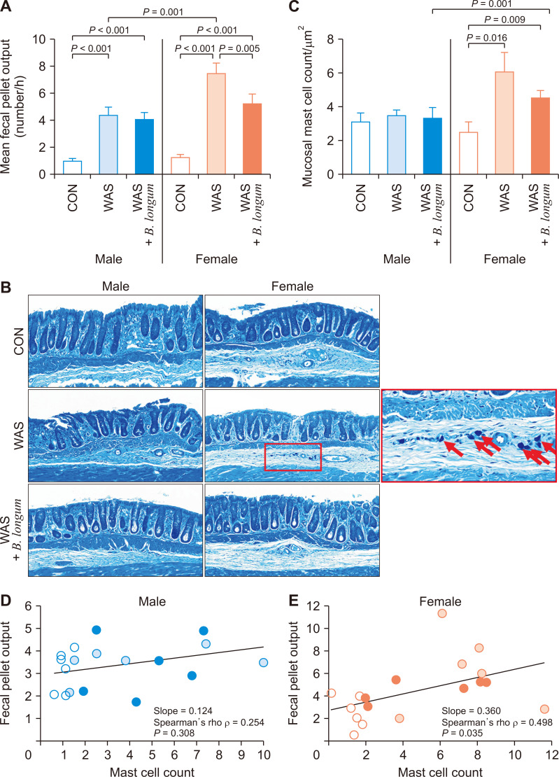
Continuous exposure to repeated WAS daily for 10 days increased FPO in a sex-dependent manner (Fig. 1A). The mean FPO was adjusted to the body weight between male and female rats. Compared with the control group animals, both male and female rats in the WAS group showed a statistically significant increase in FPO (P < 0.001). Notably, female rats in the stress group showed the significantly higher number of FPO compared to male rats in the stress group (P = 0.001). On the other hands, in case of male rats in the WAS group, B. longum administration significantly increased FPO compared to the control group (P < 0.001). However, there was no significant difference in males between the WAS group and the B. longum administrated WAS group. In the case of female rats, B. longum administration showed significantly lower FPO compared to the stress only group (P = 0.005). Similar to the FPO results, the number of mast cells infiltrated to colonic submucosa was increased significantly (P = 0.016) in the female stress group compared to the control group (Fig. 1B, red arrow and Fig. 1C). To assess the relationship between the FPO and the number of infiltrated mast cells, we employed the Spearman’s rank correlation coefficient method (also known as Spearman’s rho) (Fig. 1D, E). Only female rats showed positive correlation between the FPO and the number of mast cells with statistical significance (Fig. 1E, Spearman’s rho = 0.498, P = 0.035).
Figure 1. The fecal pellet output (FPO) and mast cell count in a repeated water avoidance stress (WAS) experiment.
FPO data represent the 10-day sessions with weight calibration for male and female rats. The difference between male and female body weight was normalized by multiplying female FPO with the ratio of male body weight to female body weight. The mean FPO (A) and the submucosal mast cell counts (C) showed a significant increase in the female WAS group compared to the female control group with statistical significance. (B) The submucosal mast cells were counted from toluidine blue stained colonic tissue slides. The mast cell count in the colonic mucosa was divided into the area of the colonic mucosa. The correlation analysis between the mean FPO and the mast cell counts in male (D) and female (E) rats indicated their positive correlation with statistical significance in female rats (P = 0.035).
Molecular responses to repeated WAS and B. longum supplement
ELISA and RT-qPCR experiments were performed on the colonic mucosa to assess the effects of repeated WAS alone and WAS with B. longum at the molecular level (Fig. 2). The colonic mucosal MPO level was not significantly different among groups in both male and female rats (Fig. 2A). Similarly, the levels of colonic mucosal mRNA of inflammatory cytokines, IL-1β (Fig. 2B) and TNF-α (Fig. 2C) were not significantly different among groups in both sexes.
Figure 2. The expression of colonic mucosal inflammation-associated markers measured in the rats subjected to repeated WAS.
The levels of myeloperoxidase (MPO) measured by ELISA (A) and mRNA transcripts of mucosal inflammatory cytokines, interleukin (IL)-1β (B) and tumor necrosis factor (TNF)-α (C) by qPCR. CON, control; WAS, water avoidance stress.
Changes of fecal microbiota in response to exposure to repeated WAS and B. longum supplement
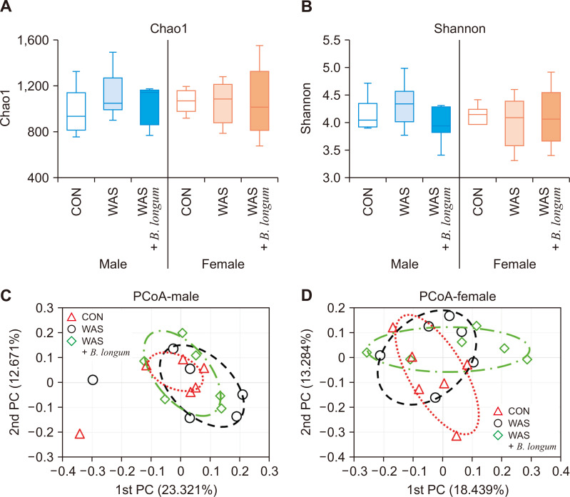
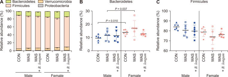
To evaluate the influence of repeated WAS and B. longum on the gut microbiota, 16s rRNA sequencing of fecal samples was performed. The α-diversity of the Chao1 (Fig. 3A) and Shannon’s index (Fig. 3B) showed no statistical differences among groups. To assess the β-diversity derived from the generalized UniFrac distance, principal coordinate analysis (PCoA) was performed on males (Fig. 3C) and females (Fig. 3D). Unlike β-diversity, the PCoA results indicated differences among the groups of the female microbial communities while male rats did not show difference. The relative abundances (%) of each group at the phylum level were represented in bar graph (Fig. 4A).
Figure 3. The α-diversity and β-diversity of fecal microbiomes from the repeated WAS exposed rats.
The α-diversity index chao1 (A), which reflects species richness and another α-diversity index Shannon (B), which reflects species evenness. β-Diversity, derived from generalized UniFrac distance, assessed through principal coordinates analysis (PCoA) (C). CON, control; WAS, water avoidance stress; PC, principal component.
Figure 4. The taxonomic composition of each group at the phylum level (A) represented in the bar graph, and Bacteroidetes (B) and Firmicutes (C) phylum represented in the scattered plot separately.
CON, control; WAS, water avoidance stress.
In the case of Bacteroidetes (Fig. 4B), there was no statistically significant difference among groups in both of males and females. Interestingly, female rats in control and WAS groups showed higher abundance of Bacteroidetes compared to those in the corresponding male groups (control male vs. female, P = 0.010; WAS male vs. female, P = 0.037). In the case of Firmicutes (Fig. 4C), no difference was found among groups in males as well as in female rats.
Predictive functional profiling of microbial communities
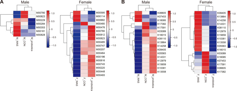
We then conducted the predictive functional profiling of microbial communities based on the KEGG modules. The representative modules and orthology in each group are indicated as heat maps (Fig. 5A and 5B). Modules were selected based on a P-value lower than 0.05 in more than two pair comparisons from the Kruskal–Wallis H test between two groups. In terms of metabolism, there was a statistically significant difference in females between control and WAS groups. However, these changes were mitigated, especially in female rats of the B. longum group with a pattern similar to the control group (Fig. 5A). However, there was no difference in case of orthology analysis at gene levels (Fig. 5B)
Figure 5. The predictive functional profiling of microbial communities, based on the Kyoto Encyclopedia of Genes and Genomes database regarding (A) module regarding metabolism pathway and (B) orthology at gene levels.
In terms of metabolism, there was statistically significant difference in females between control and WAS groups, and this difference disappeared by B. longum administration in the WAS challenged females. In contrast there was no difference in case of orthology analysis at gene levels. CON, control; WAS, water avoidance stress.
DISCUSSION
When we investigated the effects of B. longum on repeated WAS in rats, 10-day exposure of repeated WAS resulted in a significant elevation in FPO and infiltrations of submucosal mast cells. Interestingly, B. longum significantly improved repeated WAS-induced IBS-related symptoms with consistent correlation between FPO and mast cell counts. In terms of the gut microbiota, the administration of B. longum also showed different microbial community depending on sex. We suppose that this B. longum-induced microbial community improved the IBS changes in rats. Thus, B. longum could be applied to human patients with the IBS-D type after clinical trial.
The evidence supporting the use of probiotics in patients with IBS is that Lactobacillus formulations lead to interference with pathogen adhesion by binding to mucosal epithelial cells, reinforcement of epithelial barrier function, changes of mucosal response to stress, an immunomodulatory effect, reduction of visceral hypersensitivity through increased expression of opioid and cannabinoid receptors in the intestinal epithelial cells [13], and attenuation of inflammation [14]. In a recent meta-analysis with 18 randomized controlled trials, probiotics aided the treatment of IBS and were effective in one out of four patients [15]. Since the gut microbiomes of males and females differ, the male-to-female ratio of study participants would impact the effect, but no data have clarified this point yet [1].
Our recent study showed a sex difference of microbiota in human colorectal carcinogenesis [16]. According to this study, the female healthy control (HC) group had more lactate-producing bacteria (Bifidobacterium adolescentis, Bifidobacterium catenulatum, and Lactobacillus ruminis) than the male HC group [16], which partly explains the reason for the male predominance of colorectal cancer (CRC). Notably, younger subjects had more butyrate-producing bacteria (Agathobaculum butyriciproducens and Blautia faecis) than the older subjects in the HC group [16]. In addition, lactate-producing bacteria (B. catenulatum) were highly abundant in females compared to males in younger subjects of the HC group. Taken together, these results could explain why CRC develops less frequently in young females [16]. Interestingly, these sex- and age-dependent differences disappeared in the patients with adenoma or CRC [16]. These results suggest that the changes of gut microbiota could play a role in the colon carcinogenesis. As B. longum, a multi-functional probiotic is effective in alleviating the symptoms related to GI, immunological and infectious diseases. Therefore, there is a possibility that B. longum might be preventive against colon carcinogenesis, which needs further investigation.
Mast cells increase, especially, in the S colon of patients with IBS [17]. In addition, their association with hypersensitivity [18] has been reported in several human and animal studies. Furthermore, mast cells play various roles in the control of intestinal permeability [19], neuronal stimulation through the gut-brain axis [20], and visceral hypersensitivity [21]. Actually, mast cells produce various proteases as inflammatory modulators [22]. In explaining the increase of FPO as the result of mast cell infiltration by repeated WAS, we found a statistically significant positive correlation between the mast cell count and the FPO. This finding suggests that repeated WAS in a rat model has pathological similarity to human IBS.
Given the significance of the gut microbiota in human diseases, diverse omics tools have been employed to discern pivotal effectors within the gut microbiota. Metataxonomic sequencing, focusing on the highly conserved 16S ribosomal RNA, is a prevalent method used for the identification of bacterial community composition within the gut microbiota [23]. With numerous studies on the taxonomic diversity of microbiota under healthy and diseased conditions, it has been attempted to not only determine harmful bacterial strains for the host but also the microbial metabolism that influences the host, such as butyric acid-producing ability. PICRUSt can help predict the genes present in the microbiota community based on KEGG database [24]. In our experiments, repeated WAS induced different modules and orthologs, and administration of B. longum produced the profile similar to that in control rats, with sex difference after the administration of B. longum.
In spite of these interesting findings, there are some limitations in our present study. Even though we found some relationship between repeated WAS induced IBS models, the repeated WAS-induced inflammatory effects were not found. For instance, results of ELISA for measuring MPO and RT-qPCR for the transcripts of IL-1β and TNF-α in the WAS groups were not consistent. This might suggest that repeated WAS may not have been sufficient to induce the micro-inflammation in the present experiments. These variations might be dependent on strain of the animals used and other conditions such as the gender of the human researcher. It has been reported that the latter factor can influence outcomes in animal models, potentially stemming from stress activation induced provoked by the scent of a male human [25]. Next, we tried to analyze the predictive functional profiling of microbial communities based on the KEGG modules. There was a difference in the metabolism modules depending on sex by the administration of B. longum. However, in terms of orthology, there was no consistent difference.
In conclusion, repeated WAS induced FPO and mast cell infiltration into colonic submucosa, depending on sex, which was ameliorated by the administration of B. longum, especially in female rats. Moreover, in female rats, WAS changed the shape of gut microbiota, and administration of B. longum recovered repeated WAS-induced gut dysbiosis in terms of beta diversity and predictive functional profiling analysis, suggesting that the beneficial effects of B. longum on the IBS are prominent in females.
Footnotes
FUNDING
This work was supported by the Technology Innovation Program (20018499) funded by the Ministry of Trade, Industry & Energy (MOTIE, Korea).
CONFLICTS OF INTEREST
No potential conflicts of interest were disclosed.
REFERENCES
- 1.Kim N. Sex difference of gut microbiota. In: Kim N, editor. Sex/Gender-Specific Medicine in the Gastrointestinal Diseases. Springer; 2022. pp. 363–77. [DOI] [Google Scholar]
- 2.Barbara G, Cremon C, Carini G, Bellacosa L, Zecchi L, De Giorgio R, et al. The immune system in irritable bowel syndrome. J Neurogastroenterol Motil. 2011;17:349–59. doi: 10.5056/jnm.2011.17.4.349. [DOI] [PMC free article] [PubMed] [Google Scholar]
- 3.Chang L. The role of stress on physiologic responses and clinical symptoms in irritable bowel syndrome. Gastroenterology. 2011;140:761–5. doi: 10.1053/j.gastro.2011.01.032. [DOI] [PMC free article] [PubMed] [Google Scholar]
- 4.Lee JY, Kim N, Kim YS, Nam RH, Ham MH, Lee HS, et al. Repeated water avoidance stress alters mucosal mast cell counts, interleukin-1β levels with sex differences in the distal colon of wistar rats. J Neurogastroenterol Motil. 2016;22:694–704. doi: 10.5056/jnm16007. [DOI] [PMC free article] [PubMed] [Google Scholar]
- 5.Lee JY, Kim N, Nam RH, Sohn SH, Lee SM, Choi D, et al. Probiotics reduce repeated water avoidance stress-induced colonic microinflammation in Wistar rats in a sex-specific manner. PLoS One. 2017;12:e0188992. doi: 10.1371/journal.pone.0188992. [DOI] [PMC free article] [PubMed] [Google Scholar]
- 6.Abenavoli L, Scarpellini E, Colica C, Boccuto L, Salehi B, Sharifi-Rad J, et al. Gut microbiota and obesity: a role for probiotics. Nutrients. 2019;11:2690. doi: 10.3390/nu11112690. [DOI] [PMC free article] [PubMed] [Google Scholar]
- 7.Quigley EMM. Prebiotics and probiotics in digestive health. Clin Gastroenterol Hepatol. 2019;17:333–44. doi: 10.1016/j.cgh.2018.09.028. [DOI] [PubMed] [Google Scholar]
- 8.Cao F, Jin L, Gao Y, Ding Y, Wen H, Qian Z, et al. Artificial-enzymes-armed Bifidobacterium longum probiotics for alleviating intestinal inflammation and microbiota dysbiosis. Nat Nanotechnol. 2023;18:617–27. doi: 10.1038/s41565-023-01346-x. [DOI] [PubMed] [Google Scholar]
- 9.Yao S, Zhao Z, Wang W, Liu X. Bifidobacterium longum: protection against inflammatory bowel disease. J Immunol Res. 2021;2021:8030297. doi: 10.1155/2021/8030297. [DOI] [PMC free article] [PubMed] [Google Scholar]
- 10.Yun B, Song M, Park DJ, Oh S. Beneficial effect of Bifidobacterium longum ATCC 15707 on survival rate of Clostridium difficile infection in mice. Korean J Food Sci Anim Resour. 2017;37:368–75. doi: 10.5851/kosfa.2017.37.3.368. [DOI] [PMC free article] [PubMed] [Google Scholar]
- 11.Choi SI, Kim N, Nam RH, Jang JY, Kim EH, Ha S, et al. The protective effect of Roseburia faecis against repeated water avoidance stress-induced irritable bowel syndrome in a wister rat model. J Cancer Prev. 2023;28:93–105. doi: 10.15430/JCP.2023.28.3.93. Erratum in: J Cancer Prev 2023;28:219. [DOI] [PMC free article] [PubMed] [Google Scholar]
- 12.Langille MG, Zaneveld J, Caporaso JG, McDonald D, Knights D, Reyes JA, et al. Predictive functional profiling of microbial communities using 16S rRNA marker gene sequences. Nat Biotechnol. 2013;31:814–21. doi: 10.1038/nbt.2676. [DOI] [PMC free article] [PubMed] [Google Scholar]
- 13.Dai C, Zheng CQ, Jiang M, Ma XY, Jiang LJ. Probiotics and irritable bowel syndrome. World J Gastroenterol. 2013;19:5973–80. doi: 10.3748/wjg.v19.i36.5973. [DOI] [PMC free article] [PubMed] [Google Scholar]
- 14.Lee BJ, Bak YT. Irritable bowel syndrome, gut microbiota and probiotics. J Neurogastroenterol Motil. 2011;17:252–66. doi: 10.5056/jnm.2011.17.3.252. [DOI] [PMC free article] [PubMed] [Google Scholar]
- 15.Moayyedi P, Ford AC, Talley NJ, Cremonini F, Foxx-Orenstein AE, Brandt LJ, et al. The efficacy of probiotics in the treatment of irritable bowel syndrome: a systematic review. Gut. 2010;59:325–32. doi: 10.1136/gut.2008.167270. [DOI] [PubMed] [Google Scholar]
- 16.Song CH, Kim N, Nam RH, Choi SI, Jang JY, Kim EH, et al. The possible preventative role of lactate- and butyrate-producing bacteria in colorectal carcinogenesis [published online ahead of print November 30, 2023] Gut Liver. doi: 10.5009/gnl230385. doi: 10.5009/gnl230385. [DOI] [PMC free article] [PubMed] [Google Scholar]
- 17.Nasser Y, Boeckxstaens GE, Wouters MM, Schemann M, Vanner S. Using human intestinal biopsies to study the pathogenesis of irritable bowel syndrome. Neurogastroenterol Motil. 2014;26:455–69. doi: 10.1111/nmo.12316. [DOI] [PubMed] [Google Scholar]
- 18.Yu LC, Perdue MH. Role of mast cells in intestinal mucosal function: studies in models of hypersensitivity and stress. Immunol Rev. 2001;179:61–73. doi: 10.1034/j.1600-065X.2001.790107.x. [DOI] [PubMed] [Google Scholar]
- 19.Martínez C, Vicario M, Ramos L, Lobo B, Mosquera JL, Alonso C, et al. The jejunum of diarrhea-predominant irritable bowel syndrome shows molecular alterations in the tight junction signaling pathway that are associated with mucosal pathobiology and clinical manifestations. Am J Gastroenterol. 2012;107:736–46. doi: 10.1038/ajg.2011.472. [DOI] [PubMed] [Google Scholar]
- 20.Traina G. The role of mast cells in the gut and brain. J Integr Neurosci. 2021;20:185–96. doi: 10.31083/j.jin.2021.01.313. [DOI] [PubMed] [Google Scholar]
- 21.Hasler WL, Grabauskas G, Singh P, Owyang C. Mast cell mediation of visceral sensation and permeability in irritable bowel syndrome. Neurogastroenterol Motil. 2022;34:e14339. doi: 10.1111/nmo.14339. [DOI] [PMC free article] [PubMed] [Google Scholar]
- 22.Caughey GH. Mast cell proteases as protective and inflammatory mediators. Adv Exp Med Biol. 2011;716:212–34. doi: 10.1007/978-1-4419-9533-9_12. [DOI] [PMC free article] [PubMed] [Google Scholar]
- 23.Hilton SK, Castro-Nallar E, Pérez-Losada M, Toma I, McCaffrey TA, Hoffman EP, et al. Metataxonomic and metagenomic approaches vs. culture-based techniques for clinical pathology. Front Microbiol. 2016;7:484. doi: 10.3389/fmicb.2016.00484. [DOI] [PMC free article] [PubMed] [Google Scholar]
- 24.Kanehisa M, Furumichi M, Tanabe M, Sato Y, Morishima K. KEGG: new perspectives on genomes, pathways, diseases and drugs. Nucleic Acids Res. 2017;45(D1):D353–61. doi: 10.1093/nar/gkw1092. [DOI] [PMC free article] [PubMed] [Google Scholar]
- 25.Georgiou P, Zanos P, Mou TM, An X, Gerhard DM, Dryanovski DI, et al. Experimenters' sex modulates mouse behaviors and neural responses to ketamine via corticotropin releasing factor. Nat Neurosci. 2022;25:1191–200. doi: 10.1038/s41593-022-01146-x. [DOI] [PMC free article] [PubMed] [Google Scholar]


