Abstract
Objectives:
This study sought to examine the chemical profile, antioxidant, antimicrobial, antibiofilm, and anti-quorum sensing potential of two propolis ethanolic extracts (PEEs) collected from northeast Algeria.
Materials and Methods:
To achieve the main objectives of this study, multiple in vitro tests were employed. The phenolic and flavonoid contents were analyzed, and the chemical composition of both PEE was determined by high-performance liquid chromatography. The antioxidant properties of the propolis extracts were investigated using six complementary tests. The inhibitory effects of propolis extracts were evaluated against multidrug-resistant (MDR) clinical isolates using agar well diffusion and microdilution methods, whereas their antibiofilm and quorum-sensing disruption effects were determined by spectrophotometric microplate methods.
Results:
The results demonstrated that phenolic and flavonoid contents were higher in propolis from the Guelma (PEEG) region (PEEG; 188.50 ± 0.33 μg GAE/mg E, 144.23 ± 1.03 μg QE/mg E), respectively. Interestingly, different components were identified, and cynarin was the major compound detected. The PEEG sample exhibited potential antioxidant effects in scavenging ABTS•+ radicals with minimal inhibitory concentration values equal to 10.46 ± 1.40 µg/mL. Furthermore, the highest antibacterial activity was recorded by PEEG against Gram-positive Staphylococcus aureus MDR1. Similarly, PEEG effectively inhibited the biofilm formation of S. aureus MDR1 and the degradation of biofilm was up to 60%. In addition, quorum sensing disruption revealed that both extracts have a moderate capacity for violacein inhibition by the Chromobacterium violaceum ATCC 12472 strain in a concentration-dependent manner.
Conclusion:
These findings indicate that propolis can be regarded as a natural therapeutic agent for health problems associated with MDR bacteria and oxidative stress.
Keywords: Antibacterial, antioxidant, multidrug-resistant, propolis, quorum sensing
INTRODUCTION
Plant-derived natural products are considered an extraordinary source of bioactive compounds that have already proven their feasibility in the therapeutic realm.1 As one of the major beekeeping products, propolis already has well-known pharmaceutical properties. Propolis is a sticky phytocompound derived from plant exudates, it is rich in polyphenols, especially flavonoids and phenolic acids, which are known as the most active components responsible for its health benefits, including antibacterial, antioxidant, and anticancer.2 However, the chemical composition of propolis is unstable. Botanical sources and geographical areas highly influence it.3 Because of the common frequency of diseases related to drug-resistant bacteria and oxidative stress, many world scientists are searching for solutions to mitigate these issues. The sharp increase and the widespread of multidrug-resistant (MDR)bacteria compounded by the ability of bacteria to form biofilms is considered an emergent global health problem4, as bacteria in biofilms are extremely resistant and difficult to treat.5 Among the reasons that contributed to the worsening of this health problem is the overuse of antibiotics, which has increased in the last decades, especially in the current viral pandemic (coronavirus disease-2019). Furthermore, the scarcity of new antibiotics is one of the devastating purposes that contributed to the incapacity to control the spread of MDR bacteria.6 On the other hand, oxidative stress and inflammation are no less dangerous than the former issue, and many studies have proved their role in the pathogenesis of many chronic illnesses, including cardiovascular diseases, Alzheimer’s disease, and cancer.7 Dysfunction of the body’s antioxidant defense system leads to excess production of reactive oxygen species which causes inflammation via the induction of pro-inflammatory signaling pathways and the release of multiple inflammatory mediators, such as cytokines.8 The World Health Organization describes the current clinical drug pipeline as bleak and warns about the shortage of new therapeutic agents. Thus, it is important to identify new therapeutic strategies that solve these global health problems.
This study was conducted to seek more efficient natural bioactive compounds from propolis. Therefore, the total phenolics and flavonoid contents along with antioxidant activity of the Algerian propolis samples were determined.
Furthermore, in vitro studies were conducted to evaluate the antibacterial and antibiofilm activity against several MDR bacteria and the anti-quorum sensing activity of Algerian propolis.
MATERIALS AND METHODS
Propolis sampling and extraction
Propolis samples were collected in the early summer of 2019 from two different regions: Guelma and Ain-Fakroun in Northeast Algeria. 20 g propolis was extracted with 100 mL ethanol (80%). The mixture was kept for 24 hours in dark conditions before it was filtered through the Whatman no. 4 filter paper. The extract was evaporated using rotavapor and stored under dry conditions at 4 °C until use.
Total phenolic content (TPC)
TPC was calculated using the Folin-Ciocalteu (FC) reagent and gallic acid as a standard. A volume of 200 µL of propolis ethanolic extract (PEE) (0.5 mg/mL) was added to 1 mL of FC reagent (10%).
After 10 min of incubation in the dark, 800 µL of 7.5% Na2CO3 was added again, after incubation in the dark for 90 min. The absorbance was measured at 760 nm. The results are expressed as micrograms of gallic acid equivalents per milligram of extract (µg GAE/mg E).9
Total flavonoid content (TFC)
TFC was quantified using the aluminum trichloride method.10 An aliquot of 50 µL of the PEE was mixed with 130 µL methanol, 10 µL of aluminum nitrate, and 10 µL potassium acetate. After 40 min of incubation, the absorbance was measured at 415 nm. The results are expressed in micrograms of quercetin equivalents per milligram of extract (µg QE/mg E).
High-performance liquid chromatography (HPLC)
Separation and detection of propolis compounds were performed using a Shimadzu high-performance liquid chromatography (Shimadzu Cooperation, Japan) system. The detection system was a Shimadzu model SPD-M20A diode array, and the delivery system comprised a Shimadzu model LC-20AT. Separation was attained using an internal ODS-3 column (4 µm, 4.0 mm x 150 mm) and an Inertsil ODS-3 guard column. The elution program was as follows: the mobile phase included 0.1% aqueous acetic acid and methanol.
After being diluted in 1 mL of methanol (8 mg.mL-1), it was passed through a polytetrafluoroethylene filter with a 0.45 µm pore size. 20 µL was the injection volume. The HPLC-diode array detector (HPLC-DAD) system was used for quantitative and qualitative analysis using 42 standards: fumaric acid, gallic acid, p-benzoquinone, protocatechuic acid, theobromine, theophylline, catechin, 4-hydroxybenzoic acid, 6,7-dihydroxycoumarin, methyl-1,4 benzoquinone, vanillic acid, caffeic acid, vanillin, chlorogenic acid, p-coumaric acid, ferulic acid, cynarin, coumarin, propyl gallate, rutin, trans-cinnamic acid, ellagic acid, myricetin, fisetin, quercetin, trans-cinnamic acid, luteolin, rosmarinic acid, kaempferol, apigenin, chrysin, 4-hydroxy resorcinol, 14-dichlorobenzene, pyrocatechol, 4-hydroxybenzaldehyde, epicatechin, 24-dihydroxybenzaldehyde, hesperidin, oleuropein, naringenin, hesperetin, genistein, and curcumin.
Detection was performed using a DAD, and a wavelength of 254 nm was used to identify molecules. Results are expressed as micrograms per gram of dry weight.
DPPH-free radical scavenging activity
Essentially, 160 µL of DPPH solution (60 µM) was mixed with 40 µL of PEE at different concentrations. After 30 min of incubation in darkness, the prepared mixture was tested for absorbance at 517 nm. The results of DPPH scavenging effect were represented as 50% inhibition concentration (IC50) and contrasted with standards butylated hydroxyanisole (BHA) and butylated hydroxytoluene (BHT).11
Cupric ion-reducing antioxidant capacity (CUPRAC)
The CUPRAC assay was conducted using the approach described by Apak et al.12 Briefly, a mixture of 50 µL of copper (II) chloride (10 mM), 50 µL of neocuprine (7.5 mM), and 60 µL of ammonium acetate buffer solution (1M, pH: 7.0) was used. Subsequently, PEE was added to the initial mixture at a volume of 40 µL. After 60 min of incubation, the absorbance was measured at 450 nm, and the findings are represented as A0.5 (µg/mL).
Reducing power assay
A volume of 10 µL of solution was mixed with 50 µL of phosphate buffer (pH 6.6) and 50 µL of potassium ferricyanide (1%). The mixture was then incubated for 20 min at 50 °C. Following this, the initial mixture was combined with 50 µL of trichloroacetic acid (10%), 40 µL of distilled water, and 10 µL of ferric chloride solution (0.1%). At 700 nm, the absorbance was measured.13
ABTS cation radical decolorization
The ABTS•+ scavenging activity was determined using the method of Re et al.14 In short, ABTS•+ solution was prepared by the reaction between ABTS (7 mm) in water and potassium persulfate (2.45 mm) and stored for 12 hours in the dark. Prior to use, the absorbance of the ABTS solution was adjusted to 0.70 ± 0.02 (734 nm). Afterward, 40 µL of the solution was mixed with 160 µL of the dilute ABTS•+ solution. After 10 min of incubation, absorbance was measured at 734 nm. The results were established as a 50% IC50 (IC50= µg/mL).
Galvinoxyl radical (GOR) scavenging
40 µL of different concentrations of the extracts were mixed with 160 µL of a 0.1 mmol/L galvinoxyl solution. The obtained mixtures were incubated for 2 hours. The absorbance was read at 428 nm.15
Phenanthroline assay
The copper-phenanthroline assay was performed using the Szydłowska-Czerniak et al.16 method. A volume of 30 µL of 0.5% 1,10-phenanthroline solution (0.5%) was mixed with 50% FeCl3 (0.2%) and 10 µL of solution. Using methanol, the volume was made up to 200 µL. The incubation was performed in the dark for 20 min.
Antimicrobial activity determination
Selected microorganisms
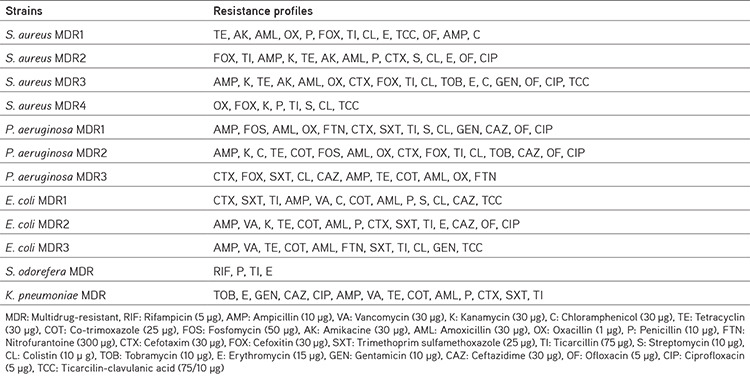
Antibacterial activity was evaluated against MDR clinical bacteria isolated from human urine samples. The strains were collected from the laboratory of bacteriology at the Public Medical Hospital in the province of Oum El Bouaghi, Algeria. Gram-negative bacteria (three MDR clinical isolates of Escherichia coli and Pseudomonas aeruginosa, one MDR clinical isolate of Klebsiella pneumoniae, and one MDR clinical isolate of Serratia odorifera), whereas only four MDR clinical isolates of S. aureus were employed in this study. The bacterial resistance profiles of each strain to antibiotics are depicted in Table 1.
Table 1. MDR profiles of different strains used in this study.
Disc diffusion assay
The disk diffusion assay was performed in accordance with the National Committee of Clinical Laboratory Standards. Culture suspensions (0.5 McFarland) were inoculated on fresh Mueller-Hinton agar plates. Afterward, 20 µL of PEE (20 mg/mL) was impregnated in sterile filter discs (Whatman paper no. 4) and deposited on the surfaces of the pre-inoculated plates. The Petri plates were incubated at 37 °C for 24 hours. Antibacterial standards included ampicillin (AMP, 10 µg/disc), kanamycin (K, 10 µg/disc), and streptomycin (S, 10 µg/disc).
Determination of minimum inhibitory (MIC) and, minimum bactericidal (MBC)
MIC and MBC were evaluated using the microdilution assay. PEE dilutions were prepared using dimethylsulfoxide (DMSO 15%), and the concentration ranged from 20 to 0.625. An aliquot of 10 µL of each bacterial strain was inoculated into the wells of a 96-well microliter plate containing 170 µL of Mueller-Hinton Broth (MHB). Then, 20 µL of different final concentrations of PEE were transferred to each well. MBC was determined by overlying 10 µL of the test dilutions from each clear well on fresh Luria-Bertani (LB) agar plates. After that, the plates were incubated for 24 hours at 37 °C. The lowest concentration with no bacterial growth was defined as MBC. Inocula and medium were used as positive controls.17
Influence of propolis extract on biofilm formation
An antibiofilm assay was employed to examine the antiadhesion activity. Only 5 strains were selected for this test namely: E. coli MDR1, P. aeruginosa MDR1, S. aureus MDR1, K. pneumoniae MDR, and S. odorifera MDR. The test was performed using the crystal violet assay.18 A volume of 20 µL of overnight isolate cultures was dispensed into the wells of 96 well microliter plates previously containing 170 µL of MHB, and then 10 µL of dissolved DMSO was added to each well at concentrations ranging from 20 to 0.625 mg/mL. Wells with bacteria and MHB served as controls. The following equation was used to estimate the percentage of biofilm inhibition:
Biofilm inhibition (%) = [optical density (OD) Control-OD Sample/OD Control x 100]
Violacein Inhibition (VI) assay
C. violaceum 12472 (CV12472) was used to test the effect of PEE on violacein production. A volume of 10 µL of an overnight broth culture of C. violaceum 12472 was dispensed into 96 well plates previously filled with 170 µL of LB broth (LBB) and incubated at 30 °C for 24 hours in the presence of various concentrations of PEE. Wells with LBB and inoculum were regarded as a positive control.19 Inhibition of violacein production was measured using a microplate reader (OD= 585 nm). Violacein repression percentage was calculated using the following formula:
Violacein inhibition (%) = [(OD Control-OD Sample)/(OD Control)] x 100
Bioassay for quorum sensing inhibition using CV026
To achieve this test, the method specified by Koh and Tham.20 was applied. The process was completed by mixing 5 mL of molten soft agar with 100 µL of C. violaceum 026 (CV026) bacterial suspension, further supplementing 20 µl of C6HSL and 10 µL of kanamycin. The latter suspension was spread across the surface of solidified LB agar (LBA) plates. Then, 6 mm wells were created through the LBA, and 50 µL different concentartions of PEE (20-2.5 mg/mL) were added to each well. The plates were incubated at 30 °C for 3 days. The presence of white or cream-colored halo around the wells signals quorum sensing (QS) inhibition, the results was measured in mm.
Statistical analysis
Graph Pad Prism 9.3.1 (Graph Pad Software, USA) was used for data analysis. One-way ANOVA followed by Tukey’s multiple comparison test was employed for statistical analyses. Results were considered statistically significant at p < 0.05.
RESULTS AND DISCUSSION
TPC and TFC
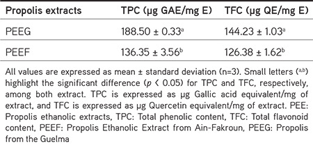
The TPC and TFC were determined as a measure of the number of propolis bioactive components. The results are displayed in Table 2. The propolis from the Guelma (PEEG) sample shows the highest TPC, followed by the Propolis Ethanolic Extract from Ain-Fakroun (PEEF) sample. Interestingly, our results present a higher TPC than previous studies conducted in different local regions in Algeria.21,22 Moreover, a more considerable variability in TPC was shown in propolis collected from several parts of the world.23 Conversely, these findings contradict the results of Bouaroura et al.24 who studied propolis from the same region (Guelma) and reported a complete lack of TPC, which emphasizes the intense variability in propolis contents. Regarding TFC, the results also displayed that the PEEG sample exhibited the highest content, greater than that reported by Boulechfar et al.25 Although the study samples were harvested in the same season and extracted using the same method, the two extracts were significantly different. This difference is mainly attributed to plant origin of the propolis and, more specifically, to the vegetation where bees gather propolis.26
Table 2. Total phenolic and flavonoid content of PEE.
Phenolic composition
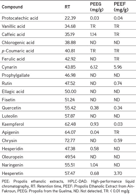
HPLC-DAD analyses were performed, and the results are illustrated in Figure 1 and Table 3. From the 42 standard compounds quantified, only 9 compounds were detected in the PEEG sample, while 7 compounds were detected in the PEEF sample. The PEEG and PEEF samples exhibited almost similar compositions but with different amounts. The most abundant flavonoid detected in the PEEG and PEEF samples was cynarin, with an amount of 6.12 and 5.96 mg/g, respectively. Interestingly, caffeic acid, apigenin, naringenin, and hesperidin were detected only in the PEEG sample, whereas rutin and chrysin were detected only in the PEEF sample. Overall, the main components identified in our propolis samples are similar to those previously described in different local regions in Algeria.27,28,29 Likewise, the phenolic compounds were approximately identical to those identified in propolis from different parts of the world.30,31 The abundance of flavonoids in both propolis samples correlates with many previous studies confirming poplar as a botanical source of propolis.24 Moreover, the botanical origin of cynarin identified in both PEEs was unknown, but it was inferred from a chemotaxonomic point of view that this compound would be collected by bees from exudates of plants belonging to the Asteraceae family, specifically Cynara cardunculus L.32 This species is in the surrounding areas of the apiaries not only in the two sites of the collection state but also in many northeast Algerian localities.33 It is worth mentioning that this report is the first on the occurrence of cynarin in Algerian propolis content.
Figure 1.
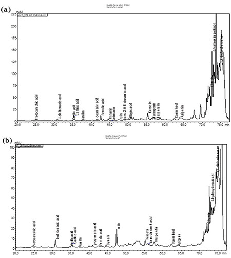
HPLC-DAD chromatogram of PEE. (a) PEEG, (b) PEEF
HPLC-DAD: High-performance liquid chromatography, PEE: Propolis ethanolic extracts, PEEF: Propolis Ethanolic Extract from Ain-Fakroun, PEEG: Propolis from the Guelma
Table 3. Chemical composition of PEE using HPLC-DAD analyses.
Antioxidant activities of PEE
As previously mentioned, excess production of free radicals leads to many disorders and may cause many chronic diseases. Therefore, antioxidant capacity of the propolis was determined, and the results are presented in Table 4. According to our DPPH results, the PEEG sample had the strongest antioxidant activity with an IC50 of 74.24 ± 1.91 µg/mL, but lower than the BHT and BHA standards. In contrast, PEEF showed no capacity to scavenge the radical DPPH. Regarding the results of the ABTS assay, the PEEG sample had a more potent scavenging capacity than the DPPH results, with an IC50 value of 10.46 ± 1.40 µg/mL, which seems important compared to BHT and BHA standards. Similarly, both extracts demonstrated high antioxidant potential in the remaining assays, except for the reducing power assay. Recently, many studies have been conducted on propolis because of its natural antioxidant potential. This potent activity is mainly related to its chemical components, which are capable of reducing radicals. This implies the beneficial efficacy of propolis for treating pathological damage caused by free radicals. Considering the employed assays, e.g., GOR and Phen, the antioxidant activity of the two tested samples was almost close to each other. This close similarity may be due to the phenolic profiles since the two extracts share some components such as cynarin, quercetin, kaemferol, hesperetin, and protocatechuic acid. The capacity of scavenging DPPH by the PEEG sample may be correlated to caffeic acid, which was absent in the remaining sample. Interestingly, this compound is well known for its high antiradical activity.34 According to Jun et al.,35 both propolis samples fall into the category of active antioxidants (IC50: 50-100 ppm).
Table 4. Antioxidant activity of propolis extracts by different assays.

Antibacterial activity of PEE
The antibacterial effects of PEE are presented in Tables 5 and 6. As can be seen, the PEEG sample showed a remarkable antibacterial effect against the tested S. aureus MDR strains. In contrast, the PEEF sample was only active against two strains of S. aureus. In contrast, PEEG demonstrated minimal efficacy against Gram-negative bacteria, whereas the PEEF sample did not show any inhibitory effects on Gram-negative strains. The microdilution approach revealed that the PEEG sample exhibited the highest bacteriostatic activity against S. aureus MDR strains with a MIC value ranging from 2.5 to 20 mg/mL. In contrast, less activity was recorded against Gram-negative bacteria. The highest bactericidal effect was observed for PEEG (MBC= 5 mg/mL) against S. aureus MDR1.
Table 5. Antimicrobial activity of the PEEG sample against MDR bacteria.
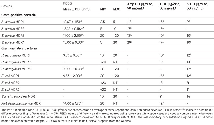
Table 6. Antimicrobial activity of the PEEF sample against MDR bacteria.
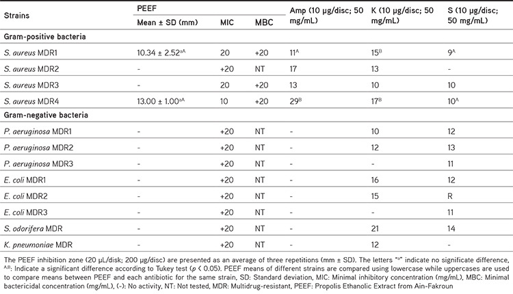
PEEF showed no bactericidal activity against all tested strains. Plants are a valuable source of bioactive compounds with various pharmacological effects. Many studies have reported the potential efficiency of plants in causing several disorders related to bacterial infections, especially those related to MDR bacteria.36 Considering that propolis is a plant-derived product and thus the abundance of several plant-bioactive compounds within its chemical content is widespread. Many researchers have focused on the possible use of propolis as an alternative antimicrobial agent for the treatment of infections caused by MDR pathogenic bacteria.
From the results mentioned above, propolis possesses significant antibacterial activity against MDR bacteria. Similarly, a study demonstrated that Palestinian propolis is active against MDR clinical isolates.37 These findings agree with previous research indicating that Gram-positive bacteria are more susceptible to propolis than Gram-negative bacteria. This sensitivity is probably related to differences in the membrane structure of bacteria. Furthermore, in some cases, the diameter zone recorded for the PEEG sample against S. aureus MDR strains was even more significant than those produced by different antimicrobial agents, which indicates the efficacy of propolis against MDR bacteria compared with the commonly used antibacterial treatment. Overall, this activity correlates with propolis bioactive contents such as flavonoids, which are known for their remarkable ability of bacterial inhibition.38 Cynarin was the major compound identified and many studies reported the antimicrobial properties of this compound39. In addition, other polyphenols, such as caffeic acid, possess highly potent antibacterial activity. However, many related reports have associated this activity with the synergistic interaction between different propolis active components.40
Antibiofilm activity of PEE
The results of the antibiofilm activity of PEE are shown in Figure 2. PEEG sample significantly inhibited biofilm formation at MIC concentration in each strain and the highest inhibition was recorded against S. aureus MDR1 (Figure 3). Lower activity was registered against the remaining strains. The PEEF sample showed eradication only against S. aureus MDR1 strain at MIC and MIC/2. Bacterial biofilms are one of the major factors that contribute to the progression and persistence of chronic infections, as the destructive effect of antibiotics is becoming more difficult.41
Figure 2.
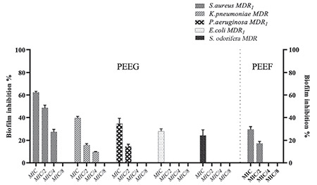
The effect of varied concentrations (MIC, MIC/2, MIC/4, and MIC/8) of PEEG and PEEF samples on biofilm formation of five MDR strains including, S. aureus MDR1, K. pneumoniae MDR, P. aeruginosa MDR1, E. coli MDR1, and S. odorifera MDR. The data represent the mean of three independent assessments. The error bars reflect standard deviations
MIC: Minimal inhibitory concentration, MDR: Multidrug-resistant,
Figure 3.

A representative image revealing the significant inhibition in bioflm formation by S. aureus MDR1 using light microscopic observation (magnification x 40): (a) before treatment with PEEG and (b) after treatment with PEEG at MIC concentration by crystal violet staining assay
MIC: Minimum inhibitory concentration
These findings agree with those of a study by Daikh et al.29 at a concentration of 300 µg/mL, Algerian propolis extract significantly inhibited the biofilm formation of virulent S. aureus. In line with these results, Brazilian green propolis has shown antibiofilm activity against the MDR strains of K. pneumoniae and P. aeruginosa.42 Many studies have highlighted the inhibitory effects of flavonoids and polyphenols on bacterial biofilms. The variability of flavonoids observed in both propolis extracts could account for their different in vitro effects. For example, the stronger activity of the PEEG extract in reducing biofilm production could be due to it is content of caffeic acid and quercetin compared with PEEF. Moreover, quercetin, kaempferol, apigenin, and naringenin were identified as biofilm inhibitors.43
VI and QSI of propolis extracts
The MIC values of the PEEG and PEEF samples against both strains were determined and shown in Table 7. It is clear from the results that both PEEs inhibited violacein production by C. violaceum 12472 in a dose-dependent manner. The PEEG sample was more potent in VI than the PEEF sample. Moreover, at lower doses of MIC/8, the PEEF sample showed no suppression of violacein synthesis. Unexpectedly, there was no inhibition of QS of C. violaceum 026, on LB Petri dish agar was observed.
Table 7. VI and anti-quorum sensing activities of PEEG and PEEF samples.
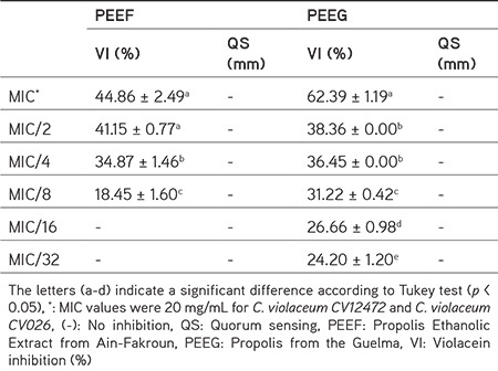
CV12472 can produce violacein pigment under a cell-to-cell communication mechanism called QS. Therefore, disruption of this phenomenon is necessary to overcome persistent infections.44 The obtained results prove that propolis inhibits the QS process. These findings correlate with the study by Sorucu and Ceylan45, which demonstrated that propolis has a high efficiency in disturbing the QS mechanism. Several types of phytochemicals, such as polyphenols and flavonoids, can affect the QS process in some bacteria by reducing the expression of several QS-controlled genes. Furthermore, recent findings have demonstrated the potent efficiency of different flavonoids, such as naringenin, kaempferol, quercetin, and apigenin, in inhibiting chemical signaling process.45,46,47,48
CONCLUSION
Recently, the widespread presence of MDR pathogens and the scarcity of novel antimicrobial agents have been considered an alarming threat to global health. To mitigate these issues, many researchers have focused on plant-derived products such as propolis. Herein, the antibacterial activity against several MDR pathogens has been reported. It was found that PEEG possessed the highest antimicrobial activity against several MDR strains. Furthermore, the antibiofilm and anti-quorum sensing activities of both extracts make them of considerable interest because they can disrupt microbial virulence factors and thus demonstrate efficacy against microbial resistance. According to the antioxidant activity results, both samples exhibited appreciable antioxidant activity, proving that propolis can eliminate the harmful effects of free radicals. Overall, these findings indicate that propolis could be used as an alternative remedy for severe pathology related to microbial resistance and oxidative stress. However, further analyses are needed to elucidate the main active compounds and mechanisms responsible for the different biological activities of propolis.
Acknowledgments
We would like to express our deepest gratitude to Prof. Mehmet Öztürk and Prof. Özgür Ceylan from the University of Muğla Sıtkı Koçman University, Türkiye, for the material support provided and for offering a collaborative and conductive platform for the current research. A further acknowledgment and recognition goes to the staff members of the biotechnology research center, Constantine, especially Dr. Chawki Bensouici, for his generous support and unconditional assistance to the study.
Footnotes
Ethics
Ethics Committee Approval: Not applicable, there are no researches conducted on animals or humans.
Informed Consent: Not required.
Authorship Contributions
Concept: W.H., A.Z., Design: W.H., A.Z., Data Collection or Processing: W.H., A.Z., M.M., C.B., Analysis or Interpretation: M.Ö., Ö.C., W.H., Literature Search: A.Z., Writing: N.G., W.H., A.Z.
Conflict of Interest: No conflict of interest was declared by the authors.
Financial Disclosure: This research was funded by the Ministry of Higher Education and Scientific Research, Algeria, through Doctoral Mobility Short Program 2019.
References
- 1.Atanasov AG, Zotchev SB, Dirsch VM. International natural product sciences taskforce; supuran CT. Natural products in drug discovery: advances and opportunities. Nat Rev Drug Discov. 2021;20:200–216. doi: 10.1038/s41573-020-00114-z. [DOI] [PMC free article] [PubMed] [Google Scholar]
- 2.Wieczorek PP, Hudz N, Yezerska O, Horčinová-Sedláčková V, Shanaida M, Korytniuk O, Jasicka-Misiak I. Chemical variability and pharmacological potential of propolis as a source for the development of new pharmaceutical products. Molecules. 2022;27:1600. doi: 10.3390/molecules27051600. [DOI] [PMC free article] [PubMed] [Google Scholar]
- 3.Zulhendri F, Chandrasekaran K, Kowacz M, Ravalia M, Kripal K, Fearnley J, Perera CO. Antiviral, antibacterial, antifungal, and antiparasitic properties of propolis: a review. Foods. 2021;10:1360. doi: 10.3390/foods10061360. [DOI] [PMC free article] [PubMed] [Google Scholar]
- 4.Minarini LADR, de Andrade LN, de Gregorio E, Grosso F, Naas T, Zarrilli R, Camargo ILBC. Editorial: antimicrobial resistance as a global public health problem: how can we address it? Front Public Health. 2020;8:612844. doi: 10.3389/fpubh.2020.612844. [DOI] [PMC free article] [PubMed] [Google Scholar]
- 5.Jiang H, Luan Z, Fan Z, Wu X, Xu Z, Zhou T, Wang H. Antibacterial, antibiofilm, and antioxidant activity of polysaccharides obtained from fresh sarcotesta of Ginkgo biloba: bioactive polysaccharide that can be exploited as a novel biocontrol agent. Evid Based Complement Alternat Med. 2021;2021:5518403. doi: 10.1155/2021/5518403. [DOI] [PMC free article] [PubMed] [Google Scholar]
- 6.Iskandar K, Murugaiyan J, Hammoudi Halat D, Hage SE, Chibabhai V, Adukkadukkam S, Roques C, Molinier L, Salameh P, van Dongen M. Antibiotic discovery and resistance: the chase and the race. Antibiotics (Basel). 2022;11:182. doi: 10.3390/antibiotics11020182. [DOI] [PMC free article] [PubMed] [Google Scholar]
- 7.Sharifi-Rad M, Anil Kumar NV, Zucca P, Varoni EM, Dini L, Panzarini E, Rajkovic J, Tsouh Fokou PV, Azzini E, Peluso I, Prakash Mishra A, Nigam M, El Rayess Y, Beyrouthy ME, Polito L, Iriti M, Martins N, Martorell M, Docea AO, Setzer WN, Calina D, Cho WC, Sharifi-Rad J. Lifestyle, oxidative stress, and antioxidants: back and forth in the pathophysiology of chronic diseases. Front Physiol. 2020;11:694. doi: 10.3389/fphys.2020.00694. [DOI] [PMC free article] [PubMed] [Google Scholar]
- 8.Mahmoud AM, Wilkinson FL, Sandhu MA, Lightfoot AP. The interplay of oxidative stress and inflammation: mechanistic insights and therapeutic potential of antioxidants. Oxid Med Cell Longev. 2021;2021:9851914. [Google Scholar]
- 9.Singleton VL, Rossi JA. Colorimetry of total phenolics with phosphomolybdic-phosphotungstic acid reagents. Am J Enol Vitic. 1965;16:144–158. [Google Scholar]
- 10.Topçu G, Ay M, Bilici A, Sarıkürkçü C, Öztürk M, Ulubelen A. A new flavone from antioxidant extracts of Pistacia terebinthus. Food Chem. 2007;103:816–822. [Google Scholar]
- 11.Blois MS. Antioxidant determinations by the use of a stable free radical. Nature. 1958;181:1199–1200. [Google Scholar]
- 12.Apak R, Güçlü K, Ozyürek M, Karademir SE. Novel total antioxidant capacity index for dietary polyphenols and vitamins C and E, using their cupric ion reducing capability in the presence of neocuproine: CUPRAC method. J Agric Food Chem. 2004;52:7970–7981. doi: 10.1021/jf048741x. [DOI] [PubMed] [Google Scholar]
- 13.Oyaizu M. Studies on products of browning reaction antioxidative activities of products of browning reaction prepared from glucosamine. Jap J Nutr Diet. 1986;44:307–315. [Google Scholar]
- 14.Re R, Pellegrini N, Proteggente A, Pannala A, Yang M, Rice-Evans C. Antioxidant activity applying an improved ABTS radical cation decolorization assay. Free Radic Biol Med. 1999;26:1231–1237. doi: 10.1016/s0891-5849(98)00315-3. [DOI] [PubMed] [Google Scholar]
- 15.Shi H, Noguchi N, Niki E. Galvinoxyl method for standardizing electron and proton donation activity. Methods Enzymol. 2001;335:157–166. doi: 10.1016/s0076-6879(01)35240-0. [DOI] [PubMed] [Google Scholar]
- 16.Szydłowska-Czerniak A, Dianoczki C, Recseg K, Karlovits G, Szłyk E. Determination of antioxidant capacities of vegetable oils by ferric-ion spectrophotometric methods. Talanta. 2008;76:899–905. doi: 10.1016/j.talanta.2008.04.055. [DOI] [PubMed] [Google Scholar]
- 17.Magina MD, Dalmarco EM, Wisniewski A Jr, Simionatto EL, Dalmarco JB, Pizzolatti MG, Brighente IM. Chemical composition and antibacterial activity of essential oils of Eugenia species. J Nat Med. 2009;63:345–350. doi: 10.1007/s11418-009-0329-5. [DOI] [PubMed] [Google Scholar]
- 18.O’Toole GA. Microtiter dish biofilm formation assay. J Vis Exp. 2011;2437. doi: 10.3791/2437. [DOI] [PMC free article] [PubMed] [Google Scholar]
- 19.Choo JH, Rukayadi Y, Hwang JK. Inhibition of bacterial quorum sensing by vanilla extract. Lett Appl Microbiol. 2006;42:637–641. doi: 10.1111/j.1472-765X.2006.01928.x. [DOI] [PubMed] [Google Scholar]
- 20.Koh KH, Tham FY. Screening of traditional Chinese medicinal plants for quorum-sensing inhibitors activity. J Microbiol Immunol Infect. 2011;44:144–148. doi: 10.1016/j.jmii.2009.10.001. [DOI] [PubMed] [Google Scholar]
- 21.Belfar ML, Lanez T, Rebiai A, Ghiaba Z. Evaluation of antioxidant capacity of propolis collected in various areas of Algeria using electrochemical techniques. Int J Electrochem Sci. 2015;10:9641–9651. [Google Scholar]
- 22.Nedji N, Loucif-Ayad W. Antimicrobial activity of Algerian propolis in foodborne pathogens and its quantitative chemical composition. Asian Pacific J Trop Dis. 2014;4:433–437. [Google Scholar]
- 23.Shehata MG, Ahmad FT, Badr AN, Masry SH, El-Sohaimy SA. Chemical analysis, antioxidant, cytotoxic and antimicrobial properties of propolis from different geographic regions. Ann Agric Sci. 2020;65:209–217. [Google Scholar]
- 24.Bouaroura A, Segueni N, Diaz JG, Bensouici C, Akkal S, Rhouati S. Preliminary analysis of the chemical composition, antioxidant and anticholinesterase activities of Algerian propolis. Nat Prod Res. 2020;34:3257–3261. doi: 10.1080/14786419.2018.1556658. [DOI] [PubMed] [Google Scholar]
- 25.Boulechfar S, Zellagui A, Bensouici C, Asan-Ozusaglam M, Tacer S, Hanene D. Anticholinesterase, anti-α-glucosidase, antioxidant and antimicrobial effects of four Algerian propolis. J Food Meas Charact. 2022;16:793–803. [Google Scholar]
- 26.Ecem Bayram N, Gerçek YC, Bayram S, Toğar B. Effects of processing methods and extraction solvents on the chemical content and bioactive properties of propolis. J Food Meas Charact. 2020;14:905–916. [Google Scholar]
- 27.Piccinelli AL, Mencherini T, Celano R, Mouhoubi Z, Tamendjari A, Aquino RP, Rastrelli L. Chemical composition and antioxidant activity of Algerian propolis. J Agric Food Chem. 2013;61:5080–5088. doi: 10.1021/jf400779w. [DOI] [PubMed] [Google Scholar]
- 28.Narimane S, Demircan E, Salah A, Ozcelik BÖ, Salah R. Correlation between antioxidant activity and phenolic acids profile and content of Algerian propolis: influence of solvent. Pak J Pharm Sci. 2017;30(4 Suppl):1417–1423. [PubMed] [Google Scholar]
- 29.Daikh A, Segueni N, Dogan NM, Arslan S, Mutlu D, Kivrak I, Akkal S, and Rhouati S. Comparative study of antibiofilm, cytotoxic activity and chemical composition of Algerian propolis. J Apic Res. 2019;59:1–10. [Google Scholar]
- 30.Alday-Provencio S, Diaz G, Rascon L, Quintero J, Alday E, Robles-Zepeda R, Garibay-Escobar A, Astiazaran H, Hernandez J, Velazquez C. Sonoran propolis and some of its chemical constituents inhibit in vitro growth of Giardia lamblia trophozoites. Planta Med. 2015;81:742–747. doi: 10.1055/s-0035-1545982. [DOI] [PubMed] [Google Scholar]
- 31.Kubina R, Kabała-Dzik A, Dziedzic A, Bielec B, Wojtyczka RD, Bułdak RJ, Wyszyńska M, Stawiarska-Pięta B, Szaflarska-Stojko E. The ethanol extract of polish propolis exhibits anti-proliferative and/or pro-apoptotic effect on HCT 116 colon cancer and Me45 malignant melanoma cells in vitro conditions. Adv Clin Exp Med. 2015;24:203–212. doi: 10.17219/acem/31792. [DOI] [PubMed] [Google Scholar]
- 32.Mandim F, Dias MI, Pinela J, Barracosa P, Ivanov M, Stojković D, Soković M, Santos-Buelga C, Barros L, Ferreira ICFR. Chemical composition and in vitro biological activities of cardoon (Cynara cardunculus L. var. altilis DC.) seeds as influenced by viability. Food Chem. 2020;323:126838. doi: 10.1016/j.foodchem.2020.126838. [DOI] [PubMed] [Google Scholar]
- 33.Issasfa B, Benmansour T, Valle V, Boukhatem M, Bouakba M. Contribution à l’étude du comportement mécanique des matériaux bio-sourcés de type composite (Cynara cardunculus/polyester) 2015. [Google Scholar]
- 34.Hachem K, Bokov D, Mahdavian L. Antioxidant capacity of caffeic acid phenethyl ester in scavenging free radicals by a computational insight. Polycycl Aromat Compd. 2022;43:1–18. [Google Scholar]
- 35.Jun M, Fu HY, Hong J, Wan X, Yang CS, Ho CT. Comparison of antioxidant activities of isoflavones from kudzu root (Pueraria lobata Ohwi) J Food Sci. 2003;68:2117–2122. [Google Scholar]
- 36.Aydın B, Yuca H, Karakaya S, Bona GE, Göger G, Tekman E, Şahin AA, Sytar O, Civas A, Canlı D, Pınar NM, Guvenalp Z. The anatomical, morphological features, and biological activity of Scilla siberica subsp. armena (Grossh.) Mordak (Asparagaceae) Protoplasma. 2023;260:371–389. doi: 10.1007/s00709-022-01784-9. [DOI] [PubMed] [Google Scholar]
- 37.Daragmeh J, Imtara H. In vitro evaluation of Palestinian propolis as a natural product with antioxidant properties and antimicrobial activity against multidrug-resistant clinical isolates. J Food Qual. 2020;2020:1–10. [Google Scholar]
- 38.Nichitoi MM, Josceanu AM, Isopescu RD, Isopencu GO, Geana EI, Ciucure CT, Lavric V. Polyphenolics profile effects upon the antioxidant and antimicrobial activity of propolis extracts. Sci Rep. 2021;11:20113. doi: 10.1038/s41598-021-97130-9. [DOI] [PMC free article] [PubMed] [Google Scholar]
- 39.Zhu X, Zhang H, Lo R. Phenolic compounds from the leaf extract of artichoke (Cynara scolymus L.) and their antimicrobial activities. J Agric Food Chem. 2004;52:7272–7278. doi: 10.1021/jf0490192. [DOI] [PubMed] [Google Scholar]
- 40.Ristivojević P, Trifković J, Andrić F, Milojković-Opsenica D. Poplar-type propolis: chemical composition, botanical origin and biological activity. Nat Prod Commun. 2015;10:1869–1876. [PubMed] [Google Scholar]
- 41.Kart D, Kuştimur AS. Investigation of gelatinase gene expression and growth of Enterococcus faecalis clinical isolates in biofilm models. Turk J Pharm Sci. 2019;16:356–361. doi: 10.4274/tjps.galenos.2018.69783. [DOI] [PMC free article] [PubMed] [Google Scholar]
- 42.Santos PBDRED, Ávila DDS, Ramos LP, Yu AR, Santos CEDR, Berretta AA, Camargo SEA, Oliveira JR, Oliveira LD. Effects of Brazilian green propolis extract on planktonic cells and biofilms of multidrug-resistant strains of Klebsiella pneumoniae and Pseudomonas aeruginosa. Biofouling. 2020;36:834–845. doi: 10.1080/08927014.2020.1823972. [DOI] [PubMed] [Google Scholar]
- 43.Slobodníková L, Fialová S, Rendeková K, Kováč J, Mučaji P. Antibiofilm activity of plant polyphenols. Molecules. 2016;21:1717. doi: 10.3390/molecules21121717. [DOI] [PMC free article] [PubMed] [Google Scholar]
- 44.Tamfu AN, Kucukaydin S, Ceylan O, Sarac N, Du r u ME. Phenolic composition, enzyme inhibitory and anti‑quorum sensing activities of cinnamon (Cinnamomum zeylanicum Blume ) and basil (Ocimum basilicum Linn) Chem Africa. 2021;4:759–767. [Google Scholar]
- 45.Sorucu A, Ceylan Ö. Determination of antimicrobial and anti-quorum sensing activities of water and ethanol extracts of propolis. Ankara Univ Vet Fak Derg. 2021;68:373–381. [Google Scholar]
- 46.Nazzaro F, Fratianni F, Coppola R. Quorum sensing and phytochemicals. Int J Mol Sci. 2013;14:12607–12619. doi: 10.3390/ijms140612607. [DOI] [PMC free article] [PubMed] [Google Scholar]
- 47.Savka MA, Dailey L, Popova M, Mihaylova R, Merritt B, Masek M, Le P, Nor SR, Ahmad M, Hudson AO, Bankova V. Chemical composition and disruption of quorum sensing signaling in geographically diverse United States propolis. Evid Based Complement Alternat Med. 2015;2015:472593. doi: 10.1155/2015/472593. [DOI] [PMC free article] [PubMed] [Google Scholar]
- 48.Asfour HZ. Anti‑quorum sensing natural compounds. J Microsc Ultrastruct. 2018;6:1–10. doi: 10.4103/JMAU.JMAU_10_18. [DOI] [PMC free article] [PubMed] [Google Scholar]


