Abstract
Genome-wide association studies (GWASs) may help inform treatments for infertility, whose causes remain unknown in many cases. Here we present GWAS meta-analyses across six cohorts for male and female infertility in up to 41,200 cases and 687,005 controls. We identified 21 genetic risk loci for infertility (P≤5E-08), of which 12 have not been reported for any reproductive condition. We found positive genetic correlations between endometriosis and all-cause female infertility (rg=0.585, P=8.98E-14), and between polycystic ovary syndrome and anovulatory infertility (rg=0.403, P=2.16E-03). The evolutionary persistence of female infertility-risk alleles in EBAG9 may be explained by recent directional selection. We additionally identified up to 269 genetic loci associated with follicle-stimulating hormone (FSH), luteinising hormone, oestradiol, and testosterone through sex-specific GWAS meta-analyses (N=6,095–246,862). While hormone-associated variants near FSHB and ARL14EP colocalised with signals for anovulatory infertility, we found no rg between female infertility and reproductive hormones (P>0.05). Exome sequencing analyses in the UK Biobank (N=197,340) revealed that women carrying testosterone-lowering rare variants in GPC2 were at higher risk of infertility (OR=2.63, P=1.25E-03). Taken together, our results suggest that while individual genes associated with hormone regulation may be relevant for fertility, there is limited genetic evidence for correlation between reproductive hormones and infertility at the population level. We provide the first comprehensive view of the genetic architecture of infertility across multiple diagnostic criteria in men and women, and characterise its relationship to other health conditions.
Introduction
Infertility, defined as the inability to achieve pregnancy within 12 months of regular unprotected sexual intercourse, affects one in six couples across the globe1. A range of demographic, environmental, and genetic factors may drive infertility, including the age-related decline of sperm and oocyte quality and quantity2,3, infectious diseases4–6, and rare Mendelian disorders such as cystic fibrosis7,8. However, the exact cause remains undetermined in up to 28% of couples and 40% of women with infertility9,10. Given that current treatments such as in vitro fertilisation pose physical, emotional, and financial burdens on couples and healthcare systems11–14, a richer understanding of the biology and pathophysiology of infertility is urgently necessary.
Heritable women’s reproductive health diseases, particularly endometriosis15 and polycystic ovary syndrome (PCOS)16, are thought to be responsible for a considerable proportion of female infertility, with PCOS in particular accounting for up to 80% of cases of anovulatory infertility17. It is hypothesised that sex-hormone dysregulation18,19 and obesity20, which often accompany reproductive diseases, may be involved in the aetiology of infertility. Yet little is known about the genetic basis of reproductive hormones and infertility, which are not well-phenotyped in men or women in large studies21,22. Moreover, negative selection against infertility naturally limits the frequency of risk alleles in the population23. Genome-wide association studies (GWASs) have thus typically queried proxy measures of fertility such as childlessness24,25, which may partly arise from socio-economic and behavioural factors.
We aggregated data from a range of sources, including primary care and hospital electronic health records (EHRs) and self-report, across six cohorts with over 1 million participants, to perform the first reported GWAS meta-analyses for male infertility and five categories of female infertility. In addition, we report results from the largest sex-specific GWASs to date for five reproductive hormones. By aggregating this data with complementary rare variant genetic association testing from the UK Biobank, we catalogue the common and rare genetic contributions to infertility and reproductive hormone levels, quantify the extent of shared genetic architecture between these traits, and prioritise genes and cell types for further functional investigation of the hormonal and non-hormonal drivers of infertility.
Results
Genome-wide meta-analyses identify novel genetic loci for female and male infertility
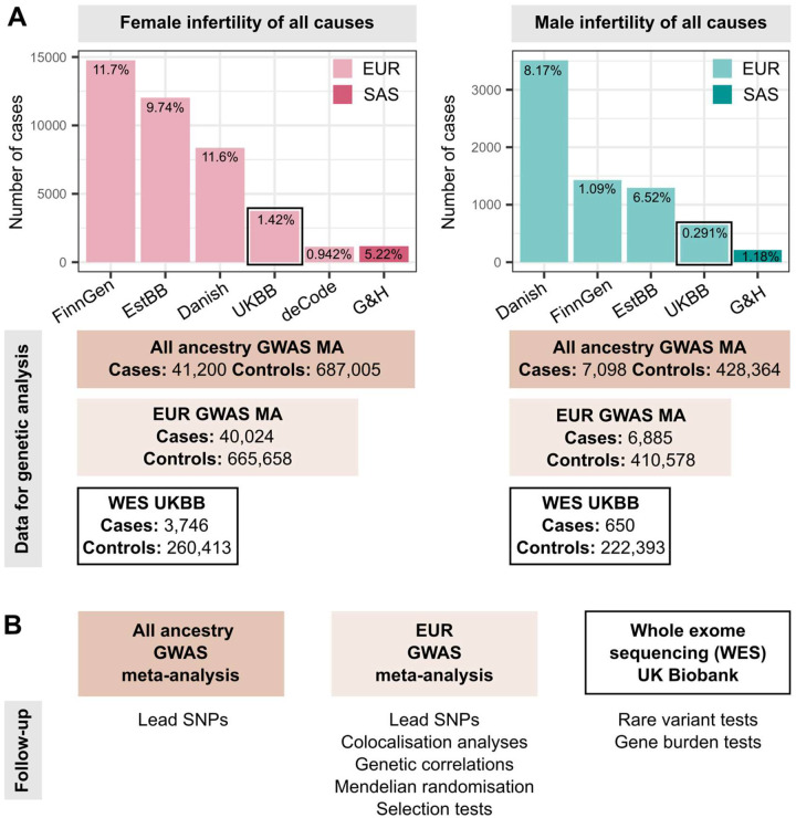
We identified female infertility of all causes (F-ALL), anatomical causes (F-ANAT), anovulation (F-ANOV), unknown causes, i.e., idiopathic infertility as defined by exclusion of known causes of infertility (anatomical or anovulatory causes, PCOS, endometriosis, or uterine leiomyomas) (F-EXCL), or idiopathic infertility defined by inclusion of diagnostic codes for idiopathic infertility (F-INCL), as well as male infertility of all causes (M-ALL) in six cohorts, primarily of European ancestry (Figure 1 and Supp. Tables 1 and 2). The case-control ratio of all-cause female infertility ranged from 0.9% in the deCODE Genetics dataset26 to 11.7% in FinnGen27, whereas the case-control ratio of male infertility was between 0.3% (UKBB) and 8.2% (Danish Biobank) (Figure 1 and Supp. Table 2). Anatomical female infertility was the least common cause of infertility in three of six cohorts (prevalence in UKBB=0.01%, EstBB=2.0%, FinnGen=0.8%). Due to varying sample ascertainment, the case-control ratio does not necessarily reflect the population prevalence of infertility.
Figure 1. Overview of study cohorts and analyses presented for infertility genetic association studies.
(A) Case numbers in each cohort contributing cases to genome-wide association study (GWAS) meta-analyses (MA) for female (left) and male (right) infertility. The prevalence of all-cause infertility in each cohort (%) is noted on the barplots. EUR=European ancestry, SAS=South Asian ancestry. EstBB=Estonian Biobank, Danish=Danish Blood Donor Study/Copenhagen Hospital Biobank, UKBB=UK Biobank, G&H=Genes and Health cohort. Total case and control counts for each type of genetic analysis: all ancestry GWAS meta-analysis (dark rectangles), EUR-only GWAS meta-analysis (light rectangles), and UK Biobank whole exome sequencing (WES) analyses (black outlined rectangles) are displayed. Male infertility in deCode, with <100 cases, was excluded from GWAS MA. Note the different Y-axis scales in each subplot. (B) Downstream analyses performed for each type of genetic analysis: lead variants were identified via distance-based pruning for all-ancestry and EUR-only GWAS meta-analyses; colocalisation, genetic correlation, and selection analyses were only performed for EUR meta-analyses due to the need for ancestry-matched linkage disequilibrium (LD) information; rare variant and gene burden tests were performed with WES data for the UK Biobank EUR-ancestry subset.
Novel genetic loci for infertility
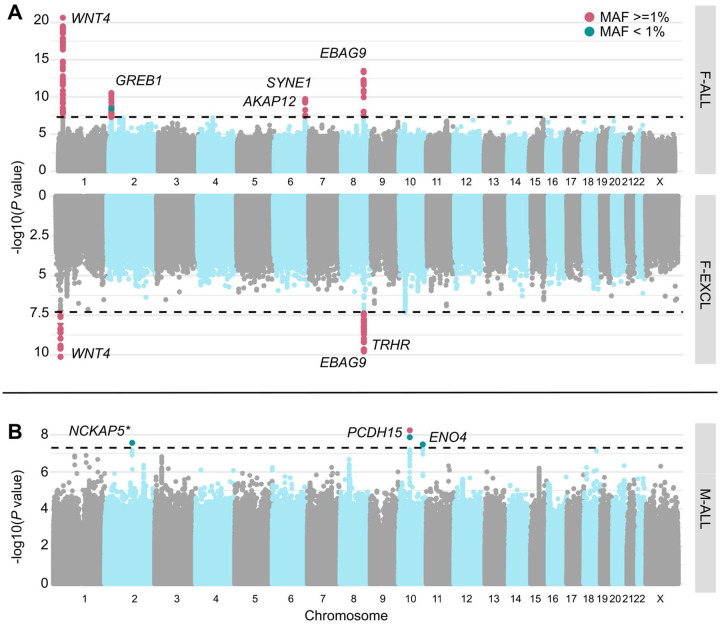
We performed GWAS meta-analyses, testing up to 28.4 million genetic variants for associations with each of the above categories of infertility, in up to 41,200 cases/687,005 controls in women, and 7,098 cases/428,364 controls in men (Figure 1 and Supp. Table 2). We identified 19 unique genome-wide significant (GWS, P<5E-8) loci associated with at least one category of female infertility and two loci for male infertility (minor allele frequency (MAF) range 0.24%−46%, lead variants reported in at least two cohorts) (Figure 2, Table 1, and Supp. Figure 1). There was no evidence for heterogeneity in lead variant effects across cohorts (Supp. Text).
Figure 2. Miami and Manhattan plots for selected infertility meta-analyses.
(A) Genetic variants associated with female infertility of all causes (F-ALL) (top) and idiopathic infertility (unknown causes) defined by exclusion of known causes such as anatomical or anovulatory causes, PCOS, endometriosis, or uterine leiomyomas (bottom). (B) Genetic variants associated with male infertility of all causes (M-ALL). Each point depicts a single SNP, with genome-wide significant (GWS) SNPs (P<5E-08, dashed line) coloured in pink for common variants with minor allele frequency (MAF)>=1% and green for those with MAF<1%. SNPs are annotated with the mapped gene. * indicates that lead variant is reported in only one cohort.
Table 1.
Lead variants associated with infertility in GWAS meta-analyses.
| RSID | chr:pos:A1 :A2 (hg38) | Mapped gene | All ancestries | EUR only | ||||
|---|---|---|---|---|---|---|---|---|
| Average MAF | OR (95% CI) | P-value | Average MAF | OR (95% CI) | P-value | |||
| Female infertility of all causes (F-ALL) | ||||||||
| rs61768001 | chr1:22139327:T:C | WNT4 | 0.166 | 0.909 (0.891–0.927) | 2.25E-21 | 0.163 | 0.911 (0.893–0.93) | 1.24E-19 |
| rs10200851 | chr2:11581956:T:C | GREB1 | 0.458 | 0.951 (0.937–0.965) | 2.90E-11 | 0.456 | 0.951 (0.936–0.965) | 5.84E-11 |
| rs6938404 | chr6:151222906:T:C | AKAP12 | 0.453 | 0.958 (0.943–0.973) | 3.88E-08 | 0.453 | 0.958 (0.943–0.973) | 3.88E-08 |
| rs17803970 | chr6:152232583:A:T | SYNE1 | 0.0836 | 1.09 (1.06–1.12) | 1.91E-10 | 0.0836 | 1.10 (1.07–1.13) | 7.50E-11 |
| rs1964514 | chr8:109463457:C:G | EBAG9 | 0.0595 | 1.13 (1.09–1.16) | 3.01E-14 | 0.0597 | 1.13 (1.09–1.16) | 6.68E-14 |
| Anatomical female infertility (F-ANAT) | ||||||||
| rs340879 | chr1:213983171:T:C | PROX1 | 0.418 | 0.906 (0.874–0.939) | 4.95E-08 | 0.418 | 0.902 (0.869–0.936) | 5.06E-08 |
| Anovulatory female infertility (F-ANOV) | ||||||||
| rs72665317 | chr1:22040580:T:G | CDC42 | 0.190 | 0.875 (0.839–0.913) | 7.76E-10 | 0.18 | 0.886 (0.847–0.927) | 1.45E-07 |
| rs72827480 | chr2:120388925:T:C | INHBB | 0.401 | 0.905 (0.873–0.938) | 4.20E-08 | 0.401 | 0.905 (0.873–0.938) | 4.20E-08 |
| rs1852684 | chr2:145068818:T:G | ZEB2 | 0.367 | 1.12 (1.08–1.16) | 9.25E-10 | 0.35 | 1.12 (1.08–1.17) | 3.44E-10 |
| rs552953683 | chr8:102898586:T:C | AZIN1 | 0.0024 | 0.341 (0.234–0.498) | 2.54E-08 | 0.0024 | 0.341 (0.234–0.498) | 2.54E-08 |
| rs9696009 | chr9:123856954:A:G | DENND1A | 0.0777 | 1.21 (1.14–1.29) | 6.87E-10 | 0.0695 | 1.24 (1.16–1.32) | 2.40E-10 |
| rs9902027 | chr17:7537667:T:C | TNFSF12 | 0.255 | 0.895 (0.86–0.931) | 4.06E-08 | 0.255 | 0.895 (0.86–0.931) | 4.06E-08 |
| rs143459581 | chr22:28068862:T:C | PITPNB | 0.0419 | 1.30 (1.19–1.43) | 1.21E-08 | 0.0419 | 1.30 (1.19–1.43) | 1.21E-08 |
| rs17879961 | chr22:28725099:A:G | CHEK2 | 0.0389 | 0.739 (0.673–0.811) | 1.55E-10 | 0.0389 | 0.739 (0.673–0.811) | 1.55E-10 |
| Idiopathic female infertility, exclusion definition (F-EXCL) | ||||||||
| rs61768001 | chr1:22139327:T:C | WNT4 | 0.165 | 0.923 (0.902–0.946) | 7.49E-11 | 0.162 | 0.928 (0.906–0.951) | 2.48E-09 |
| rs111597692 | chr8:109039973:T:C | TRHR | 0.0323 | 1.16 (1.10–1.22) | 1.51E-08 | 0.0323 | 1.16 (1.1–1.22) | 1.51E-08 |
| rs17378154 | chr8:109568721:A:G | EBAG9 | 0.059 | 1.13 (1.09–1.17) | 1.64E-10 | 0.0593 | 1.13 (1.09–1.17) | 3.36E-10 |
| Idiopathic female infertility, inclusion definition (F-INCL) | ||||||||
| rs61768001 | chr1:22139327:T:C | WNT4 | 0.170 | 0.87 (0.839–0.903) | 6.87E-14 | 0.165 | 0.872 (0.840–0.905) | 8.96E-13 |
| rs11692588 | chr2:11544358:A:G | GREB1 | 0.358 | 0.919 (0.892–0.947) | 2.98E-08 | 0.358 | 0.919 (0.892–0.947) | 2.98E-08 |
| rs190290095 | chr4:39786858:A:G | UBE2K | 0.0022 | 0.227 (0.137–0.375) | 7.60E-09 | 0.0022 | 0.227 (0.137–0.375) | 7.60E-09 |
| rs851982 | chr6:151703850:T:C | ESR1 | 0.428 | 0.921 (0.895–0.947) | 7.60E-09 | 0.437 | 0.922 (0.896–0.949) | 2.86E-08 |
| rs17378154 | chr8:109568721:A:G | EBAG9 | 0.0565 | 1.18 (1.11–1.25) | 2.47E-08 | 0.0569 | 1.18 (1.11–1.25) | 4.97E-08 |
| rs74156208 | chr10:61509370:A:G | TMEM26 | 0.184 | 1.10 (1.06–1.14) | 4.96E-08 | 0.187 | 1.10 (1.07–1.15) | 5.44E-08 |
| Male infertility of all causes (M-ALL) | ||||||||
| rs1228269928* | chr2:132923776:A:T | NCKAP5 | 0.0006 | 0.0995 (0.0441–0.224) | 2.72E-08 | 0.0006 | 0.0995 (0.0441–0.224) | 2.72E-08 |
| rs150639836 | chr10:53879806:T:C | PCDH15 | 0.0109 | 0.505 (0.402–0.636) | 5.72E-09 | 0.0109 | 0.505 (0.402–0.636) | 5.72E-09 |
| rs139862664 | chr10:116879589:C:G | ENO4 | 0.0072 | 0.388 (0.277–0.543) | 3.29E-08 | 0.0072 | 0.388 (0.277–0.543) | 3.29E-08 |
A1 is the effect allele.
lead variant is reported in only one cohort.
Among the variants associated with multiple subtypes of female infertility is rs1964514, an intronic variant in PKHD1L1 (OR (95% CI) for F-ALL=1.13 (1.09–1.16), F-EXCL=1.13 (1.09–1.17), F-INCL=1.18 (1.11–1.25)). This variant is 76 kb upstream of EBAG9, an oestrogen-responsive gene previously reported to have a recessive association with female infertility28 and thought to suppress maternal immune response during pregnancy29,30. We also identified an intronic variant in WNT4, rs61768001, associated with three categories of female infertility (F-ALL=0.909 (0.891–0.927), F-EXCL=0.923 (0.902–0.946), F-INCL=0.870 (0.839–0.903)). WNT4 is highly pleiotropic for female reproductive traits, as it is reported to associate with gestational length31, uterine fibroids32,33, endometriosis34,35, female genital prolapse27, and bilateral oophorectomy27. Such pleiotropy is expected, as WNT4 is a key regulator of female reproductive organ development in embryogenesis36–38.
The nearest gene to the idiopathic infertility-associated variant rs111597692 (F-EXCL OR=1.16 (1.10–1.22)) is TRHR, which encodes the thyrotropin-releasing hormone receptor. Mice with TRHR knockouts display a phenotype similar to primary ovarian insufficiency39,40. The F-ANOV associated variant rs72827480 (OR=0.905 (0.873–0.938)) colocalises with a testis-eQTL for INHBB in the GTEx Project41 (posterior probability (PP) of shared causal variant=91.6%) (Supp. Table 4). INHBB encodes the beta subunit of inhibin B, which regulates hypothalamic, pituitary, and gonadal hormone secretion42, and ovarian follicle and oocyte development43.
Finally, an intronic variant in ENO4, which is expressed in the testis and may play a role in sperm motility44, is associated with male infertility (rs139862664, OR=0.388 (0.277–0.543)). Male mice with ENO4 knockouts display infertility, abnormal sperm morphology and physiology, and decreased testis weight, among other altered male reproductive tract phenotypes45.
Genetic relationships between infertility and female reproductive conditions
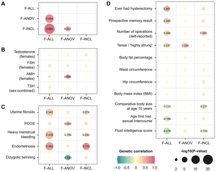
Genome-wide, we observed positive genetic correlation between endometriosis and F-ALL (rg (SE)=0.585 (0.0785), P=8.98E-14) and F-INCL (rg=0.710 (0.115), P=5.94E-10). We also observed positive correlation between F-ANOV and PCOS, the most common cause of anovulatory infertility (rg=0.403 (0.131), P=2.20E-3), and negative correlation between F-ANOV and spontaneous dizygotic twinning, a heritable metric of female fecundity that captures the propensity for multiple ovulation46 (rg=−0.740 (0.182), P=4.93E-05).
Two loci associated with both endometriosis and female infertility - WNT4 and ESR1 - may share the same putative causal variant (PP>93.6%, Supp. Table 5). Variants in both these genes have previously been associated with endometriosis-related infertility47–50. While GREB1 and SYNE1 also contain overlapping signals for infertility and endometriosis, there is strong evidence against shared causal variants (PP>75%, Supp. Table 5). Finally, three of eight loci for anovulatory infertility - INHBB, PITPNB, and CHEK2 - may share a causal variant with PCOS (PP>89.2%, Supp. Table 5).
Selection pressure may explain the persistence of some infertility-associated variants in the population
The genome-wide SNP heritability estimates (on the liability scale, accounting for disease prevalence54) for all categories of infertility are <10% (lowest for M-ALL at 1.12% (SE=0.93) and highest for F-ANOV at 9.54% (2.16)) (Supp. Table 6). This is lower than heritability estimates of two-thirds of all heritable binary phenotypes in the UK Biobank with population prevalence similar to that of infertility (64 phenotypes with Z>4 and prevalence <5%)53. We hypothesised that infertility risk-increasing alleles are subject to negative selection55, so we tested whether there was evidence for: (i) variants associated with infertility in loci under historical or recent directional selection56–58, or (ii) recent directional selection (over the last 2,000 to 3,000 years) measured by singleton density scores (SDSs)56 and balancing selection measured by standardised BetaScan2 scores (StdB2)59 at infertility loci.
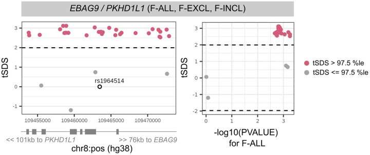
While we found no genome-wide signature of directional selection against infertility (Supp. Text), we observed extreme SDSs (in the highest 99.75th percentile (%ile) of SNPs within 10kb of a GWAS Catalog variant) at the EBAG9 locus associated with female infertility, indicating recent positive selection (Figure 4 and Supp. Table 7). EBAG9 is associated with infectious response phenotypes, suggesting that the locus may be under selection for its effects on the immune system. We additionally observed signatures of balancing selection, which maintains multiple alleles in the population through mechanisms such as heterozygote advantage or time-varying fitness60,61, at the female infertility loci GREB1 (StdB2 in the 98.6th-99.4th %ile of SNPs within 10kb of a GWAS Catalog variant) and INHBB (98.5th %ile), and the male infertility locus PCDH15 (98.7th %ile); however, variants at these loci with high probability of association with infertility did not have high balancing selection scores (Supp. Figure 2 and Supp. Table 7).
Figure 4. Directional selection scores at infertility-associated EBAG9 locus.
Recent directional selection, as measured by trait-aligned Singleton Density Scores (tSDSs) at the EBAG9 locus. The window of +/− 10 kb around the lead variant associated with female infertility of all causes (F-ALL) is displayed, along with the location of nearest gene transcription start sites (TSSs). The tSDSs are aligned to the infertility-risk increasing allele, wherein a positive tSDS indicates positive selection for infertility-risk increasing allele at the locus. Dashed lines indicate 2.5th percentile (%ile) and 97.5th %ile of SDSs, and variants below or above this threshold respectively are coloured in pink. Left: Locus plots depicting genomic position on the x-axis and tSDS on the y-axis. The lead variant rs1964514 (open circle) is not present in the tSDS dataset and thus assigned a score of 0. Right: Scatter plots depicting relationship between −log10 of the GWAS p-value for the variant association with F-ALL on the x-axis and tSDS on the y-axis.
Genetic determinants of reproductive hormone levels
Identification of novel reproductive hormone loci
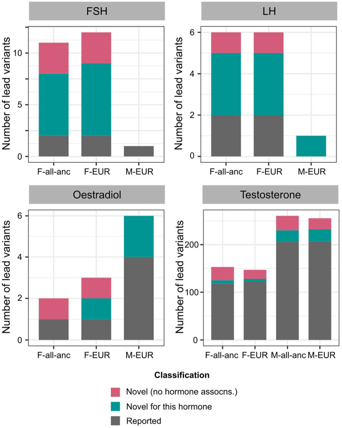
As hormone dysregulation is central to many infertility diagnoses18,19, we conducted sex-specific GWAS meta-analyses of five reproductive hormones - follicle-stimulating hormone (FSH) (Nfemale=57,890, Nmale=6,095), luteinising hormone (LH) (Nfemale=47,986, Nmale=6,769), oestradiol (Nfemale=97,887, Nmale=39,165), progesterone (Nfemale=18,368), and total testosterone (Nfemale=246,862, Nmale=243,951) - collected at assessment centre visits or identified through EHRs, in six cohorts and publicly available summary statistics (Supp. Table 9). We identified GWS loci associated with FSH (9 novel/2 previously known in females (F) and 0/1 in males (M)), LH (4/2 in F and 1/0 in M), oestradiol (1/1 in F and 2/4 in M), and testosterone (35/118 in F and 55/206 in M), but found no genetic variants associated with progesterone (Figure 5, Supp. Figure 3, and Supp. Figure 4). Several of the reported signals we replicated are near genes encoding the hormone-specific subunits themselves, such as FSHB for FSH and LHB for LH, or enzymes for steroid hormone metabolism, such as CYP3A7 for oestradiol and HSD17B13 for testosterone (Supp. Text).
Figure 5. Number of novel and reported reproductive hormone associations.
Each panel displays a different hormone (FSH=follicle-stimulating hormone, LH=luteinising hormone). Lead variants in each analysis stratum (F=female-specific, M=male-specific, all-anc=all ancestry meta-analysis, EUR=European-only meta-analysis) are classified as: (1) novel (no hormone associations) if they are not in LD (r2<0.1) with, and conditionally independent of (conditional P-value Pcond<0.05), any variants within a 1Mb window of the lead variant that are associated with 28 reproductive hormones in the GWAS Catalog62, plotted in pink, (2) novel for this hormone if they are not in LD (r2<0.1) with, and conditionally independent of (Pcond<0.05), the respective hormone-associated variants within a 1Mb window of the lead variant, plotted in green, and (3) reported otherwise, plotted in grey. Note the different Y-axis scales in each subplot. assocns.=associations.
We further classified lead variants as entirely novel hormone associations by defining a protocol based on linkage disequilibrium (LD) and conditional independence from published SNPs associated with any of 28 reproductive hormones in the GWAS Catalog62 (see Methods for detailed classification protocol).
We found 39 novel variants for testosterone in men, including those near SPOCK1 (rs1073917: β (SE)=−0.0160 (0.0029), P=4.69E-08), which is a target for the androgen receptor63, NR4A3 (rs10988865: β=−0.0161 (0.0029), P=4.33E-08), which coordinates the cellular response to corticotropin-hormone and thyrotropin-hormone releasing stimuli64,65 and regulates adipogenesis66, and obesity-associated genes ANKS1B (rs144998814: β=0.133 (0.0162), P=2.34E-16) and ANO10 (rs6809522: β=0.016 (0.0029), P=3.00E-08)67 (Supp. Table 10). The 28 novel reproductive hormone variants associated with testosterone in women include those near LAMTOR4 (rs17250196: β=−0.131 (0.0067), P=4.02E-86), associated with hyperthyroidism39 and age at menarche and menopause68, obesity-associated CCDC146 (rs138240474: β=−0.116 (0.0207), P=2.03E-08)67, which is also expressed in the fallopian tubes and endometrium69,70, and SLC8A1 (rs12611602: β=0.0163 (0.003), P=3.79E-08), which causes increased pancreatic beta cell proliferation and insulin secretion in knockout mouse models71. Finally, we report lead SNPs independent of previously published hormone variants in the HELQ locus for FSH-F (rs4235062: β=−0.046 (0.0065), P=1.50E-12), TMEM150B locus for FSH-F (rs28875253: β=−0.0599 (0.0061), P=9.90E-23) and LH-F (rs11668309: β=0.0519 (0.0071), P=3.91E-13), and in the SOX15-SAT2 locus for oestradiol-F (rs3933469: β=0.0363 (0.0051), P=1.02E-12) (Supp. Table 10).
Our results were robust to the inclusion of summary statistics from publicly available datasets, and there was no evidence for heterogeneity in variant effects across cohorts (Supp. Text).
Sex-specific genetic architecture of testosterone
Only 9.80% (of 153 total) lead variants for testosterone in females and 5.75% (of 261 total) lead variants for testosterone in males reach GWS in both sexes; and 45.9% of variants have opposing directions of effect in men and women (Supp. Figure 6). Indeed, we found no significant genetic correlation between testosterone in men and women (rg (SE)=0.0361 (0.0428), P=0.399). The heritability of testosterone in women is enriched in the adrenal gland (P=1.03E-03) and hepatocytes (P=9.36E-04); but only the latter is enriched for the heritability of testosterone in men (P=3.61E-04), as is the liver more broadly (P=1.16E-06) (Supp. Figure 10, stratified LD-score regression performed across 205 tissues and cell-types from the Genotype Tissue Expression (GTEx) Project database41 and the Franke lab single-cell database72). Finally, although testosterone regulates several traits hypothesised to be under sexual selection and may be under selection itself73, we do not find significant genome-wide directional selection for testosterone in men or women (mean genome-wide trait-SDS is not significantly different from 0, both P>0.05) (Supp. Text).
Genetic relationships between female infertility, reproductive hormones, and obesity
We observed no genome-wide genetic correlations between any category of female infertility and: (i) any reproductive hormone in this study, or (ii) thyroid stimulating hormone (TSH), or (iii) anti-Mullerian hormone (AMH), the latter two based on publicly available summary statistics74,75 (all P>0.05, Figure 3B). Mendelian randomisation (MR) analyses indicated a genetically causal protective effect of FSH on risk of F-ALL (OR (95% CI)=0.776 (0.678–0.888), P=2.15E-04) and F-EXCL (0.716 (0.604–0.850), P=1.26E-04) (Supp. Table 11).
Figure 3. Genetic correlations between female infertility and other phenotypes.
SNP-based genetic correlations (rg) between significantly heritable phenotypes (Z>4) were estimated using LD-score regression, performed using the LDSC software51 on a subset of 1 million HapMap3 SNPs52. Points are coloured by rg estimate, scaled by significance (−log10(P)), and labelled with the associated rg estimate if nominally significant without correction for multiple testing (P<0.05). (A) Genetic correlations among the three significantly heritable definitions of female infertility (all cause=F-ALL, anovulatory=F-ANOV, and idiopathic infertility defined by inclusion=F-INCL). (B) Genetic correlations between female infertility traits and reproductive hormones: testosterone, follicle stimulating hormone (FSH), and anti-Mullerian hormone (AMH, publicly available summary statistics) in female-specific analyses, and thyroid stimulating hormone (TSH, publicly available summary statistics) from sex-combined analysis. (C) Genetic correlations between female infertility traits and female reproductive conditions, with summary statistics generated from the largest available European-ancestry studies for each trait (see Methods). PCOS=polycystic ovary syndrome. (D) Genetic correlations between female infertility traits and selected heritable phenotypes (Z>4) in the UK Biobank, as generated by the Neale lab53. Correlations with all heritable phenotypes can be found in Supp. Table 12.
We found evidence for shared variants between hormones and infertility at the FSHB locus associated with FSH, LH, and testosterone (PP>84.8% for colocalisation with F-ANOV), and the ARL14EP locus associated with LH (PP=89.3% for colocalisation with F-ANOV) (Supp. Table 12). There was no evidence for colocalisation at any of the >300 other GWS loci associated with infertility or reproductive hormones in our study (Supp. Table 12). Our results suggest that while these traits are not significantly correlated at a genome-wide level, a small number of genes may drive infertility linked to hormone dysregulation.
Across 703 heritable phenotypes in the UK Biobank, we found 15 traits to be genetically correlated with female infertility, which we broadly group into: female reproductive conditions (such as having had a hysterectomy, rg (SE)=0.481 (0.0963)), general illness (such as number of operations, rg=0.266 (0.0588)), and cognitive test results (overall prospective memory test rg=0.345 (0.0736), overall fluid intelligence rg=−0.276 (0.0502)) (Figure 3D and Supp. Table 13). 24 obesity-related traits, including body mass index (BMI), waist-to-hip ratio (WHR), and body fat percentage, are correlated with testosterone and FSH, but are not genetically correlated with any category of female infertility (all P>0.05, Figure 3D, Supp. Figure 7, and Supp. Table 13). However, MR analyses using genetic instruments for BMI, WHR, and WHR adjusted for BMI (WHRadjBMI)67 indicated evidence for bi-directional causal relationships between infertility and abdominal obesity independent of overall obesity. While genetically predicted WHRadjBMI is a risk factor for F-ALL (OR (95% CI)=1.10 (1.05–1.16), P=1.71E-04) and F-ANOV (1.29 (1.16–1.45), P=4.66E-06), the latter is itself causal for increased WHRadjBMI (β (SE)=0.0547 (0.0133), P=3.74E-05) (Supp. Table 11).
Variants associated with all-cause female infertility are in genes enriched for expression in ovarian stromal cells (partitioned heritability P=2.52E-03). We did not find significant enrichment of infertility heritability in any of the 205 tissues and cell-types from the GTEx project database41 and the Franke lab single-cell database72.
Rare variant contribution to reproductive-hormone and infertility genetics
We analysed the 450k UK Biobank exome sequencing dataset to characterise the association between rare coding variation (MAF<1%) and binary traits with >100 cases (F-ALL (3,746 cases, 260,413 controls), F-EXCL (3,012 cases, 261,147 controls), and M-ALL (650 cases, 222,393 controls)), and quantitative traits with >10,000 participants (FSH-F (N=20,800), LH-F (N=16,391), oestradiol-F (N=54,609), and testosterone (Nfemale=197,038, Nmale=197,340) (Figure 1)). Gene-burden analyses implicate the PLEKHG4 gene, which is highly expressed in the testis and ovary, for F-EXCL (burden test OR (95% CI)=1.04 (1.02–1.06) when aggregated across all variant annotations with MAF<1%, Cauchy P=1.37E-08) (Supp. Table 14). Rare variants in PLEKHG4 cause cerebellar ataxia76, which is a feature of some syndromes that also cause steroid hormone deficiency and hypogonadism77,78.
Novel genes for testosterone implicated by gene burden analyses
Gene-based analyses identify 27 genes associated with testosterone-F and 24 genes for testosterone-M (P<5E-06), of which eleven have not previously been implicated in GWASs (Supp. Text). We report the first known association of HSD11B1 with testosterone-F (burden test P=1.93E-06 when aggregated across missense variants with MAF<0.01%); pathogenic variants in this gene are reported to cause hyperandrogenism due to cortisone reductase deficiency79,80 (Supp. Figure 11 and Supp. Table 14). We also report the association of testosterone-M with HSD17B2 (burden test P=1.33E-11 when aggregated across pLoF variants with MAF<0.1%), which encodes the enzyme hydroxysteroid 17β-dehydrogenase 2 that catalyses the oxidation of oestradiol, testosterone, and dihydrotestosterone to less active forms and thus regulates the biological potency of steroid hormones81,82 (Supp. Figure 11 and Supp. Table 14).
Increased risk of infertility in individuals carrying rare testosterone-associated variants
Two genes associated with testosterone in female UK Biobank participants are also associated with infertility risk (P<1.00E-03, Bonferroni adjustment for 50 unique genes): TRIM4 (F-ALL, burden test OR=1.03 (1.01–1.05), P=4.05E-04 across all variants with MAF<0.1%) and CYP3A43 (F-EXCL, burden test OR=1.02 (1.01–1.03), P=4.84E-04 across all variants with MAF<1%). The latter encodes the steroid hormone metabolic enzyme testosterone 6-beta-hydroxylase; but neither gene has previously been implicated in infertility.
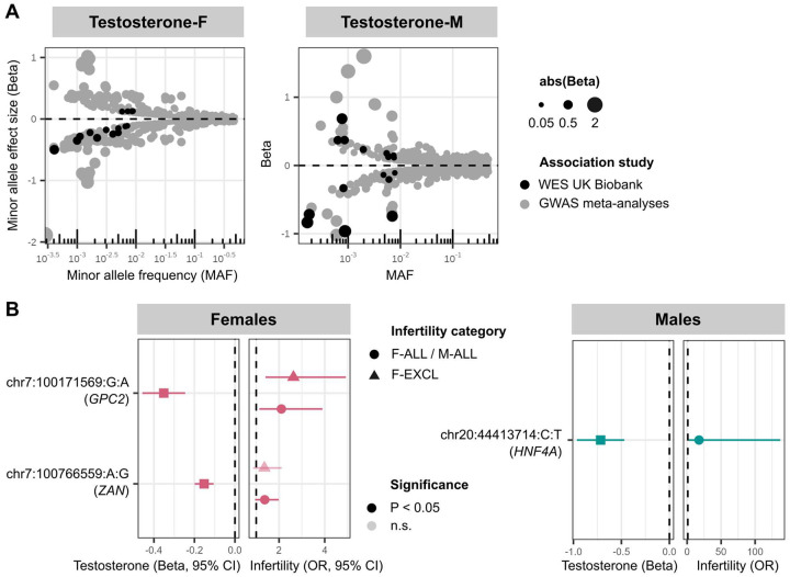
Finally, we identified 29 unique genes carrying rare variants (MAF<1%) associated with testosterone in male or female participants in the UK Biobank. Eighteen of the 29 genes also contain common testosterone-associated variants from GWASs (MAF>1%), but the rare variant has a larger absolute effect size in the majority (83%) of these (Figure 6A, Supp. Table 15, and Supp. Text).
Figure 6. Rare variants associated with testosterone and infertility in UK Biobank whole exome sequencing (WES) analyses.
(A) Effect size versus allele frequency of genetic variants associated with total testosterone. Variants discovered at genome-wide significance (P<5E-08) in GWAS meta-analyses (coloured in grey) and exome-wide significance in the UK Biobank WES analyses (coloured in black) are plotted, sized by the absolute value of their effect size. Effect sizes are aligned to the minor allele, plotted against MAF on the log x-axis. (B) Effects of testosterone-associated rare variants on infertility in females (left) and males (right). Per gene, the variant with lowest P-value of all variants that reach exome-wide significance (P<1E-07) in UK Biobank WES analyses for testosterone is displayed, for all variants with nominally significant effects on infertility. Effect sizes (β and 95% confidence intervals (CIs) for the variant effect on testosterone are to the left of each plot, and effect sizes (odds ratios (ORs) and 95% CIs) for the variant effect on infertility are to the right of each plot. Variants that reach nominal significance (P<0.05) are coloured in solid shapes.
The eleven novel testosterone associations include a female testosterone-lowering missense variant in STAG3 (chr7:100204708:C:T, β=−0.284, P=2.31E-08); STAG3 is also associated with primary ovarian insufficiency in women83,84, and induces female infertility through the absence of oocytes in knockout mouse models39. While we did not find significant association between the STAG3 variant and female infertility in the UK Biobank (P>0.05), we observed increased risk of idiopathic infertility in women carrying a novel testosterone-lowering variant in GPC2 (chr7:100171569:G:A, F-EXCL OR=2.63 (1.40–4.92), P=1.25E-03) (Figure 6B). GPC2 is highly expressed in the testis, and GPC2-knockout mouse models display reduced adrenal gland size39. The gene has not previously been reported to be associated with testosterone or infertility. Taken together, our results indicate a potential role for infertility driven by rare hormone-disrupting variants.
Discussion
Our large-scale genetic investigation of infertility and related reproductive phenotypes in over 1 million individuals identified 19 genetic loci associated with female infertility, two with male infertility, and novel variants for the reproductive hormones FSH (3 novel variants), LH (1), oestradiol (1), and total testosterone (28) in women and for total testosterone in men (39). Through rare-variant and gene-based analyses in the UK Biobank, we additionally identified PLEKHG4 associated with female infertility and 50 genes for testosterone, including the first reported hormone-associated variants in some members of the hydroxysteroid dehydrogenase enzyme family. We found evidence at non-hormonal, pleiotropic, infertility loci for recent directional selection (EBAG9) and balancing selection (GREB1, INHBB, PCDH15). Although there was evidence for distinct genetic architectures of infertility and reproductive hormones, we showed that individual genes containing rare protein-coding variants associated with testosterone (GPC2, CYP3A43, TRIM4) were also associated with higher risk of infertility in the UK Biobank.
Previous efforts to catalogue the genome-wide architecture of infertility have relied on proxy measures such as childlessness and number of children ever born24,25, which may be confounded by behavioural, socio-economic, and lifestyle factors. While we did find modest genetic correlation between female infertility and age at first sexual intercourse (−18.8%), indicating that the latter captures some shared biology with fertility, our meta-analyses did not replicate the associations of infertility proxy variables with putative behavioural loci for risk-taking85,86 or educational attainment85,87–89. Instead, we nominate genes with putative roles in both male and female gonads, such as TRHR for ovarian insufficiency39,40 and ENO4 for sperm motility44.
The strong genetic correlation of 71% between idiopathic infertility and endometriosis may indicate that some proportion of idiopathic cases are due to under-diagnosis of endometriosis, whose early treatment may prevent future infertility15,90. Our subtype-specific analyses highlight the value in dissecting heterogeneous causes of infertility. For example, PCOS is a heritable cause of up to 80% of anovulatory infertility cases that may be treated through induced ovulation17,91,92. However, as only three of eight loci for anovulatory infertility colocalise with known PCOS signals and the genetic correlation between these traits is only 40%, other hypothalamic-pituitary-ovarian disorders, endocrinopathies (hypothyroidism, hyperprolactinaemia, etc.) and ovarian insufficiency may also contribute significantly to the genetic aetiology of anovulatory infertility and require treatments different from those for PCOS-associated infertility93. Weight loss for overweight patients is often recommended as beneficial for fertility94,95, but we did not find substantial genetic correlation between obesity and infertility. Our findings add genetic support to evidence from randomised controlled trials demonstrating no fertility benefits from short-term weight loss in overweight and obese women96. Instead, we observed bi-directional causal relationships between abdominal obesity and anovulatory infertility, suggesting physiological feedback mechanisms whose complex interplay requires deeper study. Taken together, these results suggest a critical need for a richer understanding of the genetic and non-genetic contributions to infertility.
The testes and ovaries were not significantly enriched for the heritability of infertility or testosterone, despite being reproductive organs that are major sites for testosterone production97,98. However, neither organ is disaggregated into tissues or cell types in the GTEx database, so gene expression profiles may not capture cell-type specific effects. Indeed, we found enrichment of testosterone heritability in the androgen-secreting thecal cells and androgen-responsive granulosa cells of the ovary99–101, and female infertility in ovarian stromal cells. Although there are several causal roles hypothesised for stromal dysfunction in infertility, such as impaired folliculogenesis102, restricted blood flow103, and ovarian scarring104, more work is needed to robustly replicate these findings. In general, more functional studies of gonadal cell types, in both men and women, are needed to enable a mechanistic understanding of the genetic variation associated with reproductive hormones and infertility.
We employed a broad search strategy to maximise sample sizes for cases of infertility and reproductive hormone levels in our meta-analyses. Diagnostic criteria for infertility vary by country and have changed over time1, which may explain the wide spread in the prevalence of infertility across cohorts. Reproductive hormone values in this study were assayed using different methodologies, in primary care or hospital EHRs, and at different ages and stages of the menstrual cycle in women. A majority of samples in our study were derived from the UK Biobank and measured during and post-menopause (ages 40–69), whereas infertility occurs pre-menopause, so we urge caution in interpreting the lack of correlation between these traits. Although we were able to adjust for covariates such as age, which can account for some of the effect of menopause on hormone levels, we did not have the data granularity to account for hormonal fluctuations during the menstrual cycle and pregnancy. In the future, longitudinal GWASs that can incorporate mean and variance of hormone levels over the menstrual cycle, or phenotypes that calculate ratios between various hormones over time, will likely reveal fundamental biology that is missed by the broad-stroke assessments in this study.
Our results indicate that balancing selection and recent positive selection at pleiotropic loci may explain the persistence of genetic factors for infertility. For example, the EBAG9 locus associated with female infertility is under directional selection, perhaps because EBAG9, which is highly expressed in CD34-/CD41+/CD42+ megakaryocytes69,70, plays a role in T-cell mediated cytotoxicity as part of the adaptive immune memory response to infection105. However, a complementary role for EBAG9 may be in the placenta during early pregnancy, where reduction of EBAG9 levels is associated with inappropriate activation of the maternal immune system and results in foetal rejection106.
In conclusion, in this comprehensive large-scale investigation of the genetic determinants of infertility and reproductive hormones across men and women, we identified several genes associated with infertility and analysed their effects on reproductive disease and selection pressures. We did not find evidence that reproductive hormone dysregulation and obesity are strongly correlated with infertility at the population level, but instead nominate individual hormone-associated genes with effects on fertility. Other genetic and non-genetic avenues must be explored to treat complex and heterogeneous fertility disorders that impact the physical, emotional, and financial well-being of millions of individuals across the globe.
Methods
Study populations and phenotype identification
Binary traits (infertility)
Cases were identified in UK Biobank, Copenhagen Hospital Biobank and Danish Blood Donor Study, deCode, Estonian Biobank, FinnGen, and Genes and Health (Supp. Text). We defined five categories of female infertility: all causes (F-ALL), anovulatory (F-ANOV), anatomical (F-ANAT, including tubal, uterine, and cervical origins), idiopathic infertility by exclusion of known causes (anatomical and anovulatory infertility, PCOS, endometriosis, and uterine leiomyoma) (F-EXCL), and idiopathic infertility by inclusion of a diagnosis code for idiopathic infertility (F-INCL), and male infertility of all causes (M-ALL). Cases were identified through self-report (F-ALL, F-EXCL, M-ALL) and through primary-and secondary-care codes (Supp. Table 1). Within each subtype, sex-matched controls were defined as individuals not identified as cases for that subtype.
Quantitative traits (reproductive hormones)
Hormones were included from UK Biobank, Avon Longitudinal Study of Parents and Children (ALSPAC), deCode, Estonian Biobank, and Genes and Health (Supp. Text). We extracted measurements of FSH, LH, oestradiol, progesterone, and testosterone from biobank assessment centres or primary- and secondary-care records (Supp. Table 16). If repeated measurements were available for an individual, we retained the recorded hormone value closest to the individual’s median hormone value over time. Each hormone was regressed on age, age2, and cohort-specific covariates specified below; the residuals from this regression were rank-based inverse normally transformed (RINTed) prior to GWAS.
Meta-analysis of GWAS summary statistics
Genome-wide association testing
Association analyses were performed separately within each ancestry and sex stratum for all strata with at least 100 cases (binary traits) or 1,000 individuals (quantitative traits). For binary traits, each variant passing QC was tested for association under an additive model using REGENIE107 or SAIGE108, with adjustments for age, age2, and cohort-specific covariates, with the Firth correction applied to control for inflation at rare variants and traits with low case-control ratios107,108. For quantitative traits, the RINTed hormone value was tested for association under an additive model using REGENIE107 or SAIGE108, with adjustments for cohort-specific genetic covariates. Any deviations from this GWAS protocol are noted in the Supplementary Text.
Meta-analysis
Prior to meta-analysis, summary statistics from all studies underwent thorough quality control to retain variants that met the following criteria: (1) on the autosomes or X chromosome, (2) with imputation information score >0.8 (where available), (3) bi-allelic variants with A, C, G, T alleles, (4) with standard errors <10 and P-values in [0,1], and (5) without duplicate entries. Fixed-effects inverse-variance weighted meta-analysis was performed using METAL109. We report results from European-ancestry and all-ancestry meta-analyses for each trait. Genome-wide significance was established at P<5E-08.
Identification and classification of lead variants
Distance-based pruning was used to identify lead variants as the SNP with the lowest P-value within each 1Mb window at all loci with at least one GWS variant with P<5E-08.
Hormone-associated variants were classified based on conditional analysis as (1) previously reported for the hormone of interest, (2) previously reported for any of 28 reproductive hormones, or (3) novel, based on SNP associations published in the GWAS Catalog as of 27 March 202362 (Supp. Table 17). We adapted criteria developed by Benonisdottir et al. (2016)110 to classify novel variants as those that are not in LD with (r2<0.1), and conditionally independent of (Pconditional<0.05), all published hormone-associated variants within 1 Mb; all other variants are considered to be previously reported. Conditional analysis was performed in GCTA-COJO111, with LD information for European-ancestry individuals derived from the 1000 Genomes dataset112.
For lead variants on the X chromosome and those from multi-ancestry analyses, for which estimating LD is more difficult due to differences in recombination rates and selection pressures between sexes and populations113–115, we did not use the above LD-based classification system. Instead, a lead SNP was considered novel if it was not within 1 Mb of a published hormone-associated variant or if its effect was independent of published variants within a 1 Mb window (Pconditional<0.05), and reported if not.
SNP-based heritability
The following analyses, which rely on population-specific LD patterns, were restricted to European-ancestry summary statistics with pre-computed LD-scores based on European-ancestry individuals in the 1000 Genomes dataset112, restricted to HapMap3 SNPs52. We estimated the SNP-based heritability (hG2) of a trait from GWAS summary statistics using LD-score regression as implemented in the LDSC software51. For infertility traits, the observed-scale heritability (hobs2) was converted to liability-scale heritability (hliab2), which accounts for the disease prevalence in the sample (k) and population (K), under the assumption that sample prevalence equals the population prevalence54.
Genetic correlations
LDSC was used to estimate genetic correlations between infertility traits, hormone levels, and a collection of other phenotypes in the UK Biobank in European-ancestry individuals. To simplify computation of rg across a large number of traits, we used an extension of the LDSC software which allows for simultaneous estimation of multiple genetic correlations116.
We estimated genetic correlations among the three categories of female infertility with significant heritability (Z>4)51: F-ALL, F-ANOV, and F-INCL, as well as among heritable female reproductive hormones (FSH and testosterone in females). We additionally obtained summary statistics from GWASs of thyroid stimulating hormone (TSH)75 (sex-combined analysis, N=247,107 participants) and anti-Mullerian hormone (N=7,049 pre-menopausal participants)74 from the largest publicly available European-ancestry studies to date. We also tested for genetic correlations between infertility and reproductive hormones. Significant rg after multiple testing was established at 2.38E-03 (FWER controlled at 5% across 21 tests using the Bonferroni method).
We collated European-ancestry GWAS summary statistics for four female reproductive disorders: (1) endometriosis from Rahmioglu et al. (2023)35, (57,248 cases and 698,764 controls), (2) heavy menstrual bleeding by meta-analysing GWAS data from Gallagher et al. (2019)117 and FinnGen data freeze 927 (31,309 cases and 318,510 controls), (3) PCOS by meta-analysing GWAS data from Tyrmi et al. (2022)92 and a UKBB-based GWAS (14,467 cases and 430,267 controls), and (4) uterine fibroids by meta-analysing GWAS data generated by the Neale lab53 and FinnGen data freeze 927, (42,446 cases and 588,955 controls). We additionally obtained summary statistics from a GWAS of spontaneous dizygotic (DZ) twinning (8,265 cases (mothers of DZ twins) and 264,567 controls; plus 26252 DZ twins and 417,433 additional controls) from Mbarek et al. (2024), the largest European-ancestry study of female fecundity to date46. Significant rg after multiple testing was established at 2.00E-03 (FWER controlled at 5% across 25 tests using the Bonferroni method).
We downloaded LD-score formatted summary statistics for European-ancestry individuals across 703 heritable phenotypes (Z>4) from the Neale lab round 2 collection53. The number of effectively independent phenotypes estimated by the Neale lab (Meff=340) was used to establish significant rg after multiple testing at 2.45E-05 (FWER controlled at 5% across 2,040 tests using the Bonferroni method).
Mendelian randomisation
The following analyses were all performed with summary statistics from European-ancestry GWASs, using the TwoSampleMR v0.5.7 package118.
We constructed genetic instruments for BMI, WHR, and WHRadjBMI with female-specific lead variants from a recent European-ancestry GWAS meta-analysis with a maximum sample size of 434,785 female participants67. SNPs were weighted by their female-specific effect sizes. The mean F-statistic across all SNPs in each instrument indicated sufficient strength for MR (BMI=61.3, WHR=74.8, WHRadjBMI=84.7, recommended>10119). As the instrument GWASs included participants from UK Biobank, we conducted a sensitivity analysis to avoid bias from sample overlap between instrument and outcome GWASs by constructing obesity-trait instruments from an earlier release of summary statistics from the GIANT Consortium without UKBB participants120 (Supp. Table 11). As the WHRadjBMI instrument may be confounded due to adjustment for a correlated variable121, i.e. adjustment for BMI in the WHR GWAS, we performed multivariable MR with a joint instrument for BMI and WHR to estimate the BMI-adjusted causal effect of WHR on reproductive outcomes. We found no difference in effect estimates from MR conducted using an instrument for WHRadjBMI and multivariable MR (Supp. Table 19).
Hormone instruments were constructed for reproductive hormones in this study with F-statistic>10 (FSH-F=38.7, testosterone-F=66.1), using GWAS summary statistics from European-ancestry GWASs excluding UK Biobank participants to avoid sample overlap with outcome GWASs.
We also performed reciprocal MR to test the genetically predicted causal effects of infertility on obesity and reproductive hormone levels. Genetic instruments were constructed for subtypes of infertility with F-statistic>10 (F-ALL=51.0, F-ANOV=36.2), using GWAS summary statistics from European-ancestry GWASs excluding UK Biobank participants to avoid sample overlap with outcome GWASs. We assessed the causal direction between each pair of traits tested with Steiger filtering of instruments and the Steiger directionality test.
We report results from the inverse-variance weighted (IVW) method, the MR-Egger method which is robust to horizontal pleiotropy122, and the weighted median method which protects against outlier variants123 (Supp Table 11).
Colocalisation
The following analyses were all performed with summary statistics from European-ancestry GWASs, using the Bayesian framework implemented in the coloc v5.1.0 package124 under a single causal variant assumption125. Only common variants (MAF>1%) within windows of +/− 50 kb around each lead variant for an infertility or reproductive hormone trait were retained. For each pair of traits tested for colocalisation, we set the prior probabilities of variants in a locus being causally associated with trait 1 (p1) and trait 2 (p2) to 1E-04 (99% confidence in a true association), and the prior for joint association p12 to 1E-06 (assuming equal likelihood of shared and non-shared causal variants for each trait in a locus) as recommended by the developers of coloc125. We tested five hypotheses: H0=no association with either trait in region, H1=association with trait 1 in region, but not trait 2, H2=association with trait 2 in region, but not trait 1, H3=association with both traits in region, but different causal variants, and H4=association with both traits in region, and a shared causal variant. A pair of traits were considered to colocalise if posterior probability of H4>50% and the ratio of posterior probabilities of H4/H3>5124,126.
We tested for colocalisation between each female infertility category and each female-specific hormone (FSH, LH, oestradiol, and testosterone) at all genetic loci associated with at least one of the pair of traits tested. The single male infertility locus with common variants (MAF>1%) in the European-ancestry analysis did not contain enough significant associations (only 12 common variants with P<1E-06) for colocalisation analyses.
Because we noticed that some lead variants for female infertility had previously been reported as associated with endometriosis and PCOS, we estimated the posterior probability (PP) of colocalisation of genetic signals between each category of female infertility and each of these two reproductive disorders. European-ancestry summary statistics for endometriosis and PCOS were obtained as described in the genetic correlations section above.
We assessed colocalisation of genetic signals for female infertility with eQTLs for all proximal genes with transcription start sites (TSSs) within 1 Mb of an infertility lead variant. Publicly available eQTL data was downloaded from the GTEx project41.
Tissue and cell-type prioritisation
We estimated the polygenic contributions of genes with tissue-specific expression profiles to the heritability of infertility and hormones using stratified LD-score regression (partitioned heritability analyses)51. We restricted these analyses to traits with highly significant heritability in European-ancestry analyses (Z>7) (F-ALL, testosterone-F, and testosterone-M), as recommended by the developers, Finucane et al. (2015)127.
Gene sets and LD scores for 205 tissues and cell-types from the GTEx Project database41 and the Franke lab single-cell database72 were downloaded from Finucane et al. (2018)128. We established tissue-wide significance at −log10(P)>2.75, which corresponds to FDR<5%.
Ovarian cell types
As the ovary, a reproductive tissue of interest, is not well characterised in the GTEx project, we identified two publicly available single-cell gene expression datasets for ovarian cell types: (1) from Fan et al. (2019), who performed single-cell RNA sequencing on ovarian tissue from five adult women undergoing fertility preservation procedures with 20,676 cells across 19 identified cell types129, and (2) from Jin et al. (2022), who performed single-nucleus RNA sequencing on autopsy samples from four women (aged 49–54 years, with normal ovarian histology) with 42,568 cells across 8 identified cell types130. The datasets were aligned and filtered using the QC pipelines provided by the authors of each study, and clustered with identical parameters to replicate the results of each individual study. Gene sets for each cluster were identified as recommended by Finucane et al. (2018)128 - briefly, we identified differential expression between the cells in each cluster and all other clusters by using the Wilcoxon rank sum test implemented in Seurat v3.0131–133, and returned the top 10% of genes that are specifically expressed in each cluster (positive average log-fold-change values), ranked by differential expression P-value. We computed annotation-specific LD scores for these gene sets using hg38 coordinates for gene TSSs and TESs obtained from Ensembl134, across 1 million HapMap3 variants52 with LD information from European-ancestry individuals in the 1000 Genomes phase 3 dataset112.
Overlaps with genetic regions under selection
To avoid confounding by population stratification, selection look-ups were restricted to GWAS summary statistics from European-ancestry individuals.
Directional selection
Following guidelines described by Mathieson et al. (2023)25, we identified 54 genomic regions under directional selection from three previously reported genome-wide scans: (1) 39 regions from the Composite of Multiple Signals (CMS) test, which infers historical selection on the order of the past 50,000 years58, (2) 12 regions from an ancient DNA scan that uses inferences of allele frequency from ancient genomes to determine selection over the past 10,000 years57, and (3) three regions from Singleton Density Scores (SDSs), which use the pattern of singleton variants to identify recent selection in the past 2,000 to 3,000 years56. For each genomic window under directional selection, we report the infertility-associated variants with the lowest P-value.
Singleton density scores
We downloaded publicly available SDSs for SNPs in the UK10K dataset56 to report the highest SDS (positive selection of derived allele over ancestral allele in the past 2,000 to 3,000 years) and lowest SDS (negative selection) within the +/−10kb window around each infertility or hormone lead SNP. To calculate trait-SDS for each phenotype, we aligned each SDS to the trait-increasing allele rather than the derived allele56. For each lead variant window containing variants with extreme SDSs (top 97.5th %ile or bottom 2.5th %ile), we report the direction of selection with respect to the trait-increasing allele. Percentiles of SDSs were evaluated only on a subset of variants within 10kb of any variant reported in the GWAS Catalog to account for genomic context. Further, as variants that are sub-GWS for a trait may nonetheless be under selection, we calculated the genome-wide mean trait-SDS in each bin of 1000 variants, ranked by P-value for the trait association, following the protocol outlined by Field et al. (2016)56.
Balancing selection
We accessed publicly available standardised BetaScan2 scores, which detect balancing selection using polymorphism and substitution data, for all SNPs in the 1000 Genomes dataset59. We tested whether the +/−10kb window around each infertility or hormone lead variant contained SNPs with scores in the 99th %ile of standardised BetaScan2 scores. Percentiles of SDSs were evaluated only on a subset of variants within 10kb of any variant reported in the GWAS Catalog to account for genomic context. For each lead variant window, we report the highest standardised BetaScan2 score and its percentile.
Whole exome sequencing analyses in the UK Biobank
Exome sequencing quality control
Quality control outline
We first considered an initial set of “high quality” variants to evaluate the mean call rate and depth of coverage for each sample. We then ran a sample and variant level pre-filtering step and calculated sample-level QC metrics. Using these metrics, we removed sample outliers based on median absolute deviation (MAD) thresholds, and excluded sites which did not pass variant QC according to Karzcewski et al. (2022)135. We then applied a genotype-level filter using genotype quality (GQ), depth (DP), and heterozygote allele balance (AB). The resultant high-quality European call set consisted of 402,375 samples and 25,229,669 variants. For details see Supplementary Text.
Variant annotation
We annotated variants using Variant Effect Predictor (VEP) v105 (corresponding to gencode v39)136 with the LOFTEE v1.04_GRCh38137 and dbNSFP138 plugins, annotating variants with CADD v1.6139, and REVEL using dbNSFP4.3140 and loss of function confidence using LOFTEE. Complete instructions and code for this step are provided in our VEP_105_LOFTEE repository141, which contains a docker/singularity container to ensure reproducibility of annotations. We then ran SpliceAI v1.3142 using the gencode v39 gene annotation file to ensure alignment between VEP and SpliceAI transcript annotations. We defined ‘canonical’ transcripts to be used for variant-specific annotations as follows: set MANE Select143 as the canonical, where available, and if a MANE Select transcript is not present, set canonical and restrict to protein coding genes. Note that for VEP 105, this is equivalent to selecting the ‘canonical’ transcript in protein coding genes. Then, using the collection of missense, pLoF, splice metrics, and annotations of variant consequence on the ‘canonical’ transcript, we determine a set of variant categories for gene-based testing.
Variant categories for gene-based tests
High confidence pLoF: high-confidence LoF variants, as defined by LOFTEE137 (LOFTEE HC).
- Damaging missense/protein-altering: at least one of:
- Variant annotated as missense/start-loss/stop-loss/in-frame indel and (REVEL≥0.773 or CADD≥28.1 (or both)).
- Any variant with SpliceAI delta score (DS)≥0.2 where SpliceAI DS the maximum of the set {DS_AG, DS_AL, DS_DG, DS_DL} for each annotated variant (where DS_AG, DS_AL, DS_DG and DS_DL are delta score (acceptor gain), delta score (acceptor loss), delta score (donor gain), and delta score (donor loss), respectively).
- Low-confidence LoF variants, as defined by LOFTEE (LOFTEE LC)
Other missense/protein-altering: Missense/start-loss/stop-loss/in-frame indel not categorised in (2) (Damaging missense/protein-altering).
Synonymous: synonymous variants with SpliceAI DS<0.2 in the gene (our ‘control’ set).
REVEL and CADD score cut-offs are chosen to reflect the supporting level for pathogenicity (PP3) from the American College of Medical Genetics and Genomics and the Association for Molecular Pathology (ACMG/AMP) criteria144.
Variant counts and average allele counts for each annotation, split by population label and binned by MAF are displayed in Supp. Figure 13 and Supp. Figure 14, respectively.
Genetic association testing
We carried out rare variant genetic association testing in the European-ancestry subset of the UK Biobank using Scalable and Accurate Implementation of GEneralized mixed model (SAIGE)108, a mixed model framework that accounts for sample relatedness and case-control imbalance through a saddle-point approximation in binary traits. All rare-variant analysis was carried out on the UK Biobank Research Analysis Platform (RAP) using SAIGE version wzhou88/saige:1.1.9108. In the sex-combined analyses, we account for age, sex, age2, age × sex, age2 × sex, and the first 10 genetic principal components as fixed effects; and age, age2, and the first 10 principal components in sex-specific analyses. All continuous traits were inverse rank normalised prior to association testing.
For SAIGE step 0, we constructed a genetic relatedness matrix (GRM) using the UK Biobank genotyping array data. We LD pruned the genotyped data using PLINK (--indep-pairwise 50 5 0.05)145, and created a sparse GRM using 5000 randomly selected markers, with relatedness cutoff of 0.05, using the createSparseGRM.R function within SAIGE. To generate a variance ratio file for subsequent steps in SAIGE, we extracted 1000 variants each with MAC<20 and MAC>20, and combined these markers to define a PLINK file for the variance ratio determination.
In SAIGE step 1 for each trait, the curated phenotype data and sparse GRM were used to fit a null model with no genetic contribution. All parameters were set at the defaults in SAIGE, except --relatednessCutoff 0.05, --useSparseGRMtoFitNULL TRUE and --isCateVarianceRatio TRUE. Tolerance for fitting the null generalised linear mixed model was set to 0.00001.
Rare variant and gene based testing
Following null model fitting, we carried out variant and gene-based testing in SAIGE step 2 using the variant categories described above, with the --is_single_in_groupTest TRUE flag. All other parameters were set to default, except --maxMAF_in_groupTest=0.0001,0.001,0.01, --is_Firth_beta TRUE, --pCutoffforFirth=0.1, and --is_fastTest TRUE. We included the following collection of group tests, using the annotations defined in methods: variant annotation.
High confidence pLoF
Damaging missense/protein-altering
Other missense/protein-altering
Synonymous
High confidence pLoF or Damaging missense/protein-altering
High confidence pLoF or Damaging missense/protein-altering or Other missense/protein-altering or Synonymous
We then carried out Cauchy combination tests146 across these annotations for each gene.
Supplementary Material
Acknowledgements
Thanks to Prof Pier Palamara and Prof Zoltan Kutalik for helpful discussions. We are grateful to the participants of all cohorts. This research has partly been conducted using the UK Biobank Resource under Application Number 11867. Genes & Health is/has recently been core-funded by Wellcome (WT102627, WT210561), the Medical Research Council (UK) (M009017, MR/X009777/1, MR/X009920/1), Higher Education Funding Council for England Catalyst, Barts Charity (845/1796), Health Data Research UK (for London substantive site), and research delivery support from the NHS National Institute for Health Research Clinical Research Network (North Thames). Genes & Health is/has recently been funded by Alnylam Pharmaceuticals, Genomics PLC; and a Life Sciences Industry Consortium of Astra Zeneca PLC, Bristol-Myers Squibb Company, GlaxoSmithKline Research and Development Limited, Maze Therapeutics Inc, Merck Sharp & Dohme LLC, Novo Nordisk A/S, Pfizer Inc, Takeda Development Centre Americas Inc. We thank Social Action for Health, Centre of The Cell, members of our Community Advisory Group, and staff who have recruited and collected data from volunteers. We thank the NIHR National Biosample Centre (UK Biocentre), the Social Genetic & Developmental Psychiatry Centre (King’s College London), Wellcome Sanger Institute, and Broad Institute for sample processing, genotyping, sequencing and variant annotation. We thank: Barts Health NHS Trust, NHS Clinical Commissioning Groups (City and Hackney, Waltham Forest, Tower Hamlets, Newham, Redbridge, Havering, Barking and Dagenham), East London NHS Foundation Trust, Bradford Teaching Hospitals NHS Foundation Trust, Public Health England (especially David Wyllie), Discovery Data Service/Endeavour Health Charitable Trust (especially David Stables), Voror Health Technologies Ltd (especially Sophie Don), NHS England (for what was NHS Digital) - for GDPR-compliant data sharing backed by individual written informed consent. Most of all we thank all of the volunteers participating in Genes & Health. This study was funded by the European Union through the European Regional Development Fund Project No. 2014-2020.4.01.15-0012 GENTRANSMED. Data analysis was carried out in part in the High-Performance Computing Center of University of Tartu. This research has partly been conducted using the ALSPAC resource under Project Number B4568. The activities of the EstBB are regulated by the Human Genes Research Act, which was adopted in 2000 specifically for the operations of the EstBB. Individual level data analysis in the EstBB was carried out under ethical approval 1.1-12/624 from the Estonian Committee on Bioethics and Human Research (Estonian Ministry of Social Affairs), using data according to release application 3-10/GI/10790 from the Estonian Biobank. This study was funded by the Estonian Research Council grants PRG1911. We want to acknowledge the participants and investigators of the FinnGen study. The FinnGen project is funded by two grants from Business Finland (HUS 4685/31/2016 and UH 4386/31/2016) and the following industry partners: AbbVie Inc., AstraZeneca UK Ltd, Biogen MA Inc., Bristol Myers Squibb (and Celgene Corporation & Celgene International II Sàrl), Genentech Inc., Merck Sharp & Dohme LCC, Pfizer Inc., GlaxoSmithKline Intellectual Property Development Ltd., Sanofi US Services Inc., Maze Therapeutics Inc., Janssen Biotech Inc, Novartis Pharma AG, and Boehringer Ingelheim International GmbH. Following biobanks are acknowledged for delivering biobank samples to FinnGen: Auria Biobank (www.auria.fi/biopankki), THL Biobank (www.thl.fi/biobank), Helsinki Biobank (www.helsinginbiopankki.fi), Biobank Borealis of Northern Finland (https://www.ppshp.fi/Tutkimus-ja-opetus/Biopankki/Pages/Biobank-Borealis-briefly-in-English.aspx), Finnish Clinical Biobank Tampere (www.tays.fi/en-US/Research_and_development/Finnish_Clinical_Biobank_Tampere), Biobank of Eastern Finland (www.ita-suomenbiopankki.fi/en), Central Finland Biobank (www.ksshp.fi/fi-FI/Potilaalle/Biopankki), Finnish Red Cross Blood Service Biobank (www.veripalvelu.fi/verenluovutus/biopankkitoiminta), Terveystalo Biobank (www.terveystalo.com/fi/Yritystietoa/Terveystalo-Biopankki/Biopankki/) and Arctic Biobank (https://www.oulu.fi/en/university/faculties-and-units/faculty-medicine/northern-finland-birth-cohorts-and-arctic-biobank). All Finnish Biobanks are members of BBMRI.fi infrastructure (www.bbmri.fi). Finnish Biobank Cooperative -FINBB (https://finbb.fi/) is the coordinator of BBMRI-ERIC operations in Finland. The Finnish biobank data can be accessed through the Fingenious® services (https://site.fingenious.fi/en/) managed by FINBB. We are extremely grateful to all the families who took part in this study, the midwives for their help in recruiting them, and the whole ALSPAC team, which includes interviewers, computer and laboratory technicians, clerical workers, research scientists, volunteers, managers, receptionists and nurses. We acknowledge the contribution of the participants in the deCODE study. Patients and control subjects in FinnGen provided informed consent for biobank research, based on the Finnish Biobank Act. Alternatively, separate research cohorts, collected prior the Finnish Biobank Act came into effect (in September 2013) and start of FinnGen (August 2017), were collected based on study-specific consents and later transferred to the Finnish biobanks after approval by Fimea (Finnish Medicines Agency), the National Supervisory Authority for Welfare and Health. Recruitment protocols followed the biobank protocols approved by Fimea. The Coordinating Ethics Committee of the Hospital District of Helsinki and Uusimaa (HUS) statement number for the FinnGen study is Nr HUS/990/2017. The FinnGen study is approved by Finnish Institute for Health and Welfare (permit numbers: THL/2031/6.02.00/2017, THL/1101/5.05.00/2017, THL/341/6.02.00/2018, THL/2222/6.02.00/2018, THL/283/6.02.00/2019, THL/1721/5.05.00/2019 and THL/1524/5.05.00/2020), Digital and population data service agency (permit numbers: VRK43431/2017-3, VRK/6909/2018-3, VRK/4415/2019-3), the Social Insurance Institution (permit numbers: KELA 58/522/2017, KELA 131/522/2018, KELA 70/522/2019, KELA 98/522/2019, KELA 134/522/2019, KELA 138/522/2019, KELA 2/522/2020, KELA 16/522/2020), Findata permit numbers THL/2364/14.02/2020, THL/4055/14.06.00/2020, THL/3433/14.06.00/2020, THL/4432/14.06/2020, THL/5189/14.06/2020, THL/5894/14.06.00/2020, THL/6619/14.06.00/2020, THL/209/14.06.00/2021, THL/688/14.06.00/2021, THL/1284/14.06.00/2021, THL/1965/14.06.00/2021, THL/5546/14.02.00/2020, THL/2658/14.06.00/2021, THL/4235/14.06.00/2021, Statistics Finland (permit numbers: TK-53-1041-17 and TK/143/07.03.00/2020 (earlier TK-53-90-20) TK/1735/07.03.00/2021, TK/3112/07.03.00/2021) and Finnish Registry for Kidney Diseases permission/extract from the meeting minutes on 4th July 2019. The Biobank Access Decisions for FinnGen samples and data utilised in FinnGen Data Freeze 10 include: THL Biobank BB2017_55, BB2017_111, BB2018_19, BB_2018_34, BB_2018_67, BB2018_71, BB2019_7, BB2019_8, BB2019_26, BB2020_1, BB2021_65, Finnish Red Cross Blood Service Biobank 7.12.2017, Helsinki Biobank HUS/359/2017, HUS/248/2020, HUS/150/2022 § 12, §13, §14, §15, §16, §17, §18, and §23, Auria Biobank AB17-5154 and amendment #1 (August 17 2020) and amendments BB_2021-0140, BB_2021-0156 (August 26 2021, Feb 2 2022), BB_2021-0169, BB_2021-0179, BB_2021-0161, AB20-5926 and amendment #1 (April 23 2020)and it′s modification (Sep 22 2021), Biobank Borealis of Northern Finland_2017_1013, 2021_5010, 2021_5018, 2021_5015, 2021_5023, 2021_5017, 2022_6001, Biobank of Eastern Finland 1186/2018 and amendment 22 § /2020, 53§/2021, 13§/2022, 14§/2022, 15§/2022, Finnish Clinical Biobank Tampere MH0004 and amendments (21.02.2020 & 06.10.2020), §8/2021, §9/2022, §10/2022, §12/2022, §20/2022, §21/2022, §22/2022, §23/2022, Central Finland Biobank 1-2017, and Terveystalo Biobank STB 2018001 and amendment 25th Aug 2020, Finnish Hematological Registry and Clinical Biobank decision 18th June 2021, Arctic biobank P0844: ARC_2021_1001. We want to acknowledge the participants and investigators of FinnGen study. The FinnGen project is funded by two grants from Business Finland (HUS 4685/31/2016 and UH 4386/31/2016) and the following industry partners: AbbVie Inc., AstraZeneca UK Ltd, Biogen MA Inc., Bristol Myers Squibb (and Celgene Corporation & Celgene International II Sàrl), Genentech Inc., Merck Sharp & Dohme LCC, Pfizer Inc., GlaxoSmithKline Intellectual Property Development Ltd., Sanofi US Services Inc., Maze Therapeutics Inc., Janssen Biotech Inc, Novartis Pharma AG, and Boehringer Ingelheim International GmbH. Following biobanks are acknowledged for delivering biobank samples to FinnGen: Auria Biobank (www.auria.fi/biopankki), THL Biobank (www.thl.fi/biobank), Helsinki Biobank (www.helsinginbiopankki.fi), Biobank Borealis of Northern Finland (https://www.ppshp.fi/Tutkimus-ja-opetus/Biopankki/Pages/Biobank-Borealis-briefly-in-English.aspx), Finnish Clinical Biobank Tampere (www.tays.fi/en-US/Research_and_development/Finnish_Clinical_Biobank_Tampere), Biobank of Eastern Finland (www.ita-suomenbiopankki.fi/en), Central Finland Biobank (www.ksshp.fi/fi-FI/Potilaalle/Biopankki), Finnish Red Cross Blood Service Biobank (www.veripalvelu.fi/verenluovutus/biopankkitoiminta), Terveystalo Biobank (www.terveystalo.com/fi/Yritystietoa/Terveystalo-Biopankki/Biopankki/) and Arctic Biobank (https://www.oulu.fi/en/university/faculties-and-units/faculty-medicine/northern-finland-birth-cohorts-and-arctic-biobank). All Finnish Biobanks are members of BBMRI.fi infrastructure (www.bbmri.fi). Finnish Biobank Cooperative -FINBB (https://finbb.fi/) is the coordinator of BBMRI-ERIC operations in Finland. The Finnish biobank data can be accessed through the Fingenious® services (https://site.fingenious.fi/en/) managed by FINBB. This study was based on the CHB reproduction protocol and DBDS (ethical approval NVK-1805807; NVK-1700407; SJ-740).
Funding Statement
S.S.V. is supported by the Rhodes Scholarships, Clarendon Fund, and the Medical Sciences Doctoral Training Centre at the University of Oxford. L.B.L.W. was supported by the Wellcome Trust. B.M.J. is funded by an Medical Research Council (MRC) Clinical Research Training Fellowship (CRTF) jointly supported by the UK MS Society (B.M.J.; grant reference: MR/V028766/1). A.P. is supported by Alma and K.A. Snellman Foundation. Genes & Health is/has recently been core-funded by Wellcome (WT102627, WT210561), the Medical Research Council (UK) (M009017, MR/X009777/1, MR/X009920/1), Higher Education Funding Council for England Catalyst, Barts Charity (845/1796), Health Data Research UK (for London substantive site), and research delivery support from the NHS National Institute for Health Research Clinical Research Network (North Thames). A.E. and D.A.L contributions are supported by the UK Medical Research Council (MC_UU_00032/05) and the European Research Council under the European Union’s Horizon 2020 research and innovation program (grant agreements No 101021566). Genes & Health is/has recently been funded by Alnylam Pharmaceuticals, Genomics PLC; and a Life Sciences Industry Consortium of Astra Zeneca PLC, Bristol-Myers Squibb Company, GlaxoSmithKline Research and Development Limited, Maze Therapeutics Inc, Merck Sharp & Dohme LLC, Novo Nordisk A/S, Pfizer Inc, Takeda Development Centre Americas Inc. The UK Medical Research Council and Wellcome (Grant ref: 217065/Z/19/Z) and the University of Bristol provide core support for ALSPAC. A comprehensive list of grants funding is available on the ALSPAC website (http://www.bristol.ac.uk/alspac/external/documents/grant-acknowledgements.pdf). Genome-wide genotyping data was generated by Sample Logistics and Genotyping Facilities at Wellcome Sanger Institute and LabCorp (Laboratory Corporation of America) using support from 23andMe. C.M.L. is supported by the Li Ka Shing Foundation, NIHR Oxford Biomedical Research Centre, Oxford, NIH (1P50HD104224-01), Gates Foundation (INV-024200), and a Wellcome Trust Investigator Award (221782/Z/20/Z). The research was supported by the Wellcome Trust Core Award Grant Number 203141/Z/16/Z with additional support from the NIHR Oxford BRC. The views expressed are those of the authors and not necessarily those of the NHS, the NIHR or the Department of Health.
Genes & Health Research Team: Shaheen Akhtar, Mohammad Anwar, Elena Arciero, Omar Asgar, Samina Ashraf, Saeed Bidi, Gerome Breen, James Broster, Raymond Chung, David Collier, Charles J Curtis, Shabana Chaudhary, Megan Clinch, Grainne Colligan, Panos Deloukas, Ceri Durham, Faiza Durrani, Fabiola Eto, Sarah Finer, Joseph Gafton, Ana Angel, Chris Griffiths, Joanne Harvey, Teng Heng, Sam Hodgson, Qin Qin Huang, Matt Hurles, Karen A Hunt, Shapna Hussain, Kamrul Islam, Vivek Iyer, Benjamin M Jacobs, Ahsan Khan, Claudia Langenberg, Cath Lavery, Sang Hyuck Lee, Daniel MacArthur, Sidra Malik, Daniel Malawsky, Hilary Martin, Dan Mason, Rohini Mathur, Mohammed Bodrul Mazid, John McDermott, Caroline Morton, Bill Newman, Elizabeth Owor, Asma Qureshi, Shwetha Ramachandrappa, Mehru Raza, Jessry Russell, Nishat Safa, Miriam Samuel, Michael Simpson, John Solly, Marie Spreckley. Daniel Stow, Michael Taylor, Richard C Trembath, Karen Tricker, David A van Heel, Klaudia Walter, Caroline Winckley, Suzanne Wood, John Wright, Ishevanhu Zengeya, Julia Zöllner.
Estonian Biobank Research Team: Andres Metspalu, Lili Milani, Tõnu Esko, Mari Nelis and Georgi Hudjashov.
Estonian Health Information Research Team: Raivo Kolde, Sven Laur, Sulev Reisberg and Jaak Vilo.
DBDS Genomic Consortium: Agnete Lundgaard; Alexander Pil Henriksen; Bertram Dalskov Kjerulff; Bitten Aagaard Jensen; Bjarke Feenstra; Christian Erikstrup; Christina Mikkelsen; Daniel Gudbjartsson; David Westergaard; Erik Sørensen; Frank Geller; Gregor Jemec; Henrik Hjalgrim; Henrik Ullum; Hreinn Stefánsson; Ioanna Nissen; Ioannis Louloudis ; Jakob Bay; Jens Kjærgaard Boldsen; Joseph Dowsett; Kari Stefansson; Karina Banasik; Katrine Kaspersen; Khoa Manh Dinh; Klaus Rostgaard; Kristoffer Burgdorf; Lise Wegner Thørner; Lisette Kogelman; Lotte Hindhede; Margit Anita Hørup Larsen; Maria Didriksen; Mette Nyegaard; Michael Schwinn; Mie Topholm Bruun; Mona Ameri Chalmer; Ole Birger Pedersen; Palle Duun Rohde; Rikke Louise Jacobsen; Sisse Rye Ostrowski; Søren Brunak; Susan Mikkelsen; Thomas Folkmann Hansen; Thomas Werge; Thorsten Brodersen; Unnur Þorsteinsdóttir.
FinnGen: See separate document.
Funding Statement
S.S.V. is supported by the Rhodes Scholarships, Clarendon Fund, and the Medical Sciences Doctoral Training Centre at the University of Oxford. L.B.L.W. was supported by the Wellcome Trust. B.M.J. is funded by an Medical Research Council (MRC) Clinical Research Training Fellowship (CRTF) jointly supported by the UK MS Society (B.M.J.; grant reference: MR/V028766/1). A.P. is supported by Alma and K.A. Snellman Foundation. Genes & Health is/has recently been core-funded by Wellcome (WT102627, WT210561), the Medical Research Council (UK) (M009017, MR/X009777/1, MR/X009920/1), Higher Education Funding Council for England Catalyst, Barts Charity (845/1796), Health Data Research UK (for London substantive site), and research delivery support from the NHS National Institute for Health Research Clinical Research Network (North Thames). A.E. and D.A.L contributions are supported by the UK Medical Research Council (MC_UU_00032/05) and the European Research Council under the European Union’s Horizon 2020 research and innovation program (grant agreements No 101021566). Genes & Health is/has recently been funded by Alnylam Pharmaceuticals, Genomics PLC; and a Life Sciences Industry Consortium of Astra Zeneca PLC, Bristol-Myers Squibb Company, GlaxoSmithKline Research and Development Limited, Maze Therapeutics Inc, Merck Sharp & Dohme LLC, Novo Nordisk A/S, Pfizer Inc, Takeda Development Centre Americas Inc. The UK Medical Research Council and Wellcome (Grant ref: 217065/Z/19/Z) and the University of Bristol provide core support for ALSPAC. A comprehensive list of grants funding is available on the ALSPAC website (http://www.bristol.ac.uk/alspac/external/documents/grant-acknowledgements.pdf). Genome-wide genotyping data was generated by Sample Logistics and Genotyping Facilities at Wellcome Sanger Institute and LabCorp (Laboratory Corporation of America) using support from 23andMe. C.M.L. is supported by the Li Ka Shing Foundation, NIHR Oxford Biomedical Research Centre, Oxford, NIH (1P50HD104224-01), Gates Foundation (INV-024200), and a Wellcome Trust Investigator Award (221782/Z/20/Z). The research was supported by the Wellcome Trust Core Award Grant Number 203141/Z/16/Z with additional support from the NIHR Oxford BRC. The views expressed are those of the authors and not necessarily those of the NHS, the NIHR or the Department of Health.
Footnotes
Competing Interests Statement
L.B.L.W. is currently employed by Novo Nordisk Research Centre Oxford but, while she conducted the research described in this manuscript, was only affiliated to the University of Oxford. V.S., G.T., H.H., I.J., and K.S. are employees of deCODE genetics, a subsidiary of Amgen. C.M.L. reports grants from Bayer AG and Novo Nordisk and has a partner who works at Vertex. The other authors declare no conflicts of interest.
Data and code availability
Cohorts may be contacted individually for access to raw data. Summary statistics for all phenotypes will be made available through the GWAS Catalog upon publication. All code used in this study will be made available through GitHub upon publication.
References
- 1.Infertility prevalence estimates, 1990–2021. https://www.who.int/publications/i/item/978920068315 (2023).
- 2.Brandt J. S., Cruz Ithier M. A., Rosen T. & Ashkinadze E. Advanced paternal age, infertility, and reproductive risks: A review of the literature. Prenat. Diagn. 39, 81–87 (2019). [DOI] [PubMed] [Google Scholar]
- 3.Crawford N. M. & Steiner A. Z. Age-related infertility. Obstet. Gynecol. Clin. North Am. 42, 15–25 (2015). [DOI] [PubMed] [Google Scholar]
- 4.Tsevat D. G., Wiesenfeld H. C., Parks C. & Peipert J. F. Sexually transmitted diseases and infertility. Am. J. Obstet. Gynecol. 216, 1–9 (2017). [DOI] [PMC free article] [PubMed] [Google Scholar]
- 5.Passos L. G. et al. The Correlation between Chlamydia Trachomatis and Female Infertility: A Systematic Review. Rev. Bras. Ginecol. Obstet. 44, 614–620 (2022). [DOI] [PMC free article] [PubMed] [Google Scholar]
- 6.Tjahyadi D. et al. Female Genital Tuberculosis: Clinical Presentation, Current Diagnosis, and Treatment. Infect. Dis. Obstet. Gynecol. 2022, 3548190 (2022). [DOI] [PMC free article] [PubMed] [Google Scholar]
- 7.Shteinberg M., Taylor-Cousar J. L., Durieu I. & Cohen-Cymberknoh M. Fertility and Pregnancy in Cystic Fibrosis. Chest 160, 2051–2060 (2021). [DOI] [PubMed] [Google Scholar]
- 8.Jarzabek K. et al. Cystic fibrosis as a cause of infertility. Reprod. Biol. 4, 119–129 (2004). [PubMed] [Google Scholar]
- 9.Matzuk M. M. & Lamb D. J. The biology of infertility: research advances and clinical challenges. Nat. Med. 14, 1197–1213 (2008). [DOI] [PMC free article] [PubMed] [Google Scholar]
- 10.Krausz C. & Riera-Escamilla A. Genetics of male infertility. Nat. Rev. Urol. 15, 369–384 (2018). [DOI] [PubMed] [Google Scholar]
- 11.Payne N. & van den Akker O. Mental health and coping with fertility treatment cessation during the COVID-19 pandemic in the UK. J. Psychosom. Obstet. Gynaecol. 43, 550–556 (2022). [DOI] [PubMed] [Google Scholar]
- 12.Kwan I., Wang R., Pearce E. & Bhattacharya S. Pain relief for women undergoing oocyte retrieval for assisted reproduction. Cochrane Database Syst. Rev. 5, CD004829 (2018). [DOI] [PMC free article] [PubMed] [Google Scholar]
- 13.Katz P. et al. Costs of infertility treatment: results from an 18-month prospective cohort study. Fertil. Steril. 95, 915–921 (2011). [DOI] [PMC free article] [PubMed] [Google Scholar]
- 14.Lai J. D. et al. Unmet financial burden of infertility care and the impact of state insurance mandates in the United States: analysis from a popular crowdfunding platform. Fertil. Steril. 116, 1119–1125 (2021). [DOI] [PubMed] [Google Scholar]
- 15.Bonavina G. & Taylor H. S. Endometriosis-associated infertility: From pathophysiology to tailored treatment. Front. Endocrinol. 13, 1020827 (2022). [DOI] [PMC free article] [PubMed] [Google Scholar]
- 16.Hoeger K. M., Dokras A. & Piltonen T. Update on PCOS: Consequences, Challenges, and Guiding Treatment. J. Clin. Endocrinol. Metab. 106, e1071–e1083 (2021). [DOI] [PubMed] [Google Scholar]
- 17.Thessaloniki ESHRE/ASRM-Sponsored PCOS Consensus Workshop Group. Consensus on infertility treatment related to polycystic ovary syndrome. Hum. Reprod. 23, 462–477 (2008). [DOI] [PubMed] [Google Scholar]
- 18.Vannuccini S. et al. Infertility and reproductive disorders: impact of hormonal and inflammatory mechanisms on pregnancy outcome. Hum. Reprod. Update 22, 104–115 (2016). [DOI] [PMC free article] [PubMed] [Google Scholar]
- 19.Concepción-Zavaleta M. et al. Assessment of hormonal status in male infertility. An update. Diabetes Metab. Syndr. 16, 102447 (2022). [DOI] [PubMed] [Google Scholar]
- 20.Silvestris E., de Pergola G., Rosania R. & Loverro G. Obesity as disruptor of the female fertility. Reprod. Biol. Endocrinol. 16, 22 (2018). [DOI] [PMC free article] [PubMed] [Google Scholar]
- 21.A life-course approach to women’s health. Nat. Med. 30, 1 (2024). [DOI] [PubMed] [Google Scholar]
- 22.Eisenberg M. L. et al. Male infertility. Nat Rev Dis Primers 9, 49 (2023). [DOI] [PubMed] [Google Scholar]
- 23.Gardner D. K. & Sakkas D. Making and selecting the best embryo in the laboratory. Fertil. Steril. 120, 457–466 (2023). [DOI] [PubMed] [Google Scholar]
- 24.Barban N. et al. Genome-wide analysis identifies 12 loci influencing human reproductive behavior. Nat. Genet. 48, 1462–1472 (2016). [DOI] [PMC free article] [PubMed] [Google Scholar]
- 25.Mathieson I. et al. Genome-wide analysis identifies genetic effects on reproductive success and ongoing natural selection at the FADS locus. Nat Hum Behav 7, 790–801 (2023). [DOI] [PubMed] [Google Scholar]
- 26.Hakonarson H., Gulcher J. R. & Stefansson K. deCODE genetics, Inc. Pharmacogenomics 4, 209–215 (2003). [DOI] [PubMed] [Google Scholar]
- 27.Kurki M. I. et al. FinnGen provides genetic insights from a well-phenotyped isolated population. Nature 613, 508–518 (2023). [DOI] [PMC free article] [PubMed] [Google Scholar]
- 28.Heyne H. O. et al. Mono- and biallelic variant effects on disease at biobank scale. Nature 613, 519–525 (2023). [DOI] [PMC free article] [PubMed] [Google Scholar]
- 29.Wicherek L. et al. The placental RCAS1 expression during stillbirth. Reprod. Biol. Endocrinol. 3, 24 (2005). [DOI] [PMC free article] [PubMed] [Google Scholar]
- 30.Ohshima K., Nakashima M., Sonoda K., Kikuchi M. & Watanabe T. Expression of RCAS1 and FasL in human trophoblasts and uterine glands during pregnancy: the possible role in immune privilege. Clin. Exp. Immunol. 123, 481–486 (2001). [DOI] [PMC free article] [PubMed] [Google Scholar]
- 31.Zhang G. et al. Genetic Associations with Gestational Duration and Spontaneous Preterm Birth. N. Engl. J. Med. 377, 1156–1167 (2017). [DOI] [PMC free article] [PubMed] [Google Scholar]
- 32.Välimäki N. et al. Genetic predisposition to uterine leiomyoma is determined by loci for genitourinary development and genome stability. Elife 7, (2018). [DOI] [PMC free article] [PubMed] [Google Scholar]
- 33.Rafnar T. et al. Variants associating with uterine leiomyoma highlight genetic background shared by various cancers and hormone-related traits. Nat. Commun. 9, 3636 (2018). [DOI] [PMC free article] [PubMed] [Google Scholar]
- 34.Luong H. T. et al. Fine mapping of variants associated with endometriosis in the WNT4 region on chromosome 1p36. Int. J. Mol. Epidemiol. Genet. 4, 193–206 (2013). [PMC free article] [PubMed] [Google Scholar]
- 35.Rahmioglu N. et al. The genetic basis of endometriosis and comorbidity with other pain and inflammatory conditions. Nat. Genet. 55, 423–436 (2023). [DOI] [PMC free article] [PubMed] [Google Scholar]
- 36.Pellegrino M., Maiorino R. & Schonauer S. WNT4 signaling in female gonadal development. Endocr. Metab. Immune Disord. Drug Targets 10, 168–174 (2010). [DOI] [PubMed] [Google Scholar]
- 37.Bernard P. & Harley V. R. Wnt4 action in gonadal development and sex determination. Int. J. Biochem. Cell Biol. 39, 31–43 (2007). [DOI] [PubMed] [Google Scholar]
- 38.Pitzer L. M., Moroney M. R., Nokoff N. J. & Sikora M. J. WNT4 Balances Development vs Disease in Gynecologic Tissues and Women’s Health. Endocrinology 162, (2021). [DOI] [PMC free article] [PubMed] [Google Scholar]
- 39.Groza T. et al. The International Mouse Phenotyping Consortium: comprehensive knockout phenotyping underpinning the study of human disease. Nucleic Acids Res. 51, D1038–D1045 (2023). [DOI] [PMC free article] [PubMed] [Google Scholar]
- 40.Ochoa D. et al. The next-generation Open Targets Platform: reimagined, redesigned, rebuilt. Nucleic Acids Res. 51, D1353–D1359 (2023). [DOI] [PMC free article] [PubMed] [Google Scholar]
- 41.GTEx Consortium. The Genotype-Tissue Expression (GTEx) project. Nat. Genet. 45, 580–585 (2013). [DOI] [PMC free article] [PubMed] [Google Scholar]
- 42.Ying S. Y. Inhibins and activins: chemical properties and biological activity. Proc. Soc. Exp. Biol. Med. 186, 253–264 (1987). [DOI] [PubMed] [Google Scholar]
- 43.Chada M. et al. Inhibin B, follicle stimulating hormone, luteinizing hormone, and estradiol and their relationship to the regulation of follicle development in girls during childhood and puberty. Physiol. Res. 52, 341–346 (2003). [PubMed] [Google Scholar]
- 44.Nawaz S. et al. A variant in sperm-specific glycolytic enzyme enolase 4 (ENO4) causes human male infertility. J. Gene Med. e3583 (2023). [DOI] [PubMed] [Google Scholar]
- 45.Nakamura N. et al. Disruption of a spermatogenic cell-specific mouse enolase 4 (eno4) gene causes sperm structural defects and male infertility. Biol. Reprod. 88, 90 (2013). [DOI] [PMC free article] [PubMed] [Google Scholar]
- 46.Mbarek H. et al. Genome-wide association study meta-analysis of dizygotic twinning illuminates genetic regulation of female fecundity. Hum. Reprod. 39, 240–257 (2024). [DOI] [PMC free article] [PubMed] [Google Scholar]
- 47.Paskulin D. D., Cunha-Filho J. S., Paskulin L. D., Souza C. A. B. & Ashton-Prolla P. ESR1 rs9340799 is associated with endometriosis-related infertility and in vitro fertilization failure. Dis. Markers 35, 907–913 (2013). [DOI] [PMC free article] [PubMed] [Google Scholar]
- 48.Wang W. et al. Association of an oestrogen receptor gene polymorphism in Chinese Han women with endometriosis and endometriosis-related infertility. Reprod. Biomed. Online 26, 93–98 (2013). [DOI] [PubMed] [Google Scholar]
- 49.Mafra F., Catto M., Bianco B., Barbosa C. P. & Christofolini D. Association of WNT4 polymorphisms with endometriosis in infertile patients. J. Assist. Reprod. Genet. 32, 1359–1364 (2015). [DOI] [PMC free article] [PubMed] [Google Scholar]
- 50.Osiński M. et al. The assessment of GWAS - identified polymorphisms associated with infertility risk in Polish women with endometriosis. Ginekol. Pol. 89, 304–310 (2018). [DOI] [PubMed] [Google Scholar]
- 51.Bulik-Sullivan B. K. et al. LD Score regression distinguishes confounding from polygenicity in genome-wide association studies. Nat. Genet. 47, 291–295 (2015). [DOI] [PMC free article] [PubMed] [Google Scholar]
- 52.International HapMap 3 Consortium et al. Integrating common and rare genetic variation in diverse human populations. Nature 467, 52–58 (2010). [DOI] [PMC free article] [PubMed] [Google Scholar]
- 53.Neale lab round 2 GWAS blogpost. http://www.nealelab.is/blog/2019/10/24/updating-snp-heritability-results-from-4236-phenotypes-in-uk-biobank.
- 54.Dempster E. R. & Lerner I. M. Heritability of Threshold Characters. Genetics 35, 212–236 (1950). [DOI] [PMC free article] [PubMed] [Google Scholar]
- 55.Tropf F. C. et al. Human fertility, molecular genetics, and natural selection in modern societies. PLoS One 10, e0126821 (2015). [DOI] [PMC free article] [PubMed] [Google Scholar]
- 56.Field Y. et al. Detection of human adaptation during the past 2000 years. Science 354, 760–764 (2016). [DOI] [PMC free article] [PubMed] [Google Scholar]
- 57.Mathieson I. et al. Genome-wide patterns of selection in 230 ancient Eurasians. Nature 528, 499–503 (2015). [DOI] [PMC free article] [PubMed] [Google Scholar]
- 58.Grossman S. R. et al. Identifying recent adaptations in large-scale genomic data. Cell 152, 703–713 (2013). [DOI] [PMC free article] [PubMed] [Google Scholar]
- 59.Siewert K. M. & Voight B. F. BetaScan2: Standardized Statistics to Detect Balancing Selection Utilizing Substitution Data. Genome Biol. Evol. 12, 3873–3877 (2020). [DOI] [PMC free article] [PubMed] [Google Scholar]
- 60.Dobzhansky T. A review of some fundamental concepts and problems of population genetics. Cold Spring Harb. Symp. Quant. Biol. 20, 1–15 (1955). [DOI] [PubMed] [Google Scholar]
- 61.Lewontin R. C. The Units of Selection. Annu. Rev. Ecol. Syst. 1, 1–18 (1970). [Google Scholar]
- 62.Welter D. et al. The NHGRI GWAS Catalog, a curated resource of SNP-trait associations. Nucleic Acids Res. 42, D1001–6 (2014). [DOI] [PMC free article] [PubMed] [Google Scholar]
- 63.Rytinki M. et al. Dynamic SUMOylation is linked to the activity cycles of androgen receptor in the cell nucleus. Mol. Cell. Biol. 32, 4195–4205 (2012). [DOI] [PMC free article] [PubMed] [Google Scholar]
- 64.Terashima R., Tani T., Sakakibara K., Kurusu S. & Kawaminami M. Thyrotropin-releasing hormone stimulates NR4A3 expression in the pituitary thyrotrophs of proestrus rats. Endocr. J. 70, 805–814 (2023). [DOI] [PubMed] [Google Scholar]
- 65.Fernandez P. M. et al. Nuclear receptors Nor1 and NGFI-B/Nur77 play similar, albeit distinct, roles in the hypothalamo-pituitary-adrenal axis. Endocrinology 141, 2392–2400 (2000). [DOI] [PubMed] [Google Scholar]
- 66.Zhao Y. & Bruemmer D. NR4A orphan nuclear receptors: transcriptional regulators of gene expression in metabolism and vascular biology. Arterioscler. Thromb. Vasc. Biol. 30, 1535–1541 (2010). [DOI] [PMC free article] [PubMed] [Google Scholar]
- 67.Pulit S. L. et al. Meta-analysis of genome-wide association studies for body fat distribution in 694 649 individuals of European ancestry. Hum. Mol. Genet. 28, 166–174 (2019). [DOI] [PMC free article] [PubMed] [Google Scholar]
- 68.Kichaev G. et al. Leveraging Polygenic Functional Enrichment to Improve GWAS Power. Am. J. Hum. Genet. 104, 65–75 (2019). [DOI] [PMC free article] [PubMed] [Google Scholar]
- 69.Mountjoy E. et al. An open approach to systematically prioritize causal variants and genes at all published human GWAS trait-associated loci. Nat. Genet. 53, 1527–1533 (2021). [DOI] [PMC free article] [PubMed] [Google Scholar]
- 70.Ghoussaini M. et al. Open Targets Genetics: systematic identification of trait-associated genes using large-scale genetics and functional genomics. Nucleic Acids Res. 49, D1311–D1320 (2021). [DOI] [PMC free article] [PubMed] [Google Scholar]
- 71.Nguidjoe E. et al. Heterozygous inactivation of the Na/Ca exchanger increases glucose-induced insulin release, β-cell proliferation, and mass. Diabetes 60, 2076–2085 (2011). [DOI] [PMC free article] [PubMed] [Google Scholar]
- 72.Pers T. H. et al. Biological interpretation of genome-wide association studies using predicted gene functions. Nat. Commun. 6, 5890 (2015). [DOI] [PMC free article] [PubMed] [Google Scholar]
- 73.Martinez G. & Garcia C. Sexual selection and sperm diversity in primates. Mol. Cell. Endocrinol. 518, 110974 (2020). [DOI] [PubMed] [Google Scholar]
- 74.Verdiesen R. M. G. et al. Genome-wide association study meta-analysis identifies three novel loci for circulating anti-Müllerian hormone levels in women. Hum. Reprod. 37, 1069–1082 (2022). [DOI] [PMC free article] [PubMed] [Google Scholar]
- 75.Williams A. T. et al. Genome-wide association study of thyroid-stimulating hormone highlights new genes, pathways and associations with thyroid disease. Nat. Commun. 14, 6713 (2023). [DOI] [PMC free article] [PubMed] [Google Scholar]
- 76.Ishikawa K. et al. An autosomal dominant cerebellar ataxia linked to chromosome 16q22.1 is associated with a single-nucleotide substitution in the 5’ untranslated region of the gene encoding a protein with spectrin repeat and Rho guanine-nucleotide exchange-factor domains. Am. J. Hum. Genet. 77, 280–296 (2005). [DOI] [PMC free article] [PubMed] [Google Scholar]
- 77.Holmes G. AN ATTEMPT TO CLASSIFY CEREBELLAR DISEASE, WITH A NOTE ON MARIE’S HEREDITARY CEREBELLAR ATAXIA. Brain 30, 545–567 (1908). [Google Scholar]
- 78.Seminara S. B., Acierno J. S. Jr, Abdulwahid N. A., Crowley W. F. Jr & Margolin D. H. Hypogonadotropic hypogonadism and cerebellar ataxia: detailed phenotypic characterization of a large, extended kindred. J. Clin. Endocrinol. Metab. 87, 1607–1612 (2002). [DOI] [PubMed] [Google Scholar]
- 79.Lawson A. J. et al. Cortisone-reductase deficiency associated with heterozygous mutations in 11beta-hydroxysteroid dehydrogenase type 1. Proc. Natl. Acad. Sci. U. S. A. 108, 4111–4116 (2011). [DOI] [PMC free article] [PubMed] [Google Scholar]
- 80.Draper N. et al. Mutations in the genes encoding 11beta-hydroxysteroid dehydrogenase type 1 and hexose-6-phosphate dehydrogenase interact to cause cortisone reductase deficiency. Nat. Genet. 34, 434–439 (2003). [DOI] [PubMed] [Google Scholar]
- 81.Durocher F., Morissette J., Labrie Y., Labrie F. & Simard J. Mapping of the HSD17B2 gene encoding type II 17 beta-hydroxysteroid dehydrogenase close to D16S422 on chromosome 16q24.1-q24.2. Genomics 25, 724–726 (1995). [DOI] [PubMed] [Google Scholar]
- 82.Saloniemi T., Jokela H., Strauss L., Pakarinen P. & Poutanen M. The diversity of sex steroid action: novel functions of hydroxysteroid (17β) dehydrogenases as revealed by genetically modified mouse models. J. Endocrinol. 212, 27–40 (2012). [DOI] [PubMed] [Google Scholar]
- 83.Le Quesne Stabej P. et al. STAG3 truncating variant as the cause of primary ovarian insufficiency. Eur. J. Hum. Genet. 24, 135–138 (2016). [DOI] [PMC free article] [PubMed] [Google Scholar]
- 84.Caburet S. et al. Mutant cohesin in premature ovarian failure. N. Engl. J. Med. 370, 943–949 (2014). [DOI] [PMC free article] [PubMed] [Google Scholar]
- 85.Day F. R. et al. Physical and neurobehavioral determinants of reproductive onset and success. Nat. Genet. 48, 617–623 (2016). [DOI] [PMC free article] [PubMed] [Google Scholar]
- 86.Pasman J. A. et al. The CADM2 Gene and Behavior: A Phenome-Wide Scan in UKBiobank. Behav. Genet. 52, 306–314 (2022). [DOI] [PMC free article] [PubMed] [Google Scholar]
- 87.Mills M. C. et al. Identification of 371 genetic variants for age at first sex and birth linked to externalising behaviour. Nat Hum Behav 5, 1717–1730 (2021). [DOI] [PMC free article] [PubMed] [Google Scholar]
- 88.Gunnarsson B. et al. A sequence variant associating with educational attainment also affects childhood cognition. Sci. Rep. 6, 36189 (2016). [DOI] [PMC free article] [PubMed] [Google Scholar]
- 89.Okbay A. et al. Genome-wide association study identifies 74 loci associated with educational attainment. Nature 533, 539–542 (2016). [DOI] [PMC free article] [PubMed] [Google Scholar]
- 90.de Ziegler D., Borghese B. & Chapron C. Endometriosis and infertility: pathophysiology and management. Lancet 376, 730–738 (2010). [DOI] [PubMed] [Google Scholar]
- 91.Day F. et al. Large-scale genome-wide meta-analysis of polycystic ovary syndrome suggests shared genetic architecture for different diagnosis criteria. PLoS Genet. 14, e1007813 (2018). [DOI] [PMC free article] [PubMed] [Google Scholar]
- 92.Tyrmi J. S. et al. Leveraging Northern European population history: novel low-frequency variants for polycystic ovary syndrome. Hum. Reprod. 37, 352–365 (2022). [DOI] [PMC free article] [PubMed] [Google Scholar]
- 93.Crosignani P. G., Bianchedi D., Riccaboni A. & Vegetti W. Management of anovulatory infertility. Hum. Reprod. 14 Suppl 1, 108–119 (1999). [DOI] [PubMed] [Google Scholar]
- 94.Gambineri A. et al. Female infertility: which role for obesity? Int J Obes Suppl 9, 65–72 (2019). [DOI] [PMC free article] [PubMed] [Google Scholar]
- 95.Maheshwari A., Stofberg L. & Bhattacharya S. Effect of overweight and obesity on assisted reproductive technology--a systematic review. Hum. Reprod. Update 13, 433–444 (2007). [DOI] [PubMed] [Google Scholar]
- 96.Gaskins A. J. Recent advances in understanding the relationship between long- and short-term weight change and fertility. F1000Res. 7, (2018). [DOI] [PMC free article] [PubMed] [Google Scholar]
- 97.Zirkin B. R. & Papadopoulos V. Leydig cells: formation, function, and regulation. Biol. Reprod. 99, 101–111 (2018). [DOI] [PMC free article] [PubMed] [Google Scholar]
- 98.Burger H. G. Androgen production in women. Fertil. Steril. 77 Suppl 4, S3–5 (2002). [DOI] [PubMed] [Google Scholar]
- 99.Fortune J. E. & Armstrong D. T. Androgen production by theca and granulosa isolated from proestrous rat follicles. Endocrinology 100, 1341–1347 (1977). [DOI] [PubMed] [Google Scholar]
- 100.Liu Y. X. & Hsueh A. J. Synergism between granulosa and theca-interstitial cells in estrogen biosynthesis by gonadotropin-treated rat ovaries: studies on the two-cell, two-gonadotropin hypothesis using steroid antisera. Biol. Reprod. 35, 27–36 (1986). [DOI] [PubMed] [Google Scholar]
- 101.Wen X., Li D., Tozer A. J., Docherty S. M. & Iles R. K. Estradiol, progesterone, testosterone profiles in human follicular fluid and cultured granulosa cells from luteinized pre-ovulatory follicles. Reprod. Biol. Endocrinol. 8, 117 (2010). [DOI] [PMC free article] [PubMed] [Google Scholar]
- 102.Kinnear H. M. et al. The ovarian stroma as a new frontier. Reproduction 160, R25–R39 (2020). [DOI] [PMC free article] [PubMed] [Google Scholar]
- 103.Samimi M. et al. The Effects of Synbiotic Supplementation on Metabolic Status in Women With Polycystic Ovary Syndrome: a Randomized Double-Blind Clinical Trial. Probiotics Antimicrob. Proteins 11, 1355–1361 (2019). [DOI] [PubMed] [Google Scholar]
- 104.Zhou Y., Ding X. & Wei H. Reproductive immune microenvironment. J. Reprod. Immunol. 152, 103654 (2022). [DOI] [PubMed] [Google Scholar]
- 105.Rüder C. et al. The tumor-associated antigen EBAG9 negatively regulates the cytolytic capacity of mouse CD8+ T cells. J. Clin. Invest. 119, 2184–2203 (2009). [DOI] [PMC free article] [PubMed] [Google Scholar]
- 106.Basta P. et al. The immunohistochemical analysis of RCAS1, HLA-G, and B7H4-positive macrophages in partial and complete hydatidiform mole in both applied therapeutic surgery and surgery followed by chemotherapy. Am. J. Reprod. Immunol. 65, 164–172 (2011). [DOI] [PubMed] [Google Scholar]
- 107.Mbatchou J. et al. Computationally efficient whole-genome regression for quantitative and binary traits. Nat. Genet. 53, 1097–1103 (2021). [DOI] [PubMed] [Google Scholar]
- 108.Zhou W. et al. Efficiently controlling for case-control imbalance and sample relatedness in large-scale genetic association studies. Nat. Genet. 50, 1335–1341 (2018). [DOI] [PMC free article] [PubMed] [Google Scholar]
- 109.Willer C. J., Li Y. & Abecasis G. R. METAL: fast and efficient meta-analysis of genomewide association scans. Bioinformatics 26, 2190–2191 (2010). [DOI] [PMC free article] [PubMed] [Google Scholar]
- 110.Benonisdottir S. et al. Epigenetic and genetic components of height regulation. Nat. Commun. 7, 13490 (2016). [DOI] [PMC free article] [PubMed] [Google Scholar]
- 111.Yang J., Lee S. H., Goddard M. E. & Visscher P. M. GCTA: a tool for genome-wide complex trait analysis. Am. J. Hum. Genet. 88, 76–82 (2011). [DOI] [PMC free article] [PubMed] [Google Scholar]
- 112.1000 Genomes Project Consortium et al. A global reference for human genetic variation. Nature 526, 68–74 (2015). [DOI] [PMC free article] [PubMed] [Google Scholar]
- 113.Schaffner S. F. The X chromosome in population genetics. Nat. Rev. Genet. 5, 43–51 (2004). [DOI] [PubMed] [Google Scholar]
- 114.Veeramah K. R. & Novembre J. Demographic events and evolutionary forces shaping European genetic diversity. Cold Spring Harb. Perspect. Biol. 6, a008516 (2014). [DOI] [PMC free article] [PubMed] [Google Scholar]
- 115.Hinch A. G., Altemose N., Noor N., Donnelly P. & Myers S. R. Recombination in the human Pseudoautosomal region PAR1. PLoS Genet. 10, e1004503 (2014). [DOI] [PMC free article] [PubMed] [Google Scholar]
- 116.Creators Brendan Bulik-Sullivan hilaryfinucane rkwalters Tim Poterba stevengazal Oleksandr Frei1 Duncan Palmer Show affiliations 1. University of Oslo. astheeggeggs/ldsc: ldsc rg vector. doi: 10.5281/zenodo.10497662. [DOI] [Google Scholar]
- 117.Gallagher C. S. et al. Genome-wide association and epidemiological analyses reveal common genetic origins between uterine leiomyomata and endometriosis. Nat. Commun. 10, 4857 (2019). [DOI] [PMC free article] [PubMed] [Google Scholar]
- 118.Hemani G. et al. The MR-Base platform supports systematic causal inference across the human phenome. Elife 7, (2018). [DOI] [PMC free article] [PubMed] [Google Scholar]
- 119.Burgess S., Thompson S. G. & CRP CHD Genetics Collaboration. Avoiding bias from weak instruments in Mendelian randomization studies. Int. J. Epidemiol. 40, 755–764 (2011). [DOI] [PubMed] [Google Scholar]
- 120.Locke A. E. et al. Genetic studies of body mass index yield new insights for obesity biology. Nature 518, 197–206 (2015). [DOI] [PMC free article] [PubMed] [Google Scholar]
- 121.Hartwig F. P., Tilling K., Davey Smith G., Lawlor D. A. & Borges M. C. Bias in two-sample Mendelian randomization when using heritable covariable-adjusted summary associations. Int. J. Epidemiol. 50, 1639–1650 (2021). [DOI] [PMC free article] [PubMed] [Google Scholar]
- 122.Bowden J., Davey Smith G. & Burgess S. Mendelian randomization with invalid instruments: effect estimation and bias detection through Egger regression. Int. J. Epidemiol. 44, 512–525 (2015). [DOI] [PMC free article] [PubMed] [Google Scholar]
- 123.Bowden J., Davey Smith G., Haycock P. C. & Burgess S. Consistent Estimation in Mendelian Randomization with Some Invalid Instruments Using a Weighted Median Estimator. Genet. Epidemiol. 40, 304–314 (2016). [DOI] [PMC free article] [PubMed] [Google Scholar]
- 124.Giambartolomei C. et al. Bayesian test for colocalisation between pairs of genetic association studies using summary statistics. PLoS Genet. 10, e1004383 (2014). [DOI] [PMC free article] [PubMed] [Google Scholar]
- 125.Wallace C. Eliciting priors and relaxing the single causal variant assumption in colocalisation analyses. PLoS Genet. 16, e1008720 (2020). [DOI] [PMC free article] [PubMed] [Google Scholar]
- 126.Schmiedel B. J. et al. COVID-19 genetic risk variants are associated with expression of multiple genes in diverse immune cell types. Nat. Commun. 12, 6760 (2021). [DOI] [PMC free article] [PubMed] [Google Scholar]
- 127.Finucane H. K. et al. Partitioning heritability by functional annotation using genome-wide association summary statistics. Nat. Genet. 47, 1228–1235 (2015). [DOI] [PMC free article] [PubMed] [Google Scholar]
- 128.Finucane H. K. et al. Heritability enrichment of specifically expressed genes identifies disease-relevant tissues and cell types. Nat. Genet. 50, 621–629 (2018). [DOI] [PMC free article] [PubMed] [Google Scholar]
- 129.Fan X. et al. Single-cell reconstruction of follicular remodeling in the human adult ovary. Nat. Commun. 10, 3164 (2019). [DOI] [PMC free article] [PubMed] [Google Scholar]
- 130.Jin C. et al. The regulatory landscapes of human ovarian ageing. bioRxiv (2022) doi: 10.1101/2022.05.18.492547. [DOI] [Google Scholar]
- 131.Stuart T. et al. Comprehensive Integration of Single-Cell Data. Cell 177, 1888–1902.e21 (2019). [DOI] [PMC free article] [PubMed] [Google Scholar]
- 132.Butler A., Hoffman P., Smibert P., Papalexi E. & Satija R. Integrating single-cell transcriptomic data across different conditions, technologies, and species. Nat. Biotechnol. 36, 411–420 (2018). [DOI] [PMC free article] [PubMed] [Google Scholar]
- 133.Satija R., Farrell J. A., Gennert D., Schier A. F. & Regev A. Spatial reconstruction of single-cell gene expression data. Nat. Biotechnol. 33, 495–502 (2015). [DOI] [PMC free article] [PubMed] [Google Scholar]
- 134.Martin F. J. et al. Ensembl 2023. Nucleic Acids Res. 51, D933–D941 (2023). [DOI] [PMC free article] [PubMed] [Google Scholar]
- 135.Karczewski K. J. et al. Systematic single-variant and gene-based association testing of thousands of phenotypes in 394,841 UK Biobank exomes. Cell Genom 2, 100168 (2022). [DOI] [PMC free article] [PubMed] [Google Scholar]
- 136.McLaren W. et al. The Ensembl Variant Effect Predictor. Genome Biol. 17, 122 (2016). [DOI] [PMC free article] [PubMed] [Google Scholar]
- 137.Karczewski K. J. et al. The mutational constraint spectrum quantified from variation in 141,456 humans. Nature 581, 434–443 (2020). [DOI] [PMC free article] [PubMed] [Google Scholar]
- 138.Liu X., Li C., Mou C., Dong Y. & Tu Y. dbNSFP v4: a comprehensive database of transcript-specific functional predictions and annotations for human nonsynonymous and splice-site SNVs. Genome Med. 12, 103 (2020). [DOI] [PMC free article] [PubMed] [Google Scholar]
- 139.Rentzsch P., Witten D., Cooper G. M., Shendure J. & Kircher M. CADD: predicting the deleteriousness of variants throughout the human genome. Nucleic Acids Res. 47, D886–D894 (2019). [DOI] [PMC free article] [PubMed] [Google Scholar]
- 140.Ioannidis N. M. et al. REVEL: An Ensemble Method for Predicting the Pathogenicity of Rare Missense Variants. Am. J. Hum. Genet. 99, 877–885 (2016). [DOI] [PMC free article] [PubMed] [Google Scholar]
- 141.vep105_loftee. (Github; ). [Google Scholar]
- 142.Jaganathan K. et al. Predicting Splicing from Primary Sequence with Deep Learning. Cell 176, 535–548.e24 (2019). [DOI] [PubMed] [Google Scholar]
- 143.Morales J. et al. A joint NCBI and EMBL-EBI transcript set for clinical genomics and research. Nature 604, 310–315 (2022). [DOI] [PMC free article] [PubMed] [Google Scholar]
- 144.Richards S. et al. Standards and guidelines for the interpretation of sequence variants: a joint consensus recommendation of the American College of Medical Genetics and Genomics and the Association for Molecular Pathology. Genet. Med. 17, 405–424 (2015). [DOI] [PMC free article] [PubMed] [Google Scholar]
- 145.Chang C. C. et al. Second-generation PLINK: rising to the challenge of larger and richer datasets. Gigascience 4, 7 (2015). [DOI] [PMC free article] [PubMed] [Google Scholar]
- 146.Liu Y. & Xie J. Cauchy combination test: a powerful test with analytic p-value calculation under arbitrary dependency structures. J. Am. Stat. Assoc. 115, 393–402 (2020). [DOI] [PMC free article] [PubMed] [Google Scholar]
- 147.Kivisild T. et al. Patterns of genetic connectedness between modern and medieval Estonian genomes reveal the origins of a major ancestry component of the Finnish population. Am. J. Hum. Genet. 108, 1792–1806 (2021). [DOI] [PMC free article] [PubMed] [Google Scholar]
- 148.Su W. et al. Role of HSD17B13 in the liver physiology and pathophysiology. Mol. Cell. Endocrinol. 489, 119–125 (2019). [DOI] [PubMed] [Google Scholar]
- 149.Tardif S. et al. Zonadhesin is essential for species specificity of sperm adhesion to the egg zona pellucida. J. Biol. Chem. 285, 24863–24870 (2010). [DOI] [PMC free article] [PubMed] [Google Scholar]
- 150.Fry A. et al. Comparison of Sociodemographic and Health-Related Characteristics of UK Biobank Participants With Those of the General Population. Am. J. Epidemiol. 186, 1026–1034 (2017). [DOI] [PMC free article] [PubMed] [Google Scholar]
- 151.Fertility treatment 2021: preliminary trends and figures. https://www.hfea.gov.uk/about-us/publications/research-and-data/fertility-treatment-2021-preliminary-trends-and-figures/.
- 152.Leitsalu L. et al. Cohort Profile: Estonian Biobank of the Estonian Genome Center, University of Tartu. Int. J. Epidemiol. 44, 1137–1147 (2015). [DOI] [PubMed] [Google Scholar]
- 153.Sørensen E. et al. Data Resource Profile: The Copenhagen Hospital Biobank (CHB). Int. J. Epidemiol. 50, 719–720e (2021). [DOI] [PubMed] [Google Scholar]
- 154.Nisbett R. E. et al. Intelligence: new findings and theoretical developments. Am. Psychol. 67, 130–159 (2012). [DOI] [PubMed] [Google Scholar]
- 155.Rask-Andersen M., Karlsson T., Ek W. E. & Johansson Å. Modification of Heritability for Educational Attainment and Fluid Intelligence by Socioeconomic Deprivation in the UK Biobank. Am. J. Psychiatry 178, 625–634 (2021). [DOI] [PubMed] [Google Scholar]
- 156.Balbo N., Billari F. C. & Mills M. Fertility in Advanced Societies: A Review of Research: La fécondité dans les sociétés avancées: un examen des recherches. Eur. J. Popul. 29, 1–38 (2013). [DOI] [PMC free article] [PubMed] [Google Scholar]
- 157.Petersen B.-S., Fredrich B., Hoeppner M. P., Ellinghaus D. & Franke A. Opportunities and challenges of whole-genome and -exome sequencing. BMC Genet. 18, 14 (2017). [DOI] [PMC free article] [PubMed] [Google Scholar]
- 158.Park J.-H. et al. Distribution of allele frequencies and effect sizes and their interrelationships for common genetic susceptibility variants. Proc. Natl. Acad. Sci. U. S. A. 108, 18026–18031 (2011). [DOI] [PMC free article] [PubMed] [Google Scholar]
- 159.Ruth K. S. et al. Using human genetics to understand the disease impacts of testosterone in men and women. Nat. Med. 26, 252–258 (2020). [DOI] [PMC free article] [PubMed] [Google Scholar]
- 160.Tchernof A. et al. Androgens and the Regulation of Adiposity and Body Fat Distribution in Humans. Compr. Physiol. 8, 1253–1290 (2018). [DOI] [PubMed] [Google Scholar]
- 161.Loh N. Y. et al. Sex hormones, adiposity, and metabolic traits in men and women: a Mendelian randomisation study. Eur. J. Endocrinol. 186, 407–416 (2022). [DOI] [PMC free article] [PubMed] [Google Scholar]
- 162.Palmer B. F. & Clegg D. J. The sexual dimorphism of obesity. Mol. Cell. Endocrinol. 402, 113–119 (2015). [DOI] [PMC free article] [PubMed] [Google Scholar]
- 163.Schulze M. B. & Stefan N. Genetic Predisposition to Abdominal Adiposity and Cardiometabolic Risk. JAMA: the journal of the American Medical Association vol. 317 2334 (2017). [DOI] [PubMed] [Google Scholar]
- 164.Goossens G. H., Jocken J. W. E. & Blaak E. E. Sexual dimorphism in cardiometabolic health: the role of adipose tissue, muscle and liver. Nat. Rev. Endocrinol. 17, 47–66 (2021). [DOI] [PubMed] [Google Scholar]
- 165.Agarwal A., Mulgund A., Hamada A. & Chyatte M. R. A unique view on male infertility around the globe. Reprod. Biol. Endocrinol. 13, 37 (2015). [DOI] [PMC free article] [PubMed] [Google Scholar]
- 166.Sudlow C. et al. UK biobank: an open access resource for identifying the causes of a wide range of complex diseases of middle and old age. PLoS Med. 12, e1001779 (2015). [DOI] [PMC free article] [PubMed] [Google Scholar]
- 167.Bycroft C. et al. The UK Biobank resource with deep phenotyping and genomic data. Nature 562, 203–209 (2018). [DOI] [PMC free article] [PubMed] [Google Scholar]
- 168.Zhang D., Dey R. & Lee S. Fast and robust ancestry prediction using principal component analysis. Bioinformatics 36, 3439–3446 (2020). [DOI] [PMC free article] [PubMed] [Google Scholar]
- 169.Dey R. & Lee S. Asymptotic properties of principal component analysis and shrinkage-bias adjustment under the generalized spiked population model. J. Multivar. Anal. 173, 145–164 (2019). [DOI] [PMC free article] [PubMed] [Google Scholar]
- 170.randomForest: Breiman and Cutler’s Random Forests for Classification and Regression. Comprehensive R Archive Network (CRAN) https://cran.r-project.org/web/packages/randomForest. [Google Scholar]
- 171.Fraser A. et al. Cohort Profile: the Avon Longitudinal Study of Parents and Children: ALSPAC mothers cohort. Int. J. Epidemiol. 42, 97–110 (2013). [DOI] [PMC free article] [PubMed] [Google Scholar]
- 172.McCarthy S. et al. A reference panel of 64,976 haplotypes for genotype imputation. Nat. Genet. 48, 1279–1283 (2016). [DOI] [PMC free article] [PubMed] [Google Scholar]
- 173.Loh P.-R. et al. Efficient Bayesian mixed-model analysis increases association power in large cohorts. Nat. Genet. 47, 284–290 (2015). [DOI] [PMC free article] [PubMed] [Google Scholar]
- 174.Hansen T. F. et al. DBDS Genomic Cohort, a prospective and comprehensive resource for integrative and temporal analysis of genetic, environmental and lifestyle factors affecting health of blood donors. BMJ Open 9, e028401 (2019). [DOI] [PMC free article] [PubMed] [Google Scholar]
- 175.Kong A. et al. Detection of sharing by descent, long-range phasing and haplotype imputation. Nat. Genet. 40, 1068–1075 (2008). [DOI] [PMC free article] [PubMed] [Google Scholar]
- 176.Gudbjartsson D. F. et al. Large-scale whole-genome sequencing of the Icelandic population. Nat. Genet. 47, 435–444 (2015). [DOI] [PubMed] [Google Scholar]
- 177.Reisberg S. et al. Translating genotype data of 44,000 biobank participants into clinical pharmacogenetic recommendations: challenges and solutions. Genet. Med. 21, 1345–1354 (2019). [DOI] [PMC free article] [PubMed] [Google Scholar]
- 178.Finer S. et al. Cohort Profile: East London Genes & Health (ELGH), a community-based population genomics and health study in British Bangladeshi and British Pakistani people. Int. J. Epidemiol. 49, 20–21i (2020). [DOI] [PMC free article] [PubMed] [Google Scholar]
- 179.Taliun D. et al. Sequencing of 53,831 diverse genomes from the NHLBI TOPMed Program. Nature 590, 290–299 (2021). [DOI] [PMC free article] [PubMed] [Google Scholar]
- 180.Suhre K. et al. Connecting genetic risk to disease end points through the human blood plasma proteome. Nat. Commun. 8, 14357 (2017). [DOI] [PMC free article] [PubMed] [Google Scholar]
- 181.Dennis J. K. et al. Clinical laboratory test-wide association scan of polygenic scores identifies biomarkers of complex disease. Genome Med. 13, 6 (2021). [DOI] [PMC free article] [PubMed] [Google Scholar]
- 182.Thareja G. et al. Differences and commonalities in the genetic architecture of protein quantitative trait loci in European and Arab populations. Hum. Mol. Genet. 32, 907–916 (2023). [DOI] [PMC free article] [PubMed] [Google Scholar]
- 183.Pott J. et al. Genetic Association Study of Eight Steroid Hormones and Implications for Sexual Dimorphism of Coronary Artery Disease. J. Clin. Endocrinol. Metab. 104, 5008–5023 (2019). [DOI] [PubMed] [Google Scholar]
- 184.Prins B. P. et al. Genome-wide analysis of health-related biomarkers in the UK Household Longitudinal Study reveals novel associations. Sci. Rep. 7, 11008 (2017). [DOI] [PMC free article] [PubMed] [Google Scholar]
- 185.Kuhn R. M., Haussler D. & Kent W. J. The UCSC genome browser and associated tools. Brief. Bioinform. 14, 144–161 (2013). [DOI] [PMC free article] [PubMed] [Google Scholar]
- 186.hail 0.2 documentation. https://hail.is/docs/0.2/.
Associated Data
This section collects any data citations, data availability statements, or supplementary materials included in this article.
Supplementary Materials
Data Availability Statement
Cohorts may be contacted individually for access to raw data. Summary statistics for all phenotypes will be made available through the GWAS Catalog upon publication. All code used in this study will be made available through GitHub upon publication.


