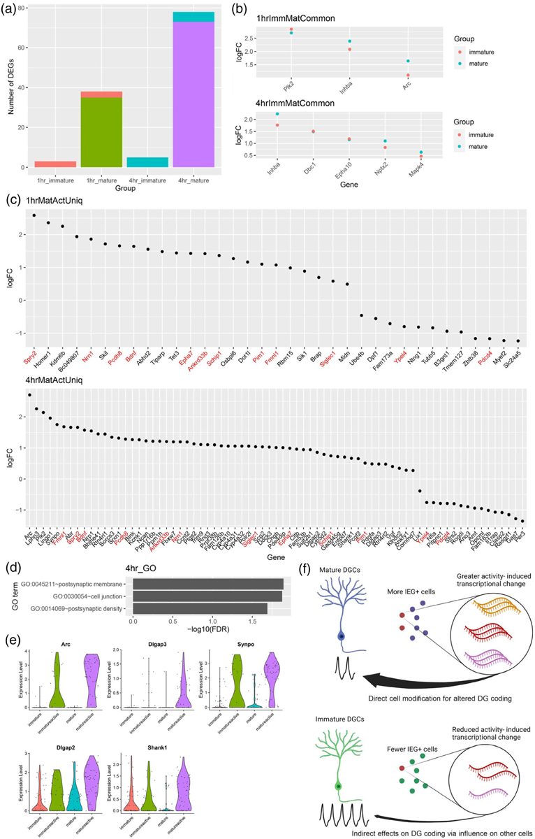
FIGURE 3.

Immature DG neurons show activity-related changes in transcription of only a subset of genes observed in mature. (a) Number of differentially expressed genes (DEGs) between Active and Inactive nuclei based on maturation status and time point, for example, 1-hr immature indicates the number of DEGs between Immature Active and Immature Inactive cells isolated at 1 hr after the start of NE. Coral bar indicates genes shared between 1-hr immature and 1-hr mature comparisons. Blue bar indicates genes shared between 4-hr immature and 4-hr mature comparisons. (b) Log2 fold change (logFC) between active and inactive nuclei of DEGs common to both immature and mature nuclei from A (ImmMatCommon genes). (c) FC of DEGs uniquely identified in mature, but not immature, active, and inactive nuclei comparisons (Uniq genes). DEGs detected in mature cells at both 1 and 4 hr after the start of NE are labeled in red. No DEGs were uniquely identified in immature cells. (d) GO terms significantly enriched among DEGs uniquely identified in mature cells 4 hr after activation. (e) Violin plots of DEGs presented in both “postsynaptic membrane” and “postsynaptic density” terms from (d). (f) Model for contribution of immature DGCs to activity-dependent changes (created with BioRender.com). Mature cells respond by engaging cell intrinsic mechanisms of transcriptional change, whereas physiologically hyperactive immature cells may contribute more indirectly by influencing other cells. DG, dentate granule; DGC, dentate granule cell; NE, novel environment.
